

Development and validation of a prediction model for lymph node metastasis in thyroid cancer: integrating deep learning and radiomics features from intra- and peri-tumoral regions
- PMID: 40771372
- PMCID: PMC12322773
- DOI: 10.21037/gs-2025-50
Development and validation of a prediction model for lymph node metastasis in thyroid cancer: integrating deep learning and radiomics features from intra- and peri-tumoral regions
Abstract
Background: Current preoperative imaging methods, such as ultrasound, are limited by operator dependency and suboptimal sensitivity for detecting central lymph node metastasis (CLNM). This study aimed to propose a method that integrates deep learning and radiomics to accurately predict lymph node metastasis in thyroid cancer by analyzing intra- and peri-tumoral imaging features, thereby improving the preoperative prediction accuracy.
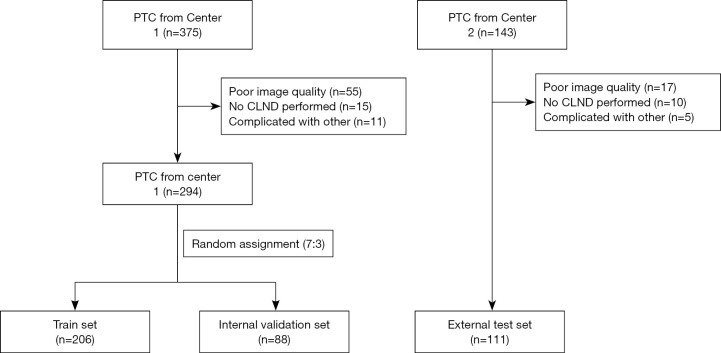
Methods: From July 2020 to June 2022, 405 patients diagnosed with PTC were enrolled from two centers: Center 1 (Shanghai Sixth People's Hospital) with 294 patients divided into a training set (n=294) and an internal validation set, and Center 2 (Tongji Hospital Affiliated to Tongji University) with 111 patients as the external test set. Postoperative pathological confirmation served as the reference standard for CLNM diagnosis. A total of 1,561 radiomics features and 2,048 deep learning features were extracted from intra- and peri-tumoral regions of each ultrasound image. Feature selection was performed using analysis of variance (ANOVA) and least absolute shrinkage and selection operator (LASSO), resulting in the selection of relevant features for constructing support vector machine (SVM) models. Additionally, radiomics-deep learning fusion models were developed by combining selected radiomics and deep learning features.
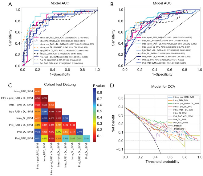
Results: Among 405 patients (mean age: 46.59±12.74 years; 68.6% female), 171 exhibited CLNM, highlighting the clinical urgency for accurate prediction. Among the 405 patients, 171 exhibited CLNM. The radiomics models demonstrated area under the curve (AUC) values of 0.760 in internal validation and 0.748 in the external test cohort. The deep learning models demonstrated improved performance with AUCs of 0.794 and 0.756 in the internal and external test sets. Notably, the highest AUC values of 0.897 (internal validation) and 0.881 (external test set) were obtained by the radiomics-deep learning fusion SVM model incorporating both intra- and peri-tumoral regions. DeLong's test confirmed statistically significant improvements (P<0.05) of the fusion model over the intra-tumoral radiomics model (P=0.008), intra-tumoral deep learning model (P=0.005), and combined intra-tumoral radiomics-deep learning model (P=0.01). However, no significant differences were observed compared to the combined intra- and peri-tumoral deep learning model (P=0.17). Decision curve analysis indicated that the fusion model offers greater clinical utility in predicting CLNM.
Conclusions: The integration of radiomics and deep learning features significantly enhances the diagnostic performance for predicting CLNM in papillary thyroid carcinoma (PTC). The radiomics-deep learning fusion SVM model outperforms individual radiomics and deep learning models, demonstrating substantial potential for clinical application in improving surgical decision-making and patient management. The fusion model could reduce unnecessary central lymph node dissections (CLNDs) and improve surgical planning by providing personalized risk stratification.
Keywords: Papillary thyroid carcinoma (PTC); central lymph node metastasis (CLNM); deep learning; intra-tumoral; peri-tumoral.
Copyright © 2025 AME Publishing Company. All rights reserved.
Conflict of interest statement
Conflicts of Interest: All authors have completed the ICMJE uniform disclosure form (available at https://gs.amegroups.com/article/view/10.21037/gs-2025-50/coif). The authors have no conflicts of interest to declare.
Figures

References
LinkOut - more resources
Full Text Sources