

Advances in ocular aging: combining deep learning, imaging, and liquid biopsy biomarkers
- PMID: 40771477
- PMCID: PMC12325013
- DOI: 10.3389/fmed.2025.1591936
Advances in ocular aging: combining deep learning, imaging, and liquid biopsy biomarkers
Abstract
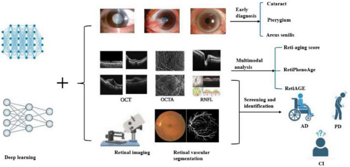
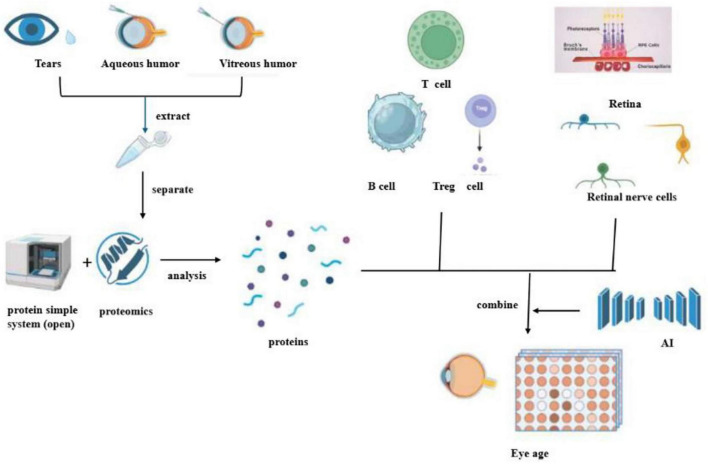
Ageing is a significant risk factor for a wide range of human diseases. Yet, its direct relationship with ocular ageing as a marker for overall age-related diseases and mortality still needs to be explored. Non-invasive and minimally invasive methods, including biomarkers detected through ocular imaging or liquid biopsies from the aqueous humour or vitreous body, provide a promising avenue for assessing ocular ageing. These approaches are particularly valuable given the eye's limited regenerative capacity, where tissue damage can result in irreversible harm. In recent years, artificial intelligence (AI), particularly deep learning, has revolutionized medical research, offering novel perspectives on the ageing process. This review highlights how integrating deep learning with advanced imaging and liquid biopsy biomarkers has become a transformative approach to understanding ocular ageing and its implications for systemic health.
Keywords: age-related eye diseases; deep learning; imaging; liquid biopsy; ocular aging.
Copyright © 2025 Zhang, Li and Li.
Conflict of interest statement
The authors declare that the research was conducted in the absence of any commercial or financial relationships that could be construed as a potential conflict of interest.
Figures
Similar articles
-
Short-Term Memory Impairment.2024 Jun 8. In: StatPearls [Internet]. Treasure Island (FL): StatPearls Publishing; 2025 Jan–. 2024 Jun 8. In: StatPearls [Internet]. Treasure Island (FL): StatPearls Publishing; 2025 Jan–. PMID: 31424720 Free Books & Documents.
-
A deep learning approach to direct immunofluorescence pattern recognition in autoimmune bullous diseases.Br J Dermatol. 2024 Jul 16;191(2):261-266. doi: 10.1093/bjd/ljae142. Br J Dermatol. 2024. PMID: 38581445
-
Recent advances in liquid biopsy for precision oncology: emerging biomarkers and clinical applications in lung cancer.Future Oncol. 2025 Aug 5:1-19. doi: 10.1080/14796694.2025.2542051. Online ahead of print. Future Oncol. 2025. PMID: 40762271 Review.
-
Can a Liquid Biopsy Detect Circulating Tumor DNA With Low-passage Whole-genome Sequencing in Patients With a Sarcoma? A Pilot Evaluation.Clin Orthop Relat Res. 2025 Jan 1;483(1):39-48. doi: 10.1097/CORR.0000000000003161. Epub 2024 Jun 21. Clin Orthop Relat Res. 2025. PMID: 38905450
-
The Lived Experience of Autistic Adults in Employment: A Systematic Search and Synthesis.Autism Adulthood. 2024 Dec 2;6(4):495-509. doi: 10.1089/aut.2022.0114. eCollection 2024 Dec. Autism Adulthood. 2024. PMID: 40018061 Review.
References
Publication types
LinkOut - more resources
Full Text Sources