Review
. 2025 Jul 23:16:1613760.
doi: 10.3389/fmicb.2025.1613760. eCollection 2025.

Advances in adhesion-related pathogenesis in Mycoplasma pneumoniae infection

Affiliations
Review

Advances in adhesion-related pathogenesis in Mycoplasma pneumoniae infection

Bingyue Sun et al. Front Microbiol. .

Abstract

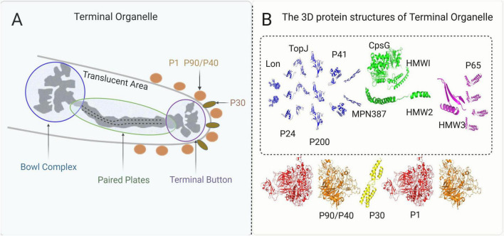
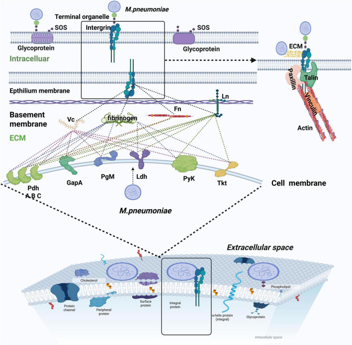
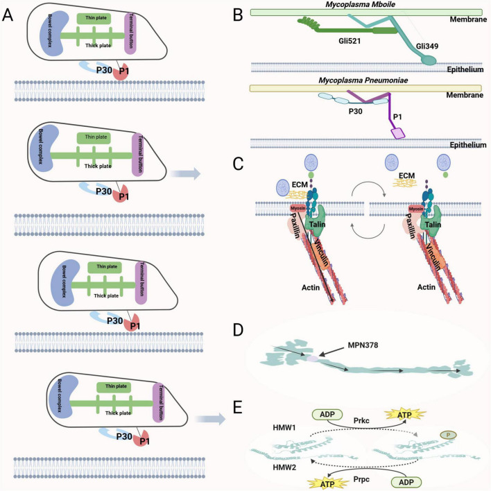
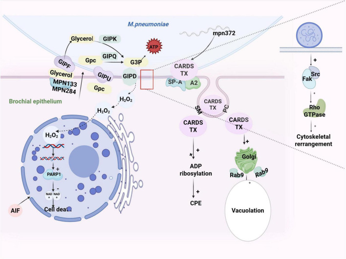
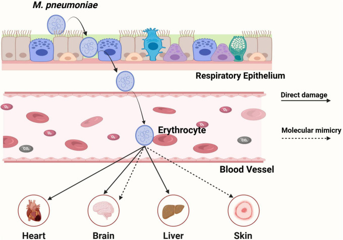
Mycoplasma pneumoniae is a leading cause of community-acquired pneumonia (CAP) and upper respiratory tract infections, particularly in children and immunocompromised individuals. The growing global prevalence of macrolide-resistant M. pneumoniae (MRMP) further emphasizes the urgent need to elucidate its pathogenic mechanisms. Among these, adhesion plays a central role, serving as a prerequisite for colonization and disease progression, and thus warrants detailed investigation. The terminal organelle of M. pneumoniae mediates both adhesion and gliding motility, facilitating colonization, tissue invasion, and potential systemic spread. In the lung, adhesion triggers cytotoxic effects through the release of hydrogen peroxide (H2O2) and CARDS toxin (CARDS TX), promotes excessive inflammatory responses, and enables immune evasion via antigenic variation. Extrapulmonary manifestations may also arise either from direct bacterial dissemination or autoimmune responses induced by molecular mimicry between bacterial and host antigens. In addition, recent advances suggest that therapies and vaccines directed at the adhesion mechanism of M. pneumoniae may offer promising strategies for combating MRMP infections. Although progress has been made, the adhesion-related pathogenesis of M. pneumoniae, as well as the prospects for therapies and vaccines targeting this mechanism, remains incompletely defined. This review synthesizes current insights into adhesion-mediated mechanisms and highlights emerging therapeutic strategies targeting adhesion, aiming to support more effective treatment and prevention of M. pneumoniae infection.

Keywords: M. pneumonia; adhesion; terminal organelle; treatment; vaccines.


Conflict of interest statement

The authors declare that the research was conducted in the absence of any commercial or financial relationships that could be construed as a potential conflict of interest.

Figures

Diagram illustrating the terminal organelle of Mycoplasma pneumoniae. Panel A shows its components, including the bowl complex, paired plates, terminal button, and proteins P1, P90/P40, and P30. Panel B presents the 3D protein structures: Lon, TopJ, P41, CpsG, HMW1, HMW2, HMW3, P65, and others, displayed in blue, green, and purple, with P90/P40, P30, and P1 highlighted below.
FIGURE 1
(A) A pattern map of the terminal organelle’s structures. (B) The structures of the proteins in the terminal organelle and their relative positions. The 3D structures of the proteins are visualized by PyMOL software based on their amino acid sequences from NCBI, while their relative positions are described based on the core image and recent mapping results from Hamaguchi (2016).
Diagram illustrating the interaction between Mycoplasma pneumoniae and human cell membranes. Elements include terminal organelles, integrins, and various proteins such as vinculin and talin. The image details processes occurring at the epithelium and basement membranes, highlighting connections with extracellular matrix components like fibronectin. Additionally, it shows intracellular components, glycoproteins, and structures like protein channels and phospholipids in the cell membrane, emphasizing the involvement of enzymes and pathways in bacterial adherence and infection.
FIGURE 2
M. pneumoniae employs specialized adherence organelles and glycolytic enzymes to establish adhesion to respiratory epithelial cells. Notably, distinct glycolytic enzymes mediate host cell attachment by interacting with different ECM proteins, as indicated by dashed lines in the schematic representation (Grundel et al., 2016). When adhering to integrins, the pathogen forms an FA-like structure. This structure achieves mechanical stability through adaptor proteins, including talin, vinculin, and paxillin, which anchor the adhesion complex to intracellular actin filaments, creating a robust host-pathogen interface.
“A scientific illustration with five panels labeled A to E. - Panel A: Shows the mechanism involving P30 and P1 proteins interacting with a membrane during various stages. - Panel B: Depicts interactions of Mycoplasma mobile and Mycoplasma pneumoniae with epithelial membranes. - Panel C: Illustrates actin and talin interaction with extracellular matrix (ECM) components across a membrane. - Panel D: Shows a structure labeled MPN378 with a directional arrow. - Panel E: Describes a process with high molecular weight (HMW) proteins interacting with ADP, ATP, and Prkc, Prpc enzymes. Each panel provides insight into cellular and molecular interactions.”
FIGURE 3
(A) The gliding mechanism of M. pneumoniae. P1 adhesin binds to the host cell surface through a catch-pull-release cycle with SOS. During the terminal organelle’s extension, P1 retracts from SOS, and during contraction, P1 binds to SOS tightly, while the iterative extension and retraction of the terminal organelle generate gliding movement. (B) The P1 and P30 proteins of M. pneumoniae may exhibit functional homology to the Gli349 and Gli521 adhesins in M. mobile, suggesting conserved molecular mechanisms underlying mycoplasma motility. (C) From the host’s perspective, when the integrin binds to the substrate, myosin generates force, actin contracts, and the substrate detaches, thereby promoting the sliding of M. pneumoniae on the cell membrane. (D) The internal force direction of the core structure. The terminal organelle forces originate in the bowl complex, travel through MPN387 to the paired plates, and then lead to extension and retraction of the terminal organelle. (E) PrkC promotes the phosphorylation of HMW1 and HMW2 proteins to enhance M. pneumoniae gliding motility, whereas PrpC functions opposite manner.
Diagram illustrating the interaction between Mycoplasma pneumoniae and bronchial epithelium. Shows processes like glycerol metabolism, CARDS toxin release, and effects like cell death, ADP ribosylation, and vacuolation. Key molecules like GIPK, GIPQ, GIPU, GIPD, hydrogen peroxide, ATP, and various proteins are labeled, indicating biochemical pathways and cellular impacts such as cytoskeletal rearrangement and cell death through AIF and PARP1.
FIGURE 4
The adhesion-associated toxin mechanism of M. pneumoniae, including H2O2 and CARDS TX.
Molecular structure illustration showing domains D1, D2, and D3, with highlighted segments. Section A depicts a complex structure with colored markers indicating specific sites. Panel B focuses on ADP ribosylation, detailing amino acids R14, D12, R1, S107, and H36. Panel C highlights a segment crucial for internalization, specifying amino acids Y571 to F591. Arrows denote the function of these regions.
FIGURE 5
The CARDS TX structure mapped by PyMoL with the main pathogenic mechanism. (A) The 3D protein structure of CARDS TX mapped by PyMOL. (B) The N-terminal Arg (R10), Asp (D12), Arg (R14), His (H36), and the mid-region Ser-Thr-Ser (S49-T50-S51) can bind with NAD+ to facilitate ADP ribosylation. (C) The C-terminal (Y571-F591) is integral to proper D3 folding for facilitating vacuolation.
Diagram showing the impact of Mycoplasma pneumoniae on the respiratory epithelium and bloodstream. The bacteria damage the epithelium and enter erythrocytes, indicating direct damage and molecular mimicry. Effects spread to the heart, brain, liver, and skin, highlighting systemic impact.
FIGURE 6
The adhesion-related mechanisms of M. pneumoniae in extrapulmonary infection.

Similar articles

References

    1. Aberg A., Gideonsson P., Bhat A., Ghosh P., Arnqvist A. (2024). Molecular insights into the fine-tuning of pH-dependent ArsR-mediated regulation of the SabA adhesin in Helicobacter pylori. Nucleic Acids Res. 52 5572–5595. 10.1093/nar/gkae188 - DOI - PMC - PubMed
    1. Andreae C. A., Sessions R. B., Virji M., Hill D. J. (2018). Bioinformatic analysis of meningococcal Msf and Opc to inform vaccine antigen design. PLoS One 13:e0193940. 10.1371/journal.pone.0193940 - DOI - PMC - PubMed
    1. Ansari S., Yamaoka Y. (2020). Role of vacuolating cytotoxin A in Helicobacter pylori infection and its impact on gastric pathogenesis. Expert. Rev. Anti. Infect. Ther. 18 987–996. 10.1080/14787210.2020.1782739 - DOI - PubMed
    1. Balasubramanian S., Kannan T. R., Baseman J. B. (2008). The surface-exposed carboxyl region of Mycoplasma pneumoniae elongation factor Tu interacts with fibronectin. Infect. Immun. 76 3116–3123. 10.1128/IAI.00173-08 - DOI - PMC - PubMed
    1. Balish M. F. (2014). Mycoplasma pneumoniae, an underutilized model for bacterial cell biology. J. Bacteriol. 196 3675–3682. 10.1128/JB.01865-14 - DOI - PMC - PubMed

LinkOut - more resources