. 2025 Jul 23:16:1617707.
doi: 10.3389/fimmu.2025.1617707. eCollection 2025.

Ex vivo- generated lymphoid progenitors encompass both T cell and innate lymphoid cell fates

Affiliations

Ex vivo- generated lymphoid progenitors encompass both T cell and innate lymphoid cell fates

Pierre Gaudeaux et al. Front Immunol. .

Abstract

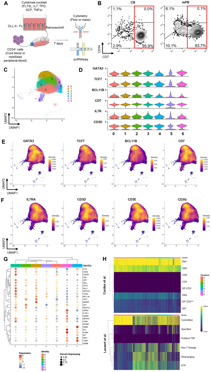
Introduction: We previously established a feeder-free cell therapy platform for the ex vivo generation of lymphoid-primed progenitors using immobilized Delta-like ligand 4 (DLL4). In vivo studies demonstrated that adoptive transfer of these progenitors accelerates T cell reconstitution following thymic engraftment.

Method: To further explore the full therapeutic potential of this cell product, we performed a comprehensive molecular and phenotypic characterization using single cell RNA sequencing and mass cytometry analysis.

Results: Our analysis revealed the presence of distinct cell subsets within the cellular product characterized mainly by commitment to lymphoid lineages. Using integrated transcriptomic analyses to compare these ex vivo-generated progenitors to in vivo human thymocytes, we revealed strong similarities with early stages of T cell development, underscoring the physiological relevance of our system. We also delineated two distinct developmental trajectories within the CD7+ progenitor population: a T cell-oriented path, marked by CD5 upregulation, and an innate lymphoid cell (ILC)-oriented branch, identified by CD161 expression and an ILC-like gene signature. Despite these lineage predispositions, both subsets demonstrated plasticity, retaining the ability to differentiate into both T cells and natural killer (NK) cells in vitro. Additionally, in our experimental setting, we observed that BCL11B, a transcription factor essential for T cell commitment, regulates negatively myeloid cell differentiation while preserving the potential for NK cell development.

Conclusion: These findings underscore the versatility of DLL4-based lymphoid progenitors in generating either T cells or ILCs in response to environmental cues. This research paves the way for innovative cell therapy approaches to treat immune deficiencies and cancer- and age-related immune dysfunctions.

Keywords: T cell progenitors; ex vivo differentiation; hematopoietic stem cell; lymphoid cell development; scRNAseq.


Conflict of interest statement

The Human Lymphohematopoiesis Laboratory and Smart Immune have contracted an industrial collaboration agreement to conduct this project. PG, is a former employee of Smart Immune. JP, RDM, MMC, HS, PR, ON, T-SS are employees of Smart Immune. IA is a co-founder and owns shares of Smart Immune. IA, T-SS, RDM are inventors of patents related to ProTcell production WO2016/055396 and WO2018/146297 which are licensed to Smart Immune. PdlG is an employee and co-founder of GenoSplice. NR is an employee of GenoSplice. FJTS is a scientific advisor for Genewity. The remaining authors declare that the research was conducted in the absence of any commercial or financial relationships that could be construed as a potential conflict of interest.

Figures

Figure 1
Figure 1
Production of ProTcell batches and their characterization at single cell level. (A) Experimental scheme of the ProTcell culture system: CD34+ cells, originated from either CB or mPB, are seeded in a culture unit coated with Retronectin and DLL4-Fc with a cocktail of cytokines in αMAM. After 7 days of culture, T cell progenitors are harvested and analyzed by flow or mass cytometry and single cell RNA sequencing. (B) Contour plot of flow cytometry analysis depicting the expression of CD7 and CD34 in a representative CB (right) or mPB (left) ProTcell product. (C–H) Further transcriptomic analyses were focused on cells positive for the CD7 transcript. (C) scRNAseq data was visualized by UMAP dimension reduction technique and 7 cell clusters were identified (Resolution 0.6). (D) Violin plots showing the normalized expression level of T lymphoid-associated genes in the 7 clusters. Density plots illustrating the gene expression of (E) T cell identity genes GATA3, TCF7, BCL11b and CD7, and (F) markers of developing T lymphoid IL7R, CD3D, CD3E and CD3G. (G) Clustered dot plot depicting gene expression of several markers in the 7 ProTcell clusters. Dot size indicates the percentage of cells expressing marker-specific genes in each cluster. Average expression levels of cluster-specific genes are depicted according to the color scale shown (blue represents low; red represents high). (H) Heatmaps illustrating the similarity scores between each individual cell from ProTcell data and annotated clusters from Cordes et al. (upper panel) and Lavaert et al. (lower panel) data. Scale bar: Z scores of the relative Spearman coefficients. Each column is representative of a single ProTcell scored across each population indicated by the row name.
Figure 2
Figure 2
ProTcell’s subsets express innate lymphoid cell markers delineating a second cell fate. ProTcells were produced from mPB and CB CD34+ cells through an ex vivo system and their transcriptome (scRNAseq) or protein expression (CyTOF) were assessed in a single cell fashion, and restricted to CD7+ progenitors, as described in Figures 1A, B . Violin plots showing the normalized expression level of (A) ILC- and (B) NK-related genes in the 7 clusters. Density plots illustrating the gene expression of (C) the main ILC markers KLRB1, ID2, NFIL3 and KIT and (D) NK-related markers GZMA, GZMB, PRF1 and CD56. (E) CyTOF data was visualized by UMAP dimension reduction technic. UMAP projections illustrating the protein expression of CD7, CD161, KIT and CD5. The green line delineates the CD161-expressing cells along with KIT enrichment, i.e. the ILC-primed cells; the red contour excludes the CD161-expressing cells and includes CD5+ progenitors. (F) UMAP projection showing the pseudotime lineages calculated by Slingshot that describes the progressive transition along CD7+ clusters.
Figure 3
Figure 3
Both CD161+ and CD161- ProTcell subsets retain T and NK cell potential as well as thymus seeding ability. (A–E, G, H) ProTcells were produced from CB CD34+ cells in 7 days as indicated in Figure 1A and sorted by FACS regarding their CD161 expression for further NK or T cell differentiation and functional in vivo experiment. To ensure the consistency, we used the same cell lot and the same sorted cell populations to perform the experiments shown in this figure, including NK cell differentiation, T cell differentiation, and the in vivo engraftment assay. (A) Experimental scheme for CD7+ CD161- (red frame) and CD7+ CD161+ (green frame) cell subset sorting before NK or T cell differentiation. Differentiating cell phenotype was assessed by flow cytometry. (B, C) CD7+ CD161+/- lymphoid progenitors were cultured for 14 days in the presence of IL15 without any feeder cell for NK differentiation. Bar plots represent the mean ± SD of (B) the proportion and (C) number of CD56+ NK-primed cells in the culture. (D, E) CD7+ CD161+/- lymphoid progenitors were co-cultured for 4 weeks with OP9-DLL1 to advance T cell differentiation. Line plots depicting the mean ± SD of the (D) proportion and (E) number of CD3+ developing T cell throughout the culture. *p<0.05 values are for multiple paired Student’s t-test. (F) ProTcells were produced from mPB and CB CD34+ cells and their protein expression (CyTOF) was assessed in a single cell fashion, and restricted to CD7+ progenitors, as described in Figures 1A, B . CyTOF data was visualized by UMAP dimension reduction technic. UMAP projections illustrate the protein expression of several adhesion/homing molecules CCR9, CCR7, CXCR4, CD44, CD62L, SELPLG, ITGA4 and ITGB4. The black line separates the CD161-enriched from the CD161-devoid cells. (G) CD7+ CD161- or CD7+ CD161+ cell subset has been injected separately into NSG neonates (<4-day-old). T cell reconstitution was analyzed by flow cytometry in the thymus at 6-week-post-transplantation. Bar plot representing the human chimerism (human CD45+ cells/human + murine CD45+ cells) and (H) the proportion of mature CD3+ TCRαβ+ T cells in the thymus of mice injected either CD161+ or CD161- ProTcells. ns, non significant for (B, C) Wilcoxon signed-rank test, (D) multiple paired Student's t-test, (G, H) Mann-Whitney U test.
Figure 4
Figure 4
Activation of BCL11B regulatory elements does not restrict NK cell potential. (A) Experimental design for assessing BCL11B role in human T cell commitment. EGFP-BAC reporter was integrated at exon 1 of endogenous BCL11B gene in hPSC, resulting in a monoallelic disruption of this gene and the creation of BCL11B-EGFP reporter cell line. BCL11B-EGFP hPSCs were differentiated into hematopoietic progenitors (HP) for 9 days. The generated HP cells were isolated for early T cell differentiation on OP9-DLL4 for 14 more days. At this time, T9+T14, BCL11B-EGFP negative and positive progenitor cells had been sorted by FACS and secondary cultures were performed to assess their myeloid, NK and T cell potential. Phenotypes were assessed at each stage by flow cytometry for myeloid, lymphoid, NK or T membrane markers. (B) Representative dot plot of flow cytometry analysis of BCL11B-EGFP hPSC-derived progenitors upon sorting at day T9+T14 after culture on OP9-DLL4 for 14 days. At this stage, CD45+ CD56- CD7+ cells are clearly distributed into two populations, according to BCL11B-EGFP expression. (C, D) BCL11B-EGFP neg/+ progenitors were co-cultured for 10 days with OP9-DLL4 with IL15 for NK differentiation. (C) Representative contour plots of flow cytometry analysis illustrating the expression of CD56, delineating NK differentiation, and CD16, translating NK maturation. (D) Bar plot representing the mean ± SD of CD56-expressing NK-primed cells in three independent experiments. *p<0.05 values are for paired Student’s t-test. (E, F) BCL11B-EGFPneg/+ progenitors were co-cultured 5 days with OP9 for myeloid differentiation. (E) Representative contour plots of flow cytometry analysis illustrating CD11b expression translating myeloid priming. (F) Bar plot representing the mean ± SD of CD11b-expressing myeloid progenitors in three independent experiments. *p<0.05 values are for paired Student’s t-test. (G, H) BCL11B-EGFPneg/+ progenitors were co-cultured 7 days with OP9-DLL4 devoid of IL15 for T cell differentiation. (G) Representative contour plots of flow cytometry analysis illustrating the expression of CD7 and CD5 translating T cell differentiation. (F) Bar plot representing the mean ± SD of CD7 and CD5-expressing T cell progenitor cells in four independent experiments.

Similar articles

References

    1. Bosch M, Khan FM, Storek J. Immune reconstitution after hematopoietic cell transplantation. Curr Opin Hematol. (2012) 19:324–35. doi: 10.1097/MOH.0b013e328353bc7d, PMID: - DOI - PubMed
    1. Storek J, Geddes M, Khan F, Huard B, Helg C, Chalandon Y, et al. Reconstitution of the immune system after hematopoietic stem cell transplantation in humans. Semin Immunopathol. (2008) 30:425–37. doi: 10.1007/s00281-008-0132-5, PMID: - DOI - PubMed
    1. Bartelink IH, Belitser SV, Knibbe CA, Danhof M, de Pagter AJ, Egberts TC, et al. Immune reconstitution kinetics as an early predictor for mortality using various hematopoietic stem cell sources in children. Biol Blood Marrow Transplant. (2013) 19:305–13. doi: 10.1016/j.bbmt.2012.10.010, PMID: - DOI - PubMed
    1. Chaudhry MS, Velardi E, Malard F, van den Brink MR. Immune reconstitution after allogeneic hematopoietic stem cell transplantation: time to T up the thymus. J Immunol. (2017) 198:40–6. doi: 10.4049/jimmunol.1601100, PMID: - DOI - PubMed
    1. Clave E, Lisini D, Douay C, Giorgiani G, Busson M, Zecca M, et al. Thymic function recovery after unrelated donor cord blood or T-cell depleted HLA-haploidentical stem cell transplantation correlates with leukemia relapse. Front Immunol. (2013) 4:54. doi: 10.3389/fimmu.2013.00054, PMID: - DOI - PMC - PubMed

MeSH terms

LinkOut - more resources