. 2025 Jul 23:16:1571074.
doi: 10.3389/fimmu.2025.1571074. eCollection 2025.

Peripheral monocyte transcriptional signatures of inflammation and oxidative stress in Parkinson's disease

Affiliations

Peripheral monocyte transcriptional signatures of inflammation and oxidative stress in Parkinson's disease

Aaron D Thome et al. Front Immunol. .

Abstract

Parkinson's disease (PD) is a progressive neurodegenerative disorder characterized by dopaminergic neuron loss in the substantia nigra, which is accompanied by immune dysfunction and chronic inflammation. Peripheral monocytes, key players in systemic inflammation, cross the blood-brain barrier and alter PD etiology and progression. To define the role of peripheral monocytes, cross-sectional studies of RNA transcripts isolated from PD monocytes were compared with age- and sex-matched control monocytes. After stratification by Hoehn & Yahr (H&Y) stage, inflammatory transcripts IL-6, IL-1β, ARG1, CD163, and CCR2 were upregulated in PD monocytes and increased with disease burden. Furthermore, PPARGC1A (PGC-1α), GPX4, NFE2L2 (NRF2), and SIRT3 decreased with increasing disease burden, while only SIRT1 expression increased, reflecting oxidative stress and mitochondrial dysregulation. Overall, the PD monocyte transcripts correlated with PD disease burden as monitored by H&Y, UPDRS total, UPDRS Part 3, ADL, and disease duration. This study demonstrated that dysregulation of inflammation and oxidative stress pathways contributed to disease progression in PD. Monocytes may serve as biomarkers for tracking clinical symptoms and could be leveraged as targets for therapeutic intervention.

Keywords: Parkinson’s disease; inflammation; monocytes; neurodegeneration; neuroimmunology; neuroinflammation; oxidative stress; peripheral biomarkers.


Conflict of interest statement

AT is a consultant for Coya Therapeutics, Inc. JT transitioned employment from Houston Methodist to Director of Clinical Development at Coya Therapeutics, Inc. during preparation of the manuscript. AF is a consultant for Coya Therapeutics, Inc. SHA is Chair of the Coya Scientific Advisory Committee. Coya Therapeutics, Inc. has licensed research from Houston Methodist Research Institute. The remaining authors declare that the research was conducted in the absence of any commercial or financial relationships that could be construed as a potential conflict of interest.

Figures

Figure 1
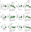
Figure 1
Differential expression of inflammatory and immunoregulatory transcripts in peripheral monocytes from PD patients and through disease progression. Peripheral pan monocytes were isolated from blood of PD patients and controls. (A–C) Increased IL-6 and IL-1β pro-inflammatory transcripts in PD monocytes and with increased H&Y stage scoring. (D) Cell migration and chemokine receptor CCR2 transcripts increased in PD monocytes. (E, F) Increased levels of immunoregulatory markers in PD monocytes compared to controls. Decreasing MRC1/CD206 transcripts with increased H&Y scoring while CD163 transcripts increase with advancing H&Y PD staging. Data shown in Figure 1 represent log2FC transcript values depicted via box plot summary statistics and analyzed using Welch’s t test with Holm-Sidak p-value correction (PD n=60-62; C n= 16). H&Y data analyzed using ordinary one-way ANOVA with Sidak’s multiple comparisons testing (C n=16; H&Y1-1.5 n=14-17; H&Y2-2.5 n=12-13; H&Y3 n=12-15; H&Y4 n=6-9; H&Y5 n=5-6). Statistical significance read as *p<0.05, **p<0.01, and ***p<0.001).
Figure 2
Figure 2
Alterations in metabolic, oxidative stress, and mitochondrial regulatory gene expression in peripheral monocytes of Parkinson’s disease patients. (A) Changes in PD monocyte PPARGC1A/PGC-1α gene transcripts compared with controls and through H&Y disease progression. (B) GPX4 transcripts in PD vs control monocytes that increased early in PD progression staging. (C, D) NRF1 and NRF2 transcripts in PD monocytes and through disease progression staging. Antioxidant NRF2 increased early in PD staging but significantly decreased in later stages. (E) Increased SIRT1 expression in PD monocytes with elevations early in PD progression and remaining elevated through intermediate disease. (F) SIRT3 RNA is slightly decreased early in PD with a significant drop in late stage of disease. Data shown in Figure 2 represent log2FC transcript values depicted via box plot summary statistics and analyzed using Welch’s t test with Holm-Sidak p-value correction for PD vs Control analysis (PD n=60-62; C n= 16). Ordinary one-way ANOVA with Sidak’s multiple comparisons testing utilized for H&Y staging of disease analysis (C n=16; H&Y1-1.5 n=14-17; H&Y2-2.5 n=12-13; H&Y3 n=12-15; H&Y4 n=6-9; H&Y5 n=5-6). Statistical significance read as *p<0.05, **p<0.01, and ***p<0.001).
Figure 3
Figure 3
PD monocyte transcript correlation matrix. Correlation matrix of PD monocyte inflammatory and oxidative stress transcripts as a function of common disease burden assessments done in PD patients. Spearman r correlation values calculated using PD monocyte log2FC expression against patient testing parameters of H&Y, UPDRS, disease duration, and age of the patient. Exact correlation values and significance shown in Table 1 .

Similar articles

References

    1. Armstrong MJ, Okun MS. Diagnosis and treatment of parkinson disease: A review. JAMA. (2020) 323:548–60. doi: 10.1001/jama.2019.22360, PMID: - DOI - PubMed
    1. Dehay B, Bourdenx M, Gorry P, Przedborski S, Vila M, Hunot S, et al. Targeting alpha-synuclein for treatment of Parkinson's disease: mechanistic and therapeutic considerations. Lancet Neurol. (2015) 14:855–66. doi: 10.1016/S1474-4422(15)00006-X, PMID: - DOI - PMC - PubMed
    1. Goedert M. Alpha-synuclein and neurodegenerative diseases. Nat Rev Neurosci. (2001) 2:492–501. doi: 10.1038/35081564, PMID: - DOI - PubMed
    1. Appel SH. Inflammation in Parkinson's disease: cause or consequence? Mov Disord. (2012) 27:1075–7. doi: 10.1002/mds.25111, PMID: - DOI - PubMed
    1. Fathi M, Vakili K, Yaghoobpoor S, Qadirifard MS, Kosari M, Naghsh N, et al. Pre-clinical studies identifying molecular pathways of neuroinflammation in parkinson's disease: A systematic review. Front Aging Neurosci. (2022) 14:855776. doi: 10.3389/fnagi.2022.855776, PMID: - DOI - PMC - PubMed