Review
. 2025 Jul 23:16:1627161.
doi: 10.3389/fimmu.2025.1627161. eCollection 2025.

Immunosuppressive cells in acute myeloid leukemia: mechanisms and therapeutic target

Affiliations
Review

Immunosuppressive cells in acute myeloid leukemia: mechanisms and therapeutic target

Mengnan Liu et al. Front Immunol. .

Abstract

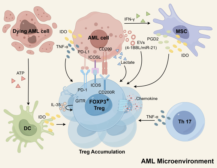
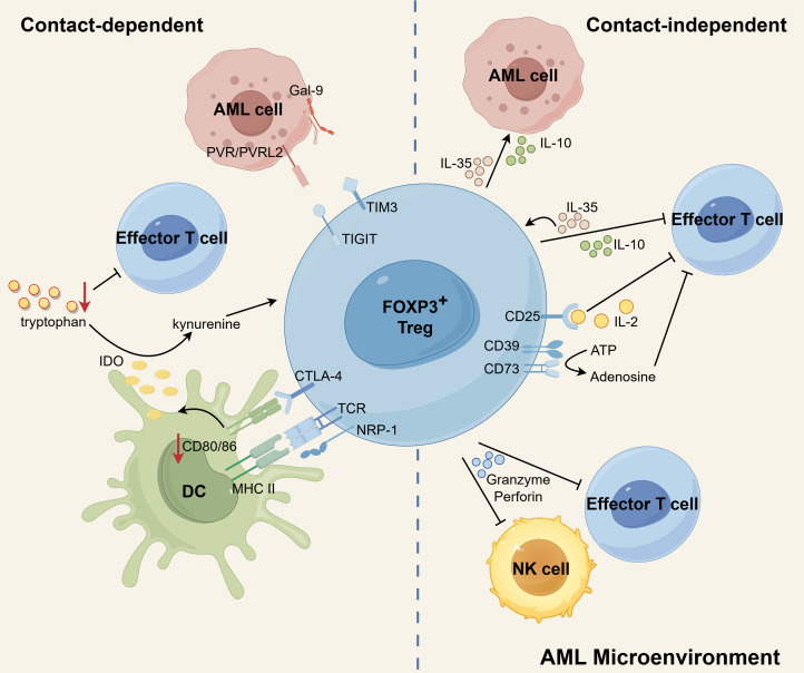
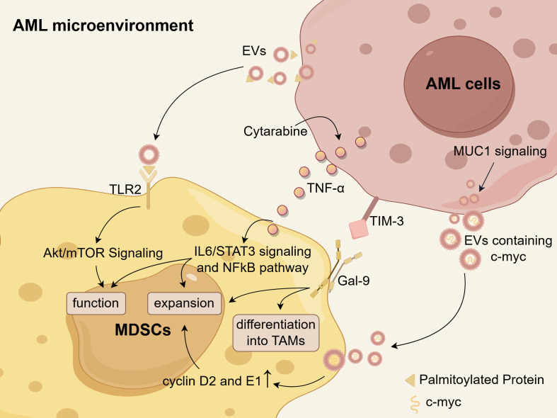
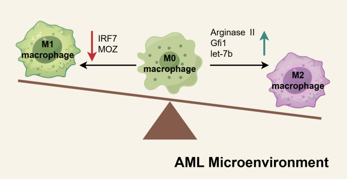
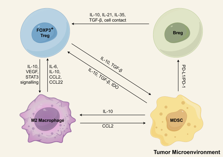
Immunotherapy has emerged as a cornerstone strategy for augmenting therapeutic efficacy in acute myeloid leukemia (AML). The immunosuppressive AML microenvironment, characterized by profound immune dysfunction, critically impairs anti-leukemic immune surveillance. This immunologically hostile niche is principally governed by specialized immunosuppressive cell populations-notably regulatory T cells (Tregs), myeloid-derived suppressor cells (MDSCs), leukemia-associated macrophages (LAMs), and regulatory B cells (Bregs)-which collectively establish an immune-privileged sanctuary for leukemic cells. This review critically examines three fundamental aspects of these immunosuppressive regulators in AML pathogenesis: (1) their recruitment dynamics within the leukemic niche, (2) the molecular mechanisms underlying their immunosuppressive functions, and (3) current and emerging therapeutic approaches designed to neutralize their inhibitory effects. Through this comprehensive analysis, we aim to provide a mechanistic framework for developing more effective immunotherapeutic interventions against AML.

Keywords: acute myeloid leukemia; leukemia-associated macrophages; leukemia-associated neutrophils; myeloid-derived suppressor cells; regulatory B cells; regulatory T cells.


Conflict of interest statement

The authors declare that the research was conducted in the absence of any commercial or financial relationships that could be construed as a potential conflict of interest.

Figures

Figure 1
Figure 1
The mechanisms of Treg cells accumulation in the AML microenvironment. The secretion of EVs by AML cells plays a role in increasing Tregs, as these EVs contain molecules such as miR-21 and 4-1BBL that promote Treg expansion. Additionally, AML cells, DCs, and MSCs can produce IDO, which induces proliferation of Tregs. MSCs also release PGD2 to enhance Treg numbers. Both Th17 cells and AML cells express TNF-α, which supports the expansion of Tregs. Furthermore, Tregs themselves express high levels of IL-35, which can further amplify Treg proliferation. The interaction between AML cells and Tregs through receptor-ligand interactions, including PD-L1/PD-1, ICOSL/ICOS, and CD200/CD200R, also promotes Treg expansion. Tregs possess enhanced chemokine receptors, facilitating robust migration and contributing to their aggregation. Moreover, Tregs have a metabolic advantage as they can utilize lactate for metabolism, indirectly contributing to their accumulation. Schematic figure was drawn by Figdraw (www.figdraw.com).
Figure 2
Figure 2
The immunosuppressive mechanisms of Tregs in AML microenvironment. CTLA-4 expressed by Tregs binds to CD80/86 on DCs, leading to inhibition of co-stimulation of Teffs, downregulation of CD80/86 on DCs, and elevated expression of IDO in DCs. By degrading tryptophan to kynurenines, IDO contributes to the induction of Tregs and the suppression of T-cell responses. Additionally, NRP1 prolongs the MHC-II molecule-dependent interactions between Tregs and DCs, which effectively restricts the recruitment of MHC-II peptides to immune synapses, ultimately inhibiting immune responses. Treg-derived IL-10 diminishes anti-leukemia immunity by suppressing the activity of Teffs. IL-35 released by Treg can suppress Teff functions and proliferation while also expanding a population of inducible Tregs. IL-10 and IL-35 also stimulate the proliferation of AML blasts. Additionally, Tregs can induce cell death in NK and Teff cells by utilizing granzyme and perforin. CD25, expressed on Tregs, allows for continuous uptake of IL-2, leading to the cytokine deprivation-induced apoptosis of Teff cells. Tregs express membrane surface enzymes CD39 and CD73, which can hydrolyze ATP to generate adenosine. Adenosine, in turn, inhibits cytokine production and proliferation of Teff cells, further contributing to the suppressive function of Tregs. In addition, the possible existence of TIGIT-PVR/PVRL2 and TIM3-Gal9 signaling pathways between Tregs and AML cells may contribute to a propensity for leukemia progression. Schematic figure was drawn by Figdraw (www.figdraw.com).
Figure 3
Figure 3
The mechanisms of MDSC accumulation in the AML microenvironment. Palmitoylated proteins present on the surface of AML-derived EVs activate TLR2, triggering the Akt/mTOR-dependent induction of MDSCs. Cytarabine-induced TNF-α secretion from AML cells leads to an expansion of MDSCs and enhances their functions and survival by activating IL-6/STAT3 signaling and NFκB pathways. AML cells secrete EVs containing c-myc in a MUC1-dependent manner, which facilitates MDSC proliferation through upregulation of cyclin D2 and E1. There is a hypothesis that the Tim-3/Gal-9 pathway may promote the expansion of MDSCs and their differentiation into TAMs in AML. Schematic figure was drawn by Figdraw (www.figdraw.com).
Figure 4
Figure 4
The mechanisms of M2-like LAMs accumulated in AML microenvironment. In the AML microenvironment, the factors regulating M1 or M2 macrophage polarization are dysregulated, creating an imbalance in macrophage differentiation. Pro-M1 factors such as IRF7 and MOZ are downregulated in AML macrophages, resulting in diminished M1 activation. Conversely, elevated levels of pro-M2 factors, including arginase II, Gfi1, and let-7b, drive increased polarization toward the M2 phenotype. These shifts culminate in the accumulation of M2-like macrophages within the AML microenvironment, fostering an immunosuppressive milieu that supports leukemia progression. Schematic figure was drawn by Figdraw (www.figdraw.com).
Figure 5
Figure 5
The positive feedback loops of immunosuppressive cells in tumor microenvironment. Tregs, MDSCs, M2 macrophages, and Bregs form interlinked positive feedback loops that reinforce immune suppression and drive immune evasion and AML progression. Key interactions include: (1) Breg-mediated enhancement of Treg function via IL-10, IL-21, IL-35, TGF-β, and direct cell contact; (2) Treg-induced monocyte-to-M2 differentiation through IL-10, VEGF, and STAT3 signaling; (3) M2 macrophage secretion of IL-6/IL-10 for Treg activation and CCL2/CCL22 for Treg recruitment; (4) Reciprocal TGF-β/IL-10-mediated activation between Tregs and MDSCs; (5) MDSC-driven Treg differentiation via IDO upregulation; (6) PD-1/PD-L1-dependent MDSC induction of PD-L1+ Bregs; and (7) IL-10-mediated MDSC promotion of M2 polarization. M2-derived CCL2 further recruits MDSCs to the TME. Schematic figure was drawn by Figdraw (www.figdraw.com).

Similar articles

References

    1. Döhner H, Weisdorf DJ, Bloomfield CD. Acute myeloid leukemia. N Engl J Med. (2015) 373:1136–52. doi: 10.1056/NEJMra1406184, PMID: - DOI - PubMed
    1. Shallis RM, Wang R, Davidoff A, Ma X, Zeidan AM. Epidemiology of acute myeloid leukemia: recent progress and enduring challenges. Blood Rev. (2019) 36:70–87. doi: 10.1016/j.blre.2019.04.005, PMID: - DOI - PubMed
    1. Yi M, Li A, Zhou L, Chu Q, Song Y, Wu K. The global burden and attributable risk factor analysis of acute myeloid leukemia in 195 countries and territories from 1990 to 2017: estimates based on the global burden of disease study 2017. J Hematol Oncol. (2020) 13:72. doi: 10.1186/s13045-020-00908-z, PMID: - DOI - PMC - PubMed
    1. Dong Y, Shi O, Zeng Q, Lu X, Wang W, Li Y, et al. Leukemia incidence trends at the global, regional, and national level between 1990 and 2017. Exp Hematol Oncol. (2020) 9:14. doi: 10.1186/s40164-020-00170-6, PMID: - DOI - PMC - PubMed
    1. Kelly LM, Gilliland DG. Genetics of myeloid leukemias. Annu Rev Genomics Hum Genet. (2002) 3:179–98. doi: 10.1146/annurev.genom.3.032802.115046, PMID: - DOI - PubMed

MeSH terms

LinkOut - more resources