

Three-Dimensional Curved Workflow-Based Optical Coherence Tomography Angiography for Enhancing Atopic Dermatitis Theranostics
- PMID: 40771838
- PMCID: PMC12327028
- DOI: 10.34133/research.0778
Three-Dimensional Curved Workflow-Based Optical Coherence Tomography Angiography for Enhancing Atopic Dermatitis Theranostics
Abstract
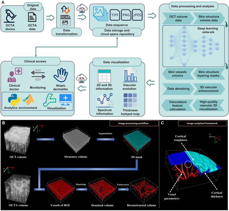
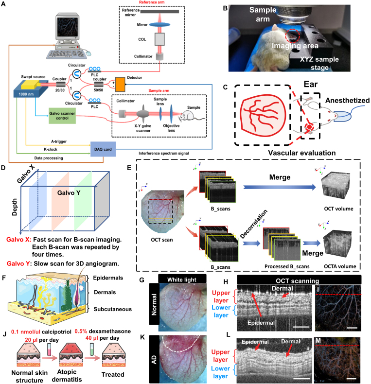
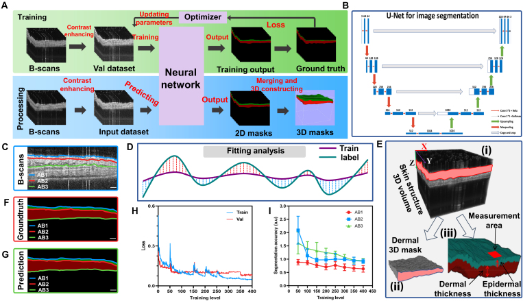
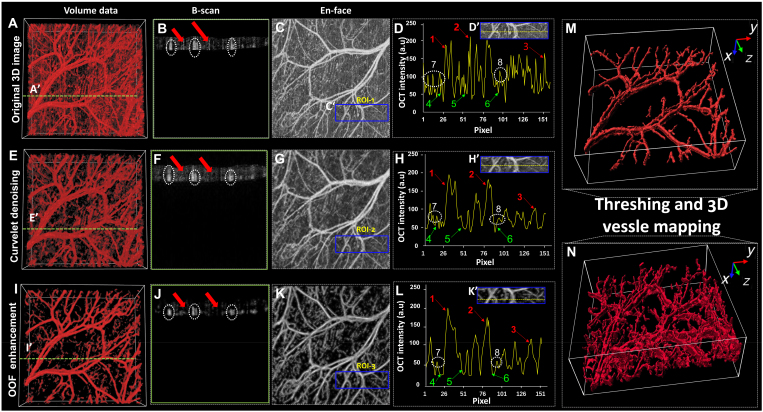
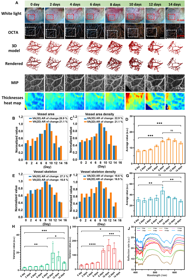
Optical coherence tomography angiography (OCTA) is a major advancement in imaging, offering high-resolution microvascular volumetric images crucial for diagnosing and studying dermatological diseases. However, current data analysis and clinical evaluation criteria primarily rely on 2-dimensional (2D) imaging results, resulting in imprecise diagnoses due to the substantial loss of 3D curved structures and microvascular details. To address this issue, we propose a high-fidelity 3D curved processing workflow that integrates an artificial neural network (ANN) with a 3D denoising algorithm based on the curvelet transform and optimal orientation flow (OOF). This innovative workflow enables precise 3D segmentation and accurate quantification of dermal layer microvasculature in atopic dermatitis (AD) in vivo. Furthermore, the use of 3D multiparametric microvasculature quantitative metrics establishes a robust framework for assessing the efficacy of AD treatments in 3D images. Our study results demonstrate that skin structure imaging and the dynamic evolution of 3D microvasculature align with observed pathological changes. Compared to traditional 2D analysis, the maximum variation rate of 3D curved multiparametric information is approximately 10%. Consequently, our research marks a significant advancement in the accurate quantification of microvasculature in AD development and theranostics, paving the way for the clinical application of OCTA in dermatology.
Copyright © 2025 Junwei Li et al.
Conflict of interest statement
Competing interests: The authors declare that they have no competing interests.
Figures
Similar articles
-
Long-term investigation of 3D tissue detailed structure and multiparametric vascular network properties using OCT/OCTA for guiding atopic dermatitis theranostics.J Transl Med. 2025 Jun 19;23(1):687. doi: 10.1186/s12967-025-06228-5. J Transl Med. 2025. PMID: 40537798 Free PMC article.
-
MarkVCID cerebral small vessel consortium: II. Neuroimaging protocols.Alzheimers Dement. 2021 Apr;17(4):716-725. doi: 10.1002/alz.12216. Epub 2021 Jan 21. Alzheimers Dement. 2021. PMID: 33480157 Free PMC article.
-
Artificial intelligence for diagnosing exudative age-related macular degeneration.Cochrane Database Syst Rev. 2024 Oct 17;10(10):CD015522. doi: 10.1002/14651858.CD015522.pub2. Cochrane Database Syst Rev. 2024. PMID: 39417312
-
Abrocitinib, tralokinumab and upadacitinib for treating moderate-to-severe atopic dermatitis.Health Technol Assess. 2024 Jan;28(4):1-113. doi: 10.3310/LEXB9006. Health Technol Assess. 2024. PMID: 38343072 Free PMC article.
-
Non-orthogonal kV imaging guided patient position verification in non-coplanar radiation therapy with dataset-free implicit neural representation.Med Phys. 2025 Jul;52(7):e17885. doi: 10.1002/mp.17885. Epub 2025 May 19. Med Phys. 2025. PMID: 40387508
References
-
- Guttman-Yassky E, Renert-Yuval Y, Brunner PM. Atopic dermatitis. Lancet. 2025;405(2):583–596. - PubMed
-
- Oykhman P, Dookie J, Al-Rammahy H, de Benedetto A, Asiniwasis RN, LeBovidge J, Wang J, Ong PY, Lio P, Gutierrez A, et al. Dietary elimination for the treatment of atopic dermatitis: A systematic review and meta-analysis. J Allergy Clin Immunol Pract. 2022;10:2657–2666. - PubMed
-
- Ferrara F, Andrea Z, Maurizio C, Roberto L. Atopic dermatitis: Treatment and innovations in immunotherapy. Inflammopharmacology. 2024;32(3):1777–17893. - PubMed
LinkOut - more resources
Full Text Sources
