Review
. 2025 Jul 23:16:1630475.
doi: 10.3389/fphar.2025.1630475. eCollection 2025.

Harnessing nanotechnology for stem-cell therapies: revolutionizing neurodegenerative disorder treatments - a state-of-the-art update

Affiliations
Review

Harnessing nanotechnology for stem-cell therapies: revolutionizing neurodegenerative disorder treatments - a state-of-the-art update

Neevashini Chengebroyen et al. Front Pharmacol. .

Abstract

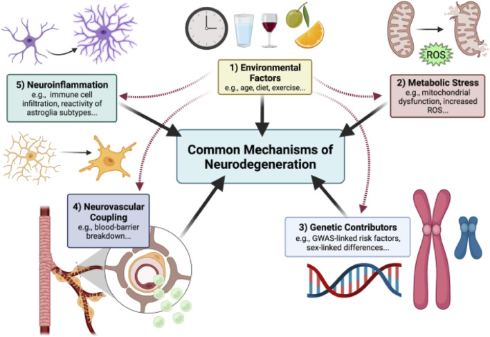
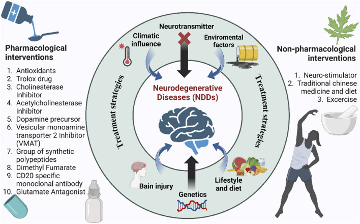
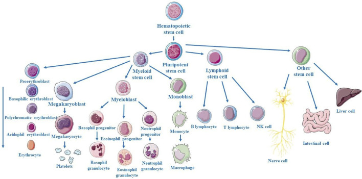
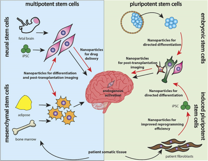
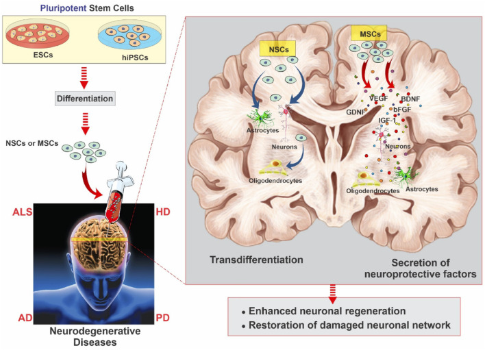
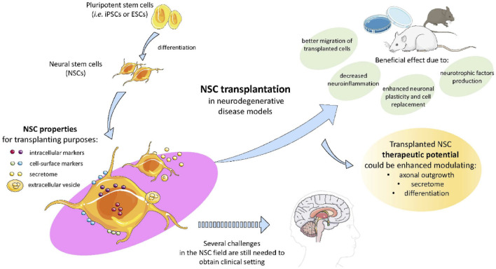
Neurodegenerative disorders, marked by the gradual degeneration and dysfunction of neurons, pose substantial clinical challenges due to the paucity of effective therapeutic strategies and the intricate and multifactorial nature of their underlying pathophysiology. On the other hand nanotechnology, Recent advancements in nanotechnology-driven interventions have significantly augmented the therapeutic potential of stem-cell therapies for the treatment of these complex conditions. Critical limitations in current therapeutic approaches have been highlighted, while potential future directions for their therapy have been outlined. Stem cell types-embryonic, induced pluripotent, and adult neural stem cells-are categorized, with a focus on their unique biological properties and therapeutic potentials in addressing neurodegenerative conditions. The role of nanomaterials in augmenting stem cell generation, scaffold fabrication, and targeted delivery mechanisms is examined, with particular emphasis on the capacity of nanotechnology to enhance regenerative processes and neuroprotective interventions. Nanomaterial-conjugated stem cell therapies are specifically addressed, focusing on their applications in neuronal recovery and treatment monitoring. Challenges associated with stem cell therapies, including ethical considerations, immunogenicity, and the necessity for stringent clinical validation, are critically examined. The integration of nanomedicine with stem cell research is proposed as a promising strategy to overcome these challenges and facilitate the development of novel therapeutic approaches. A comprehensive framework for future research is proposed, focusing on the synergistic integration of nanotechnological advancements with stem cell therapies to improve clinical outcomes and drive innovation in the treatment of neurodegenerative disorders. By integrating existing knowledge and highlighting critical gaps, this review seeks to foster continued research and interdisciplinary collaboration, accelerating progress in this rapidly evolving field.

Keywords: nanomaterial-conjugated regenerative therapy; nanomedicine; nanotechnology; neurodegenerative disorders; neuroprotective-nanotechnology stem-cell therapy; scaffold.


Conflict of interest statement

The authors declare that the research was conducted in the absence of any commercial or financial relationships that could be construed as a potential conflict of interest.

Figures

FIGURE 1
FIGURE 1
Outlined Mechanism of Neurodegenerative disease progression. Neurodegeneration is a result of misfolded protein accumulation due to genetic contributors, environmental factors, metabolic stress causing neuroinflammation and death of neuronal cells, reproduced with permission from ref. (Wareham et al., 2022), copyright 2022, BMC Springer Nature.
FIGURE 2
FIGURE 2
Causes and conventional treatment strategies for NDDs. Central factors include genetics, brain injury, diet, and environmental influences. Pharmacological (left) and non-pharmacological (right) interventions aim to mitigate disease progression.
FIGURE 3
FIGURE 3
Schematic representation of hematopoietic stem cell (HSC) progression from multipotent HSCs to common progenitors, differentiation into various blood cell lineages of distinct myeloid and lymphoid cell types.
FIGURE 4
FIGURE 4
Overview of stem cell categories explored for neurodegenerative therapies. Multipotent neural and mesenchymal stem cells (left) are delivered into the brain alongside nanoparticle systems, while pluripotent embryonic and induced pluripotent stem cells (right) utilize nanoparticles for reprogramming, differentiation, and targeted drug delivery, reproduced with permission from Ref. (Vissers et al., 2019), Copyright 2019, Elsevier.
FIGURE 5
FIGURE 5
Illustrative schematic showing key stem cell types and their differentiation potential into multiple cellular lineages, reproduced with permission from ref (Sivandzade and Cucullo, 2021a). Copyright 2021, MDPI.
FIGURE 6
FIGURE 6
Schematic of neurodegenerative disease modeling using hiPSCs and ESCs. These cells differentiate into neuronal progenitors and MSCs, giving rise to neurons, astrocytes, oligodendrocytes, and other glial lineages. MSCs further support neural repair via secretion of growth and neurotrophic factors that promote angiogenesis, neurogenesis, and immunomodulation, reproduced with permission from ref (Sivandzade and Cucullo, 2021a). Copyright 2021, MDPI.
FIGURE 7
FIGURE 7
The figure illustrates the potential of pluripotent stem cells like iPScs and ESCs in therapeutic potential modulating axonal growth, secretory activity, and differentiation in NDDs. These NSCs transplantation proved to decrease neuroinflammation, enhanced neuronal plasticity and regeneration due to NSCs secretome and extracellular vesicle characteristics, reproduced with permission from ref (De Gioia et al., 2020). Copyright 2020, MDPI.
FIGURE 8
FIGURE 8
MSC-exosomes diminish Aβ plaque accumulation, suppress astrocyte activation, and enhance the expression of memory- and synapse-associated genes. (i) Transgenic or wild-type mice were administered MSC-exosomes or PBS for 4 weeks, after which homogenised brain tissues were subjected to analysis using SDS-PAGE and immunoblotting. A representative Western blot of soluble Aβ, immunoprobed with Aβ antibody and visualised using ECL, is presented. Internal control: β-actin. Quantitative RT-PCR findings of plasticity-associated genes from extracted hemispheres. Representative confocal micrographs of sagittal brain slices, immunolabeled with GFAP, within the hippocampus. Red indicates GFAP; Blue denotes DAPI. Scale bar: 100 μm (top), 50 μm (bottom). Quantification of GFAP + cells within a designated area of interest (Area = 0.05 mm2). Data are presented as mean ± SEM (p = 0.0043; **p < 0.01 according to Student’s t-test) (Chen et al., 2021). (ii) (a) The DiI-labeled OE-MSCs prior to transplantation (red cells), (b) The micrograph of rat calvaria for the transplantation of DiI-labeled stem cells using stereotaxic injection. DiI, 1,1‐dioctadecyl‐3,3,3′3′‐tetramethyl indocarbocyanine perchlorate; OE‐MSCs, olfactory ectomesenchymal stem cells (Simorgh et al., 2019). (iii) (a) Assessment of TH, DAT, PITX3, PaX2, and PaX5 expression using immunohistochemistry. The nuclei were labelled with DAPI (blue), whereas the cells exhibited green staining upon exposure to dopaminergic marker antibodies. The control group (Ctl) exhibited normal expression of dopaminergic markers in the right SNpc region. In contrast, the lesion group demonstrated a significant reduction in the expression of these markers compared to other groups within the same region. Conversely, the experimental group (Exp), which received injections of OE‐MSCs DiI+ (red cells), showed an increase in dopaminergic marker expression relative to the lesion group, with arrowheads indicating double-labeled cells. The proportion of positive responses to midbrain dopaminergic neurone markers (TH, DAT, Pax2, Pax5, and PITX3) was analysed across the groups (control, lesion, and experiment) in the right SNpc, republished with permission from Simorgh et al., 2019, Chen et al., 2021. Copyright 2019, Wiley.
FIGURE 9
FIGURE 9
(i) Immune cells modulate neuropathic pain. After a peripheral nerve injury, immune cells release proinflammatory cytokines (e.g., TNF-α, IL-1β) that interact with receptors and cause pain. Opioid peptides, which help to relieve pain, can also be produced by immune cells. Peripheral opioid receptors are found in the dorsal root ganglia and go to the nerve injury site. Once there, opioid peptides activate receptors and reduce neuropathic pain. (ii) A schematic depicting the mechanism of neuropathic pain healing facilitated by mesenchymal stem cells (MSCs). Several distinct mechanisms are involved: (1) Growth factor secretion; MSCs produce neurotrophic growth factors such as GDNF, VEGF, and BDNF. Neurotrophic growth factors have been shown to promote neuronal survival in cases of neuropathic pain. (2) Reduced neuroinflammation; MSCs significantly influence the immune system and help in wound repair. Interestingly, MSCs can be anti-inflammatory or proinflammatory depending on the environment in which they dwell. In an inflammatory environment, MSCs produce TGF-β1, IDO, and PEG, converting macrophages/microglia from proinflammatory to anti-inflammatory M2 phenotypes. Furthermore, MSCs can stimulate the upregulation of T cells, which are known to play an important role in pain modulation (3), as well as the production of exosomes and microRNAs. MSCs produce biological substances via extracellular vesicles (EVs), which include microvesicles and exosomes. EVs include thousands of proteins, messenger RNA, and/or microRNA, which have been shown to promote neuronal development. (iii) A schematic picture of how stem cells work in peripheral neuropathic pain. (a) Anti-Inflammatory Regulation: Stem cells drive macrophage polarization to anti-inflammatory phenotypes. M2 macrophages increased following MSC therapy, but genes associated with M1 macrophages decreased. (b) Neuroprotection and axonal myelin regeneration. Stem cells also have anti-inflammatory properties via the mitogen-activated protein kinase (MAPK) pathway. After nerve injury, signals from damaged axons activate the extracellular signal-related MAPK signal pathway in Schwann cells. MSCs inhibited the expression of pERK1/2 in CCI-induced dorsal root ganglion (DRG) cells. Furthermore, VEGF, GDNF, and NGF are essential nerve regeneration regulators that can help and stimulate the formation of newly formed nerve fibres. (iv) Mechanisms linked with nerve damage at the synapse of peripheral nerves and spinal cord dorsal horn neurones. (a) Weakened and reversed central sensitisation. Following nerve damage, the release of excitatory amino acids (glutamate) in the spinal dorsal horn significantly increased, and the excitatory N-methyl-d-aspartate (NMDA) receptor (NMDAR) is continuously activated. According to reports, bone marrow stromal cells (BMSCs) can decrease the production of NMDA receptors and protect them from glutamate excitotoxicity, reducing mechanical hyperalgesia. (b) The inhibition of glial cell activation. Stem cells can effectively prevent the activation of glial cells like microglia and astroglia. They also prevent MAPK signal pathway activation in activated glial cells. (c) Reduced apoptosis and autophagy in spinal cord cells. The stimulation of intermediate inhibitory neurons causes the release of GABA, a neurotransmitter that inhibits postsynaptic neurons via membrane hyperpolarization, reproduced with permission from ref. (Joshi et al., 2021), copyright 2021 MDPI.
FIGURE 10
FIGURE 10
Schematic illustration defining the significance of different scaffold architectures in identifying the specific lineage of stem cells. Stem cells cultured on different nanostructured scaffolds produce distinct differentiated cell types, such as (a) bone marrow stem cells grown on nanofibrous PCL scaffold promotes osteogenic fate, (b) embryonic stem cell cultured on nanoscale ridge or groove promote neural fate, (c) tendon stem cells cultured on aligned and random PLLA scaffolds guide tendon and stellate lineage, respectively, (d) mesenchymal stem cells on PDMS promote osteogenic and adipogenic fate, reproduced with permission from ref (Krishna et al., 2016). copyright 2021 MDPI.
FIGURE 11
FIGURE 11
Summarization of key challenges associated with stem cell therapy and nanomedicine highlighting barriers such as clinical translation, ethical and practical conerns in the treatment of NDDs.

Similar articles

References

    1. Abad M., Mosteiro L., Pantoja C., Cañamero M., Rayon T., Ors I., et al. (2013). Reprogramming in vivo produces teratomas and iPS cells with totipotency features. Nature 502 (7471), 340–345. 10.1038/nature12586 - DOI - PubMed
    1. Abdul N., Bai Q., Khan T., Husn M., Khan M., Khan A., et al. (2024). Advances in nanotechnology-assisted photodynamic therapy for neurological disorders: a comprehensive review. Artif. Cells Nanomed Biotechnol. 52 (1), 84–103. 10.1080/21691401.2024.2304814 - DOI - PubMed
    1. Afzal O., Altamimi A. S. A., Nadeem M. S., Alzarea S. I., Almalki W. H., Tariq A., et al. (2022). Nanoparticles in drug delivery: from history to therapeutic applications. Nanomaterials 12 (24), 4494. 10.3390/nano12244494 - DOI - PMC - PubMed
    1. Ahani-Nahayati M., Shariati A., Mahmoodi M., Zekiy A. O., Javidi K., Shamlou S., et al. (2021). Stem cell in neurodegenerative disorders; an emerging strategy. Int. J. Dev. Neurosci. 81, 291–311. 10.1002/jdn.10101 - DOI - PubMed
    1. Ahmad F., Salem-Bekhit M. M., Khan F., Alshehri S., Khan A., Ghoneim M. M., et al. (2022). Unique properties of surface-functionalized nanoparticles for bio-application: functionalization mechanisms and importance in application. Nanomaterials 12 (8), 1333. 10.3390/nano12081333 - DOI - PMC - PubMed

LinkOut - more resources