

Minimally Invasive Surgical Approach for Parotid Sialolith Removal
- PMID: 40772217
- PMCID: PMC12327925
- DOI: 10.7759/cureus.87393
Minimally Invasive Surgical Approach for Parotid Sialolith Removal
Abstract
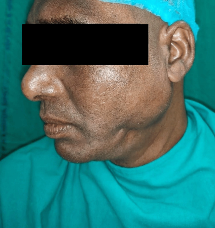
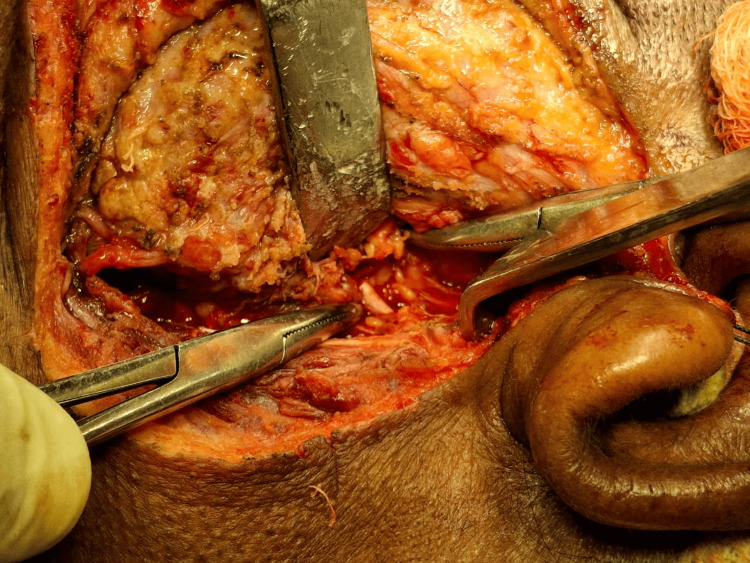
Sialolithiasis is one of the most common diseases that affects the salivary glands and develops stones inside the salivary glands. We describe the case of a 36-year-old man who had been experiencing pain and swelling in the left cheek area for a month, along with discharge in the left posterior buccal vestibular region. A 3.5 mm calculus in the major parotid duct was discovered during the ultrasound scan. Under general anesthesia, the patient had a superficial parotidectomy, and the calculus was successfully removed. This case study illustrates the value of early detection and treatment of parotid sialolithiasis and shows how well superficial parotidectomy works to treat the problem.
Keywords: minimally invasive surgery; parotid duct calculus; parotid sialolithiasis; salivary gland stones; sialolithiasis treatment.
Copyright © 2025, Burman et al.
Conflict of interest statement
Human subjects: Informed consent for treatment and open access publication was obtained or waived by all participants in this study. Conflicts of interest: In compliance with the ICMJE uniform disclosure form, all authors declare the following: Payment/services info: All authors have declared that no financial support was received from any organization for the submitted work. Financial relationships: All authors have declared that they have no financial relationships at present or within the previous three years with any organizations that might have an interest in the submitted work. Other relationships: All authors have declared that there are no other relationships or activities that could appear to have influenced the submitted work.
Figures


Similar articles
-
Spontaneous cutaneous-parotid fistula of the cheek caused by sialolithiasis in a child: a case report.Eur Arch Otorhinolaryngol. 2025 Jan;282(1):543-547. doi: 10.1007/s00405-024-08914-4. Epub 2024 Sep 6. Eur Arch Otorhinolaryngol. 2025. PMID: 39242409
-
Standardization of Basket Use in Sialendoscopy: A Ten-Year Retrospective Study.J Vis Exp. 2025 Jun 6;(220). doi: 10.3791/68055. J Vis Exp. 2025. PMID: 40549674
-
A Comprehensive Study of Combined Approach Sialendoscopy in Managing Salivary Gland Sialolithiasis.Turk Arch Otorhinolaryngol. 2025 Jun 27;63(2):69-74. doi: 10.4274/tao.2025.2024-8-13. Epub 2025 Jun 24. Turk Arch Otorhinolaryngol. 2025. PMID: 40552906 Free PMC article.
-
Does Minimally Invasive Surgery Provide Better Clinical or Radiographic Outcomes Than Open Surgery in the Treatment of Hallux Valgus Deformity? A Systematic Review and Meta-analysis.Clin Orthop Relat Res. 2023 Jun 1;481(6):1143-1155. doi: 10.1097/CORR.0000000000002471. Epub 2022 Nov 4. Clin Orthop Relat Res. 2023. PMID: 36332131 Free PMC article.
-
Endoscopic retrograde cholangiopancreatography versus intraoperative cholangiography for diagnosis of common bile duct stones.Cochrane Database Syst Rev. 2015 Feb 26;2015(2):CD010339. doi: 10.1002/14651858.CD010339.pub2. Cochrane Database Syst Rev. 2015. PMID: 25719222 Free PMC article.
References
-
- Parotid sialolithiasis. Bodner L. J Laryngol Otol. 1999;113:266–267. - PubMed
-
- Sialolithiasis. Williams M. https://www.oto.theclinics.com/article/S0030-6665(05)70175-4/pdf. Otolaryngol Clin North Am. 1999;32:819–834. - PubMed
-
- Parotid sialolithiasis in Stensen´s duct (Article in Spanish) Torres-Lagares D, Barranco Piedra S, Serrera Figallo MA, Iglesias PH, Martínez-Sahuquillo Márquez A, Gutiérrez Pérez JL. https://pubmed.ncbi.nlm.nih.gov/16388301/ Med Oral Patol Oral Cir Bucal. 2006;11:0–4. - PubMed
-
- Sioalolithiasis: a survey on 245 patients and review of the literatura. Lustran J, Regev E, Melamed Y. https://pubmed.ncbi.nlm.nih.gov/2114453/ Int J Oral Maxillofac Surg. 1990;19:135–138. - PubMed
-
- Salivary gland calculi: pain, swelling associated with eating. Levy DM, ReMine WH, Devine KD. JAMA. 1962;181:1115–1119. - PubMed
Publication types
LinkOut - more resources
Full Text Sources