. 2025 Jul 23:19:1558227.
doi: 10.3389/fnins.2025.1558227. eCollection 2025.

Accumulation of TDP-43 causes karyopherin-α4 pathology that characterises amyotrophic lateral sclerosis

Affiliations

Accumulation of TDP-43 causes karyopherin-α4 pathology that characterises amyotrophic lateral sclerosis

Manpreet Singh Atwal et al. Front Neurosci. .

Abstract

Cytoplasmic mislocalisation and nuclear depletion of TDP-43 are pathological hallmarks of amyotrophic lateral sclerosis (ALS), including mutations in the C9ORF72 gene that characterise the most common genetic form of ALS (C9ALS). Studies in human cells and animal models have associated cytoplasmic mislocalisation of TDP-43 with abnormalities in nuclear transport receptors, referred to as karyopherins, that mediate the nucleocytoplasmic shuttling of TDP-43. Yet the relationship between karyopherin abnormalities and TDP-43 pathology are unclear. Here we report karyopherin-α4 (KPNA4) pathology in the spinal cord of TDP-43-positive sporadic ALS and C9ALS patients. Structural analyses revealed the selective interaction between KPNA subtypes, especially KPNA4, with the nuclear localisation signal (NLS) of TDP-43. Targeted cytoplasmic mislocalisation and nuclear depletion of TDP-43 caused KPNA4 pathology in human cells. Similar phenotypes were observed in Drosophila whereby cytoplasmic accumulation of the TDP-43 homolog, TBPH, caused the nuclear decrease and cytosolic mislocalisation of the KPNA4 homolog, Importin-α3 (Impα3). In contrast, induced accumulation of Impα3 was not sufficient to cause TBPH mislocalisation. Instead, targeted gain of Impα3 in the presence of accumulating cytosolic TBPH, restored Impα3 localisation and partially rescued nuclear TBPH. These results demonstrate that cytoplasmic accumulation of TDP-43 causes karyopherin pathology that characterises ALS spinal cord. Together with earlier reports, our findings establish KPNA4 abnormalities as a molecular signature of TDP-43 proteinopathies and identify it as a potential therapeutic target to sustain nuclear TDP-43 essential for cellular homeostasis affected in ALS and frontotemporal dementia.

Keywords: C9ORF72; KPNA4; TDP-43; amyotrophic lateral sclerosis; karyopherin; nuclear import.


Conflict of interest statement

The authors declare that the research was conducted in the absence of any commercial or financial relationships that could be construed as a potential conflict of interest. The author(s) declared that they were an editorial board member of Frontiers, at the time of submission. This had no impact on the peer review process and the final decision.

Figures

Figure 1
Figure 1
KPNA4 pathology in ALS patient spinal cord. (A) DAB conjugated immunohistochemistry of KPNA4 in spinal motor neuron (arrow) of control, sporadic ALS (sALS) and C9ALS cases. KPNA4 immunolabelling reveals nuclear staining with minimal cytoplasmic labelling in control, however nuclear decrease and cytoplasmic accumulation of KPNA4 can be detected in spinal motor neurons (arrows) of sporadic ALS and C9ALS cases. Note, dystrophic neurites or inclusions were not detected by KPNA4 immunohistochemistry in all spinal cord samples; scale bars: 20 μm. (B) Decrease in anti-KPNA4 nuclear staining intensity in sporadic ALS and C9ALS as compared to control cases (*p < 0.05). (C) Severity of reduced nuclear KPNA4 immunostaining is significantly increased in sALS and C9ALS (*p < 0.05; **p < 0.01). (D) Significantly increased cytoplasmic localisation of KPNA4 in sALS and C9ALS cases, compared to control. For each human post-mortem case (control, n = 5; sALS, n = 8; C9ALS, n = 8) a minimum of 25 anterior horn motor neurons were assessed and scored (see Table 1) on a minimum of 3 consecutive sections with anti-KPNA4 immunolabelling. Statistical analyses were performed using one-way ANOVA with Tukey’s multiple comparison post-hoc test; *p < 0.05; **p < 0.01; mean ± SEM shown. Scale bars, 20 μm.
Figure 2
Figure 2
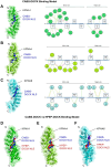
Structural analysis of KPNA binding to TDP-43-NLS. Computational CABS-DOCK analysis reveals TDP-43 nuclear localisation signal (NLS, blue) and KPNA binding (left) together with corresponding molecular contact maps (right) for (A) KPNA1 (PDB: 6WX9, green), (B) KPNA4 (PDB: 5XZX, chartreuse) and (C) KPNA6 (PDB: 4UAD, cyan). The corresponding 2D contact map shows the intermolecular interaction between the minor and major binding site of the NLS (squares) and the active residues of the KPNA proteins (coloured spheres) which is mediated via conserved Tryptophan (W) residues. Interaction types are, electrostatic and salt-bridge (orange), hydrogen bonds (green), hydrophobic contact (pink). Note, all models generated reveal the bipartite binding pattern, with the N-terminal P1´- P5´ residues (KRKMD) interfacing with the minor groove residues (ARM6-8) of the KPNA protein, and the C-terminal P1’ – P5’ residues (VKRAV) dovetailing with the major groove (ARM2-4). (D–F) Comparison between CABS-DOCK (blue peptide) and HPEP-DOCK (red peptide) computational models reveals correspondences for binding prediction between TDP-43-NLS and (D) KPNA1, (E) KPNA4 or (F) KPNA6.
Figure 3
Figure 3
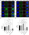
Cytoplasmic accumulation of TDP-43 causes KPNA4 pathology in human cells. (A) HEK293 cells expressing the reporter construct mScarletI-myc either on its own or fused to human wildtype TDP-43 (mScarletI-myc-wtTDP-43) or fused to the nuclear localisation signal depleted form (mScarletI-myc-TDP-43ΔNLS). Imaging of mScarletI and immunolabeling with anti-TDP-43 and anti-KPNA4 reveals nuclear depletion of TDP-43 and cytoplasmic accumulation of KPNA4 and TDP-43 in TDP-43-ΔNLS, but not in wildtype TDP-43 expressing cells. (B) Quantification of the nuclear-cytoplasmic ratio of TDP-43 in mScarletI-myc expressing control cells (CTRL); in mScarletI-myc-wt-TDP-43 expressing cells (wtTDP-43) and in mScarletI-myc-∆NLS-TDP-43 expressing cells TDP-43ΔNLS. (C) Quantification of the nuclear-cytoplasmic ratio of KPNA4 in CTRL, wtTDP-43 and TDP-43ΔNLS expressing cells. Examined samples sizes, mScarletI-myc cells (n = 831), mScarletI-myc-wt-TDP-43 cells (n = 826) and mScarletI-myc-TDP-43ΔNLS cells (n = 1,081). Statistical analyses were performed using one-way ANOVA with Tukey’s multiple comparison post-hoc test; ns, not significant; *p < 0.05; **p < 0.01; mean ± SEM shown. Scale bars, 10 μm.
Figure 4
Figure 4
Accumulating Impα3 does not mislocalise but partially restores nuclear TBPH. (A) Confocal images of L3 salivary gland cells immunolabelled with nuclear marker DAPI (blue), anti-Importin-α3 (green) and anti-MAB414 (red) detecting nuclear pore complex proteins to mark the nuclear rim. In FKH-Gal4/+ and w1118 control flies, anti-Impα3 immunostaining reveals predominantly nuclear localisation of Importin-α3. In FKH>Impα3, Importin-α3 labelling appears amplified in the nucleus whereas cytoplasmic accumulation of TBPH in TBPH-ΔNLS reveals nuclear depletion and cytoplasmic accumulation of Importin-α3, which is partially restored when overexpressing it in TBPH-ΔNLS; FKH>Impα3, flies. (B) Quantification of the nuclear-cytoplasmic ratio of Impα3 in FKH-Gal4/+ (n = 4); w1118 (n = 8); FKH>Impα3 (n = 6); TBPH-ΔNLS (n = 20); TBPH-ΔNLS; FKH>Impα3 (n = 15). (C) Confocal images of salivary gland cells immunolabelled with nuclear marker DAPI (blue), anti-TBPH (green) and anti-MAB414 (red). In FKH-Gal4/+ and w1118 controls, immunostaining reveals predominant nuclear localisation of TBPH. In FKH>Impα3 flies overexpressing Importin-α3 nuclear labelling of anti-TBPH appears slightly amplified. However, in flies expressing TBPH-ΔNLS, immunostaining reveals nuclear depletion (asterisk) and cytoplasmic accumulation of TBPH. Note, overexpressing Importin-α3 in the presence of accumulating cytoplasmic TBPH (ΔNLS; FKH>Impα3) results in partial restoration of nuclear TBPH (asterisk). (D) Quantification of the nuclear-cytoplasmic ratio of anti-TBPH immunolabelling in FKH-Gal4/+ (n = 27); w1118 (n = 31); FKH>Impα3 (n = 20); TBPH-ΔNLS (n = 36); ΔNLS; FKH>Impα3 (n = 9). Statistical analyses were performed using one-way ANOVA with Tukey’s multiple comparison post-hoc test; ns, not significant; *p < 0.05; **p < 0.01; ***. Scale bars: A, 10 μm; C, 5 μm.

References

    1. Al-Sarraj S., King A., Troakes C., Smith B., Maekawa S., Bodi I., et al. (2011). p62 positive, TDP-43 negative, neuronal cytoplasmic and intranuclear inclusions in the cerebellum and hippocampus define the pathology of C9orf72-linked FTLD and MND/ALS. Acta Neuropathol. 122, 691–702. doi: 10.1007/s00401-011-0911-2, PMID: - DOI - PubMed
    1. Arai T., Hasegawa M., Akiyama H., Ikeda K., Nonaka T., Mori H., et al. (2006). TDP-43 is a component of ubiquitin-positive tau-negative inclusions in frontotemporal lobar degeneration and amyotrophic lateral sclerosis. Biochem. Biophys. Res. Commun. 351, 602–611. doi: 10.1016/j.bbrc.2006.10.093, PMID: - DOI - PubMed
    1. Ayala Y. M., Pantano S., D'Ambrogio A., Buratti E., Brindisi A., Marchetti C., et al. (2005). Human, Drosophila, and C. elegans TDP-43: nucleic acid binding properties and splicing regulatory function. J. Mol. Biol. 348, 575–588. doi: 10.1016/j.jmb.2005.02.038, PMID: - DOI - PubMed
    1. Blaszczyk M., Ciemny M. P., Kolinski A., Kurcinski M., Kmiecik S. (2019). Protein–peptide docking using CABS-dock and contact information. Brief. Bioinform. 20, 2299–2305. doi: 10.1093/bib/bby080, PMID: - DOI - PMC - PubMed
    1. Boeynaems S., Bogaert E., Michiels E., Gijselinck I., Sieben A., Jovičić A., et al. (2016a). Drosophila screen connects nuclear transport genes to DPR pathology in c9ALS/FTD. Sci. Rep. 6:20877. doi: 10.1038/srep20877, PMID: - DOI - PMC - PubMed

LinkOut - more resources