. 2025 Aug 15;39(15):e70919.
doi: 10.1096/fj.202500961RR.

Prostaglandin E2 and Progesterone Receptor Coordinately Regulate Primary Cilia for Proper Decidualization

Affiliations

Prostaglandin E2 and Progesterone Receptor Coordinately Regulate Primary Cilia for Proper Decidualization

Chih-Jhen Lee et al. FASEB J. .

Abstract

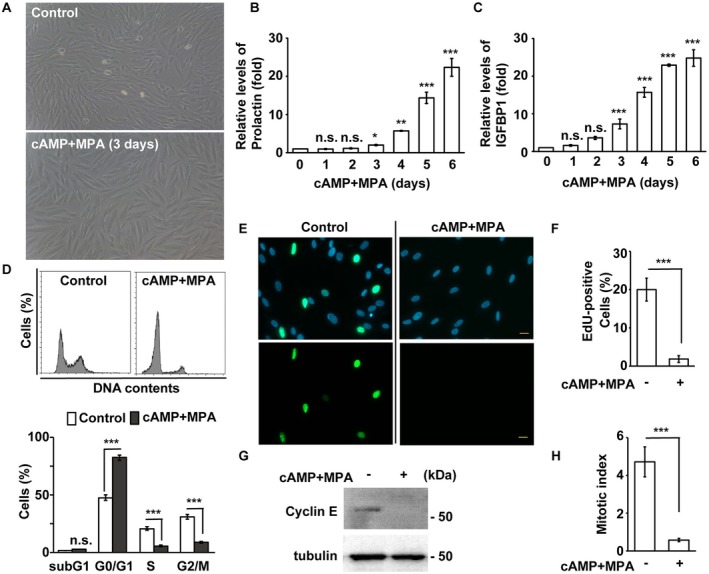
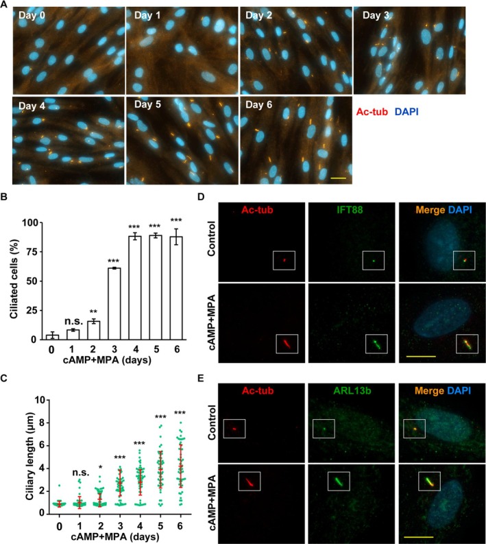
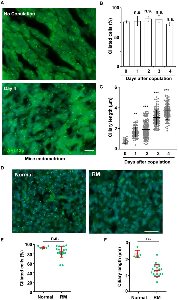
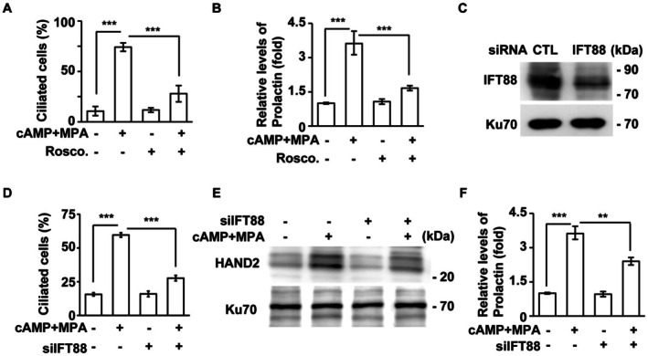
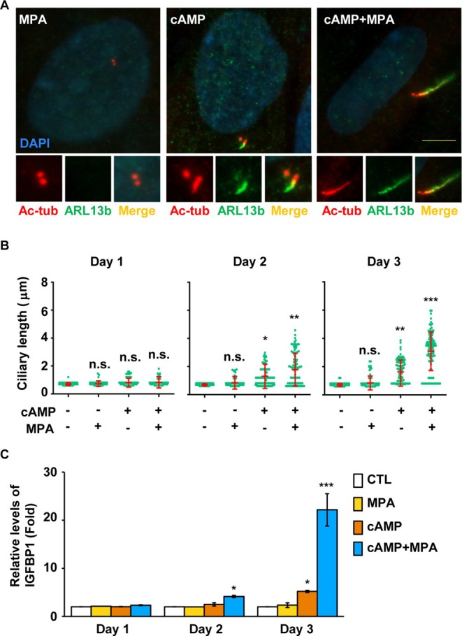
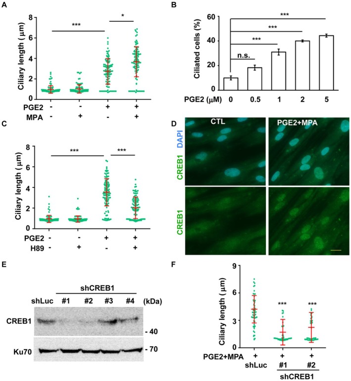
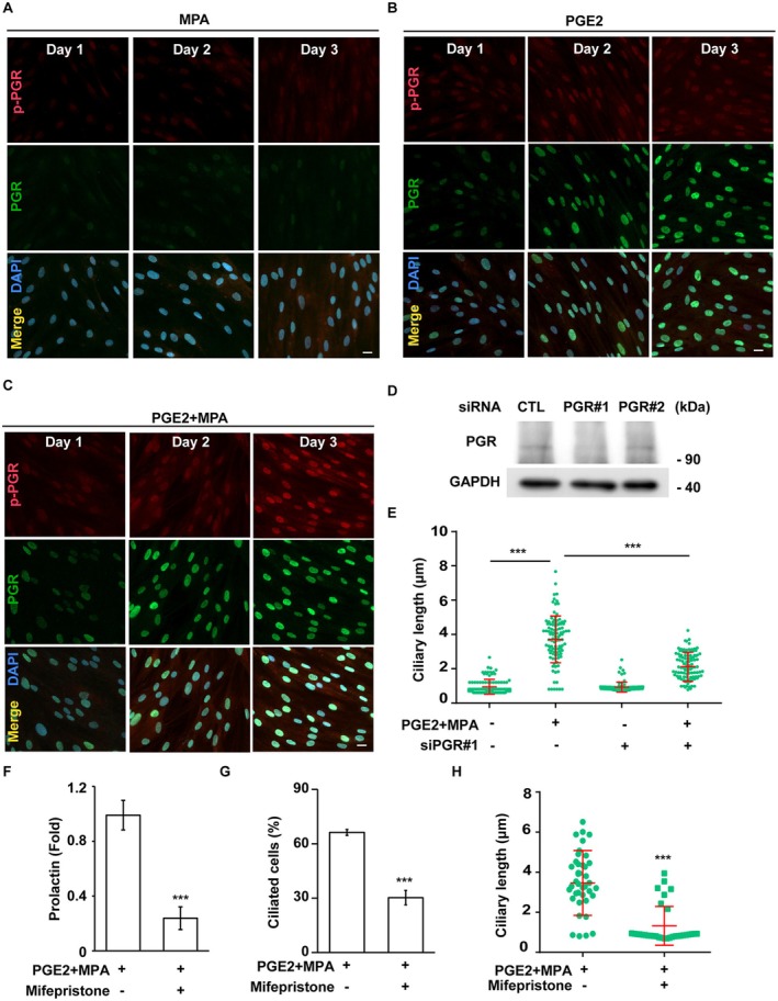
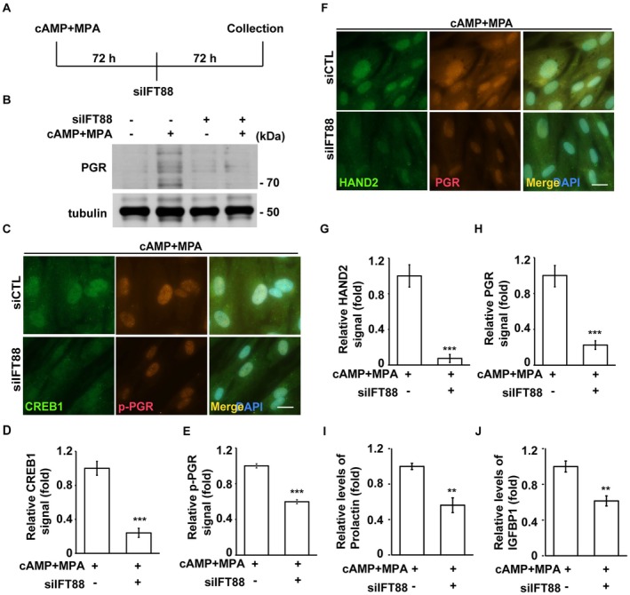
Decidualization, the process by which endometrial stromal cells differentiate into decidual cells in response to prostaglandin E2 (PGE2) and progesterone receptor (PGR) signaling, is essential for proper implantation and placentation. Primary cilia, microtubule-based cellular antennae, contribute to various differentiation processes, including decidualization. In this study, we demonstrated that both the proportion of ciliated cells and ciliary length increased in a time-dependent manner during in vitro decidualization. In a mouse model, after copulation, the proportion of ciliated cells fluctuated, but ciliary length progressively increased over time. Additionally, we observed defective primary cilia in the endometrium of women with recurrent miscarriages. Mechanistically, we found that primary cilia were present before the expression of decidual markers under decidual stimulation. Depletion or inhibition of primary cilia impaired decidualization, highlighting their critical role in this process. Furthermore, we identified the PGE2-PKA-CREB1 axis as a key regulator of ciliary growth and PGR upregulation. Upon progesterone stimulation, active PGR further increased ciliary length, thereby facilitating decidualization. Thus, our study not only establishes a link between ciliary length and decidualization but also elucidates the sequential regulation of ciliary dynamics by PGE2 and PGR in a coordinated manner.

Keywords: cAMP; decidualization; primary cilium; progesterone receptor; prostaglandin E2.


Conflict of interest statement

The authors declare no conflicts of interest.

Figures

FIGURE 1
FIGURE 1
Decidualization induces cell cycle arrest in the G0/G1 phase. (A) The morphology changed in the absence (upper panel, control) or presence (lower panel, cAMP+MPA) of decidual stimulation in T‐HESC cells as shown by light microscopy. Scale bar, 40 μm. (B, C) Increased expressions of decidual markers, Prolactin (B) and IGFBP1 (C), in a time‐dependent manner. (D–H) Decidualization induces cell cycle arrest in the G0/G1 phase. (D) Cell cycle profiles were analyzed using flow cytometry. (E, F) Reduced S phase entry as shown by EdU assay. (E) Lower EdU‐positive cells were observed in cells treated with cAMP and MPA. (F) Quantitative result of (E). (G) The expression of cyclin E was reduced during decidualization. Cells were treated with MPA and cAMP, followed by Western blotting using antibodies against cyclin E and tubulin. (H) The proportion of mitotic cells (mitotic index) was reduced upon cAMP and MPA treatment. n.s.: no significance, *p < 0.05, **p < 0.01, ***p < 0.001. These results are mean ± SD from three independent experiments. At least 300 cells were counted in each individual group.
FIGURE 2
FIGURE 2
Decidual stimulation induces primary cilia formation in T‐HESC cells. (A–C) Co‐treatment of cAMP and MPA promoted primary ciliogenesis in a time‐dependent manner. (A) Primary ciliogenesis was induced by decidual stimulation. Primary cilia were observed by immunofluorescence staining with antibodies against acetylated tubulin (Ac‐tub, red). DNA were stained with DAPI (blue). Scale bar, 20 μm. (B) Quantitative result of the proportion of ciliated cells upon co‐treatment of cAMP and MPA. (C) Quantitative result of the length of primary cilia upon co‐treatment of cAMP and MPA. (D, E) Ciliary structures were examined by immunofluorescence staining. (D) Primary cilia were examined by immunofluorescence staining with antibodies against acetylated tubulin (Ac‐tub, D and E), IFT88 (D) and ARL13b (E). DNA were stained with DAPI (blue). Scale bar, 10 μm. n.s. no significance, *p < 0.05, **p < 0.01, ***p < 0.001. These results are mean ± SD from three independent experiments. At least 300 cells were counted in each individual group.
FIGURE 3
FIGURE 3
Endometrial stroma grows primary cilia in vivo. (A–C) The length of primary cilia increased after mice copulation. (A) Primary cilia in the endometrial stroma after copulation were observed by immunofluorescence staining with an antibody against ARL13b (green). DNA were stained with DAPI. Scale bar, 20 μm. (B) Quantitative result of the proportion of ciliated cells in (A). (C) Quantitative result of the length of primary cilia in (A). (D–F) Defective primary cilia were shown in women with recurrent miscarriages. (D) Primary cilia in normal proliferative phase endometrium (N = 6) or the endometrium of women with recurrent miscarriages (N = 20) were observed by immunofluorescence staining with an antibody against ARL13b (green). DNA were stained with DAPI. Scale bar, 20 μm. (E) Quantitative result of the proportion of ciliated cells in (D). (F) Quantitative result of the length of primary cilia in (D). n.s.: no significance, *p < 0.05, **p < 0.01, ***p < 0.001. These results are mean ± SD from three independent experiments. At least 300 cells were counted in each individual group.
FIGURE 4
FIGURE 4
Disruption of primary cilia inhibits decidualization. (A, B) Inhibition of primary cilia by treating cells with roscovitine (Rosco.) reduced primary cilia and decidualization. (A) Quantitative results of the proportion of ciliated cells in the absence or presence of decidual stimulation (cAMP+MPA) or roscovitine. (B) The expression of the decidual marker Prolactin was reduced in roscovitine‐treated cells. (C–F) Depletion of ciliary gene IFT88 reduced decidualization. (C) IFT88 was successfully depleted by transfecting T‐HESC cells with siRNA against IFT88. Extracts of siRNA‐transfected cells were analyzed by Western blotting with antibodies against IFT88 and Ku70 (loading control). (D) The proportion of ciliated cells were reduced in IFT88‐deficient cells. (E, F) Depletion of primary cilia impaired decidualization. (E) The expressions of decidual marker HAND2 (E) and Prolactin (F) were reduced in IFT88‐deficient T‐HESC cells. **p < 0.01, ***p < 0.001. These results are mean ± SD from three independent experiments. At least 300 cells were counted in each individual group.
FIGURE 5
FIGURE 5
The ciliary length was coordinately regulated by cAMP and MPA. (A–C) MPA treatment did not induce primary cilia, whereas cAMP induced primary cilia growth. Co‐treatment with cAMP and MPA resulted in longer primary cilia. (A) Primary cilia were shown by immunofluorescence staining with antibodies against acetylated tubulin (Ac‐tub, red) and ARL13b (green) in MAP‐, cAMP‐, or cAMP+MPA‐treated T‐HESC cells. DNA were stained with DAPI (blue). Scale bar, 10 μm. (B) Quantitative result of the length of primary cilia of cells in the absence or presence of cAMP or MPA for one, two, or three days. (C) Quantitative result of decidual marker IGFBP1 in the absence or presence of cAMP or MPA for one, two, or three days. CTL: control. n.s.: no significance, *p < 0.05, **p < 0.01, ***p < 0.001. These results are mean ± SD from three independent experiments. At least 300 cells were counted in each individual group.
FIGURE 6
FIGURE 6
PGE2 promotes primary ciliogenesis to facilitate decidualization. (A) PGE2 promoted primary cilia formation. Quantitative result of the length of primary cilia when cells were treated with PGE2, MPA, and PGE2 + MPA. (B) PGE2 promoted primary ciliogenesis in a dose‐dependent manner. Quantitative result of the proportion of ciliated cells in the presence of PGE2 at different doses. (C) Inhibition of PKA by treating cells with H89 reduced the length of primary cilia. Quantitative result of the length of primary cilia in the absence or presence of PGE2 or H89. (D) Co‐treating cells with PGE2 and MPA activated CREB1. Nuclear localization of CREB1 was detected by immunofluorescence staining with antibodies against CREB1 (green). DNA were stained with DAPI (blue). Scale bar, 20 μm. (E, F) CREB1 induced primary cilia. (E) Depletion of CREB1 by infecting cells with lentivirus containing shRNA against CREB1 (shCREB1#1, #2, #3, and #4). Extracts of CREB1 deficient cells were analyzed by Western blotting with antibodies against CREB1 and Ku70. shLuc: shRNA against luciferase, negative control. (F) Depletion of CREB1 reduced primary cilia. Quantitative result of ciliary length of control or CREB1‐deficient cells. n.s.: no significance, *p < 0.05, ***p < 0.001. These results are mean ± SD from three independent experiments. At least 300 cells were counted in each individual group.
FIGURE 7
FIGURE 7
Activation of PGR promotes the elongation of primary cilia during decidualization. (A) Treatment of MPA alone had no effect on PGR. (B) Treatment of cAMP induced, but not activated, the expression of PGR. (C) Co‐treatment of cAMP and MPA induced and activated the PGR. (A–C) Active PGR (phosphorylated PGR, p‐PGR) and PGR were examined by immunofluorescence staining with antibodies against p‐PGR (red) and PGR (green). DNA were stained with DAPI (blue). Scale bar, 20 μm. (D, E) Depletion of PGR reduced the primary cilia upon decidualization. (D) The expression of PGR was successfully depleted. Extracts of PGR‐deficient (T‐HESC cells were transfected with two siRNA against PGR: siPGR#1 and siPGR#2) were analyzed by Western blotting with antibodies against PGR and GAPDH. (E) Depletion of PGR reduced the length of primary cilia. Quantitative result of the length of primary cilia. (F–H) Inhibition of PGR by treating cells with mifepristone reduced primary cilia and decidualization. (F) Quantitative result of the expression of decidual marker prolactin under decidualization. (G) Quantitative result of the proportion of ciliated cells with or without mifepristone under decidualization. (H) Quantitative result of the length of primary cilia with or without mifepristone under decidualization. ***p < 0.001. These results are mean ± SD from three independent experiments. At least 300 cells were counted in each individual group.
FIGURE 8
FIGURE 8
Primary cilia maintain decidualization. (A) The schematic model of experimental designs. (B–J) Depletion of IFT88 impairs decidualization. (B) Western blot analysis showing reduced expression of progesterone receptor (PGR) in cells transfected with siRNA targeting IFT88 (siIFT88), with or without cAMP+MPA treatment. Tubulin was used as a loading control. (C) Immunofluorescence staining of CREB1 and phosphorylated PGR (p‐PGR) in control and siIFT88‐transfected cells treated with cAMP+MPA. Nuclei were counterstained with DAPI (blue). Scale bar, 20 μm. (D, E) Quantification of fluorescence intensity for (D) CREB1 and (E) p‐PGR, measured using ImageJ. (F) Immunofluorescence staining of HAND2 and PGR under the same conditions as (C). Scale bar, 20 μm. (G, H) Quantification of fluorescence intensity for (G) HAND2 and (H) PGR. (I, J) ELISA analysis of (I) prolactin and (J) IGFBP1 secretion following IFT88 depletion. Quantitative data are presented as mean ± SD from three independent experiments. At least 300 cells were analyzed per condition for immunofluorescence quantification. Fluorescence intensities were measured using ImageJ. **p < 0.01, ***p < 0.001.

Similar articles

References

    1. Chao Y. Y., Lin R. C., Su P. J., et al., “Melanophilin‐Induced Primary Cilia Promote Pancreatic Cancer Metastasis,” Cell Death and Disease 16 (2025): 22. - PMC - PubMed
    1. Chao Y. Y., Huang B. M., Peng I. C., et al., “ATM‐ and ATR‐Induced Primary Ciliogenesis Promotes Cisplatin Resistance in Pancreatic Ductal Adenocarcinoma,” Journal of Cellular Physiology 237 (2022): 4487–4503. - PubMed
    1. Tsai Y. C., Kuo T. N., Lin R. C., et al., “MicroRNA‐155‐5p Inhibits Trophoblast Cell Proliferation and Invasion by Disrupting Centrosomal Function,” Molecular Medicine Reports 29 (2024): 85. - PMC - PubMed
    1. Lin R. C., Chao Y. Y., Su M. T., Tsai H. L., Tsai P. Y., and Wang C. Y., “Upregulation of miR‐20b‐5p Inhibits Trophoblast Invasion by Blocking Autophagy in Recurrent Miscarriage,” Cellular Signalling 113 (2024): 110934. - PubMed
    1. Mill P., Christensen S. T., and Pedersen L. B., “Primary Cilia as Dynamic and Diverse Signalling Hubs in Development and Disease,” Nature Reviews Genetics 24 (2023): 421–441. - PMC - PubMed

LinkOut - more resources