Case Reports
. 2025 Aug;13(8):e70126.
doi: 10.1002/mgg3.70126.

Functional Characterization of a Novel Intronic Variant in PIEZO2 in a Recessive Form of Distal Arthrogryposis With Impaired Proprioception and Touch (DAIPT)

Affiliations
Case Reports

Functional Characterization of a Novel Intronic Variant in PIEZO2 in a Recessive Form of Distal Arthrogryposis With Impaired Proprioception and Touch (DAIPT)

Michela Bellardita et al. Mol Genet Genomic Med. 2025 Aug.

Abstract

Background: Distal arthrogryposis with impaired proprioception and touch (DAIPT) is a rare autosomal recessive neurological disease characterized by progressive alteration of mechanosensation. DAIPT is caused by loss of function variants in the PIEZO2 gene that encodes an ionic channel involved in mechanotransduction signaling. Our study started from the case of an 11-year-old boy with skeletal and neuromuscular features suggestive of DAIPT.

Methods: Exome sequencing was performed on the trio. The identified variants in PIEZO2 were validated by Sanger sequencing. Functional assays of the variants were performed by minigene assay in HEK-293 cells and on patient-derived cells using NMD inhibitors.

Results: Trio exome sequencing revealed the presence of two novel variants in the PIEZO2 gene: a nonsense variant (c.1924G>T; p.Glu642*) and an intronic variant of uncertain significance (c.2170-15A>G). Functional analysis demonstrated that the intronic variant disrupts splicing, leading to premature stop codon formation and possible mRNA targeting to nonsense-mediated mRNA decay (NMD). Molecular study in patient-derived fibroblasts with specific NMD inhibitors shows that transcripts derived from both alleles are degraded by NMD, thus confirming the effect of the nonsense variant and enabling reclassification of the VUS.

Conclusion: We present the phenotypic and genetic description of a patient with features suggestive of DAIPT carrying novel biallelic variants in PIEZO2, one of which could be reclassified as pathogenic after functional assays. This study also provides a detailed review of all the published patients with DAIPT and expands the phenotypic and genetic understanding of DAIPT, aiding in diagnosis, genetic counseling, and clinical management.

Keywords: PIEZO2; DAIPT; intronic variant; splicing.


Figures

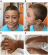
FIGURE 1
FIGURE 1
Clinical presentation of the patient at the age of 5 years old. (a) facial appearance characterized by bitemporal narrowing of the forehead, ptosis, protruding columella, thin upper lip vermilion, pointed chin; (b) flat facial profile with zygomatic hypoplasia, and low‐set ears. (c) right hand showing contractures of fingers; (d) feet with talipes equinovarus, more pronounced to the right. The appearance of hands and feet deformities has worsened over time.
FIGURE 2
FIGURE 2
Open muscle biopsy performed at 11 months. (a) Hematoxylin and Eosin (H&E) staining shows a moderate variability in fiber caliber, with the presence of several hypotrophic fibers with a rounded shape (white arrows). The percentage of centralized nuclei is within normal limits, although some nuclei with a vesicular appearance are noted (black arrow caps). Connective tissue is moderately increased, particularly in the perimysial area; (b) Gomori Trichrome (GT) staining reveals an increased fuchsinophilic punctate pattern in a considerable percentage of fibers (black arrow caps); (c) Nicotinamide adenine dehydrogenase (NADH)‐tetrazolium reductase staining reveals minimal alterations in the myofibrillar network of several fibers, with no specific significance (black arrow caps).
FIGURE 3
FIGURE 3
Identification of two novel variants of PIEZO2. Details of Sanger sequences of the two variants identified in the PIEZO2 gene in the proband and his parents.
FIGURE 4
FIGURE 4
The c.2170‐15A>G variant alters the splicing of PIEZO2 exon 16. (a) Schematic representation of the PCR products obtained by the amplification of the cDNA retro‐transcribed from RNA extracted of Hek‐293 cells transfected 24 h with the following pSPL3 vectors: EV, empty vector; WT, pSLP3 vector with wild type PIEZO2 exon 16 and Var, pSLP3, the vector carrying the variant in PIEZO2 intron 15. Vector exons SD and SA were indicated in light blue, PIEZO2 exon 16 is shown in sky blue and the 14 nucleotides of introns 15 are represented by a red rectangle. The agarose gel electrophoresis of PCR products obtained by the amplification with SD6 and SA2 oligonucleotides is reported in the bottom panel, the corresponding sizes are indicated on the left. (b) Details of the Sanger sequences of minigenes transcripts at the junction between SD/exon16, and exon 16/SA in the WT and Var minigene. MW, molecular weight marker ladders, 1 kb on the left and 100 kb on the right; − no template control. Image created with Biorender.com.
FIGURE 5
FIGURE 5
The identified variants target the PIEZO2 mRNA to NMD in patient's primary fibroblasts. (a) PCR products obtained by the amplification of cDNA derived from patient's fibroblasts treated with 100 μg/mL of Cycloheximide (Chx) or 200 μg/mL of Puromycin (Puro). CTR+ cDNA derived from RNA extracted from human dermal fibroblasts used as positive control. GAPDH was used as housekeeping gene to check the efficiency of the cDNA synthesis. (b) Details of Sanger sequencing of the cDNA, focusing on exon 15 and of the region spanning from exons 15 and 16. Blue asterisk indicates the variant inherited from the mother and the red area represent the insertion of 14 nt of intron 15 into the transcript due to the splicing variant inherited from the father. MW, Molecular Weight 1 Kb. Image created with Biorender.com.
FIGURE 6
FIGURE 6
PIEZO2 variants associated with DAIPT. The “lollipop” diagram illustrates the position of all the variants in PIEZO2 associated with DAIPT so far. Transmembrane domains are indicated in gray and the topological domains in blue. The variants reported are nonsense (light blue), frameshift (green), intronic (red) and missense (pink). The mutations identified in this study are circled in red. Numbers reported on the black line indicate amino acid positions. Data used to generate this chart are provided in the Table S3.

References

    1. al Balushi, A. , al Hinai M., Al Hosni A., et al. 2023. “Distal Arthrogryposis With Impaired Proprioception and Touch: A Novel Variant in PIEZO2 Gene in Omani Patients and a Genotype‐Phenotype Review From a Single‐Center Experience.” Journal of Pediatric Genetics 13, no. 3: 175–180. 10.1055/s-0043-1764127. - DOI - PMC - PubMed
    1. Alhazmi, W. , Qurban A., and Alrashidi E.. 2023. “Case Report of Generalized Lymphatic Dysplasia With PIEZO1 Mutation and Review of the Literature.” Respiratory Medicine Case Reports 44: 101872. 10.1016/j.rmcr.2023.101872. - DOI - PMC - PubMed
    1. Behunova, J. , Gerykova Bujalkova M., Gras G., et al. 2019. “Distal Arthrogryposis With Impaired Proprioception and Touch: Description of an Early Phenotype in a Boy With Compound Heterozygosity of PIEZO2 Mutations and Review of the Literature.” Molecular Syndromology 9, no. 6: 287–294. 10.1159/000494451. - DOI - PMC - PubMed
    1. Burn, T. C. , Connors T. D., Klinger K. W., and Landes G. M.. 1995. “Increased Exon‐Trapping Efficiency Through Modifications to the pSPL3 Splicing Vector.” Gene 161, no. 2: 183–187. 10.1016/0378-1119(95)00223-s. - DOI - PubMed
    1. Carter, M. S. , Doskow J., Morris P., et al. 1995. “A Regulatory Mechanism That Detects Premature Nonsense Codons in T‐Cell Receptor Transcripts In Vivo Is Reversed by Protein Synthesis Inhibitors in Vitro.” Journal of Biological Chemistry 270, no. 48: 28995–29003. 10.1074/jbc.270.48.28995. - DOI - PubMed

Publication types

LinkOut - more resources