. 2025 Aug 1;66(11):19.
doi: 10.1167/iovs.66.11.19.

Artesunate Treats Aspergillus fumigatus Keratitis by Inhibiting Fungal Activity and Activating Autophagy Pathway to Reduce Corneal Inflammation

Affiliations

Artesunate Treats Aspergillus fumigatus Keratitis by Inhibiting Fungal Activity and Activating Autophagy Pathway to Reduce Corneal Inflammation

Yuchen Liu et al. Invest Ophthalmol Vis Sci. .

Abstract

Purpose: The purpose of this study was to investigate the therapeutic effect of artesunate (ART) on fungal keratitis (FK).

Methods: The antifungal properties of ART against Aspergillus fumigatus (A. fumigatus) were confirmed by minimum inhibitory concentration (MIC), biofilm formation inhibition test, propidium iodide fluorescence, and calcium fluorescence white test. The levels of HSP90, BrlA, AbaA, WetA, CnaA, and CrzA were detected by quantitative reverse transcription PCR (qRT-PCR). Cell counting kit 8 (CCK-8) was used to detect the cytotoxicity of ART on RAW 264.7 cells and human corneal epithelial cells (HCECs). Clinical corneal score, hematoxylin and eosin (H&E) staining, and corneal fungal plate counts were used to evaluate the therapeutic effect of ART on FK in mice. The qRT-PCR, ELISA, and Western blot were used to investigate the effect of ART on inflammatory mediator expression during fungal infection.

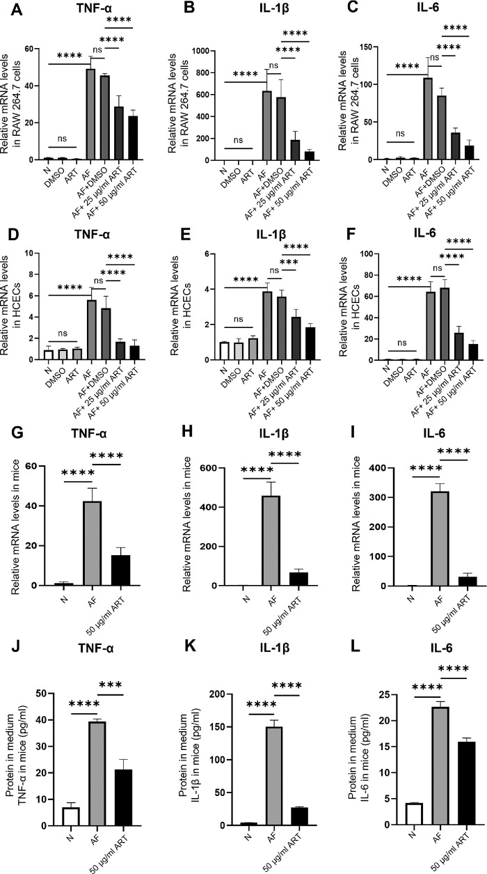
Results: ART inhibited the growth of A fumigatus, biofilm formation, and conidium adhesion in vitro, and destroyed fungal cell walls and cytomembrane. In vivo, ART effectively reduced corneal fungal load. ART could reduce the inflammation of the cornea by reducing the accumulation of inflammatory cells and down-regulating the expression of TNF-α, IL-1β, and IL-6. ART could activate the autophagy pathway to play an anti-inflammatory role in FK by increasing the expression of Beclin-1 and LC3B.

Conclusions: ART inhibits fungal growth by inhibiting biofilm formation, destroying fungal cell walls and membranes, and reducing the expression of HSP90 and calcineurin-related factors, and activates the autophagy pathway to reduce the expression of TNF-α, IL-1β, and IL-6 in FK by upregulating the protein expression of Beclin-1 and LC3B. ART has therapeutic potential for FK.


Conflict of interest statement

Disclosure: Y. Liu, None; X. Tian, None; C. Li, None; J. Lin, None; L. Zhang, None; Q. Wang, None; H. Li, None; Y. Li, None; X. Liu, None; G. Zhao, None

Figures

Figure 1.
Figure 1.
Analysis of antifungal action and drug safety of ART. (A, B) CCK-8 test indicated the viability of RAW 264.7 cells and HCECs treated for 24 hours with ART at concentration gradient (10, 25, 50, 100, 150, 200, and 250 µg/mL). (C) The inhibitory effect of ART on the growth of Aspergillus fumigatus for 36 hours in vitro at concentration gradient (10, 25, 50, 100, 150, 200, and 250 µg/mL). (D) Fluorescein staining of mice ocular surface and (E) scores at 1, 3, and 5 days post-ART (50, 100, and 150 µg/mL) treatment (n = 5/group/time). Mean ± SD (ns, no significance; ****P < 0.0001; DMSO, 0.1%DMSO; AF, Aspergillus fumigatus).
Figure 2.
Figure 2.
The antifungal mechanism of ART against Aspergillus fumigatus. (A) ART affected fungal biofilm formation. Effect of ART on Aspergillus fumigatus conidium (B) without sorbitol and (C) with sorbitol. (D) ART damaged the morphology and integrity of the cytoderm and cytomembrane of Aspergillus fumigatus. PI staining images and calcium fluorescent white staining images of 0.1% DMSO and ART (10, 25, 50, and 100 µg/mL; magnification × 200; Scale = 100 µm). (E, F) H&E staining (400 × magnification) of conidium adherent to HCECs in the 0.1% DMSO group and the 50 µg/mL ART group. Effects of ART on phagocytosis and killing function of RAW 264.7 cells. (G, H) CFU of extracellular active Aspergillus fumigatus. (I, J) Intracellular survival of Aspergillus fumigatus after phagocytosis by RAW 264.7 cells. (K) The mRNA levels of HSP90, BrlA, AbaA, WetA, CnaA, and CrzA in Aspergillus fumigatus exposed to 50 µg/mL ART were detected by qRT-PCR. The above-mentioned values were displayed as mean ± SD (ns, no significance; **P < 0.01; ***P < 0.001; ****P < 0.0001; AF, Aspergillus fumigatus).
Figure 3.
Figure 3.
ART alleviated FK in mice. (A) Therapeutic effect of ART in mice model of infection. (B) Corneal clinical scores of experimental mice (n = 6/group/times) in the 0.1% DMSO group, the 50 µg/mL ART group, and the 100 µg/mL ART group at days 1, 3, and 5. (C, D) On day 3, representative agar plates and the number of fungal colonies of 3 groups mice cornea (n = 3/group). (E) H&E staining of corneal sections of the healthy, AF, 50 µg/mL ART, and 100 µg/mL ART four groups. Corneal sections of mice (n = 3/group) on day 5 were observed by using a microscope (× 400). The above-mentioned values were displayed as mean ± SD (**P < 0.01, ***P < 0.001, ****P < 0.0001; AF, Aspergillus fumigatus).
Figure 4.
Figure 4.
ART reduced the inflammatory expression induced by Aspergillus fumigatus. (AC) Twenty-five or 50 µg/mL ART treated RAW 264.7 cells exposed to Aspergillus fumigatus and co-cultured for 8 hours and the mRNA expressions of TNF-α, IL-1β, and IL-6 in RAW 264.7 cells were detected by qRT-PCR. (DF) The 25 or 50 µg/mL of ART treated HCECs exposed to Aspergillus fumigatus and co-cultured for 8 hours and the mRNA expressions of TNF-α, IL-1β, and IL-6 were detected by qRT-PCR. The corneas of mice were collected 5 days after infection. (GI) The mRNA expressions of TNF-α, IL-1β, and IL-6 in the corneas of mice were detected by qRT-PCR and (JL) the proteins expressions of TNF-α, IL-1β, and IL-6 in corneal homogenate were detected by ELISA. The above-mentioned values were displayed as mean ± SD (ns, no significance, ***P < 0.001, ****P < 0.0001; AF, Aspergillus fumigatus).
Figure 5.
Figure 5.
ART activated the autophagy pathway to exert anti-inflammatory effect in RAW 264.7 cells. RAW 264.7 cells stimulated with inactivated Aspergillus fumigatus mycelia were treated with 50 µg/mL ART or pretreated with MHY1485 and then treated with 50 µg/mL ART for 24 hours. Protein levels of (A, C) Beclin-1, (B, D) LC3B, and β-actin were detected by Western blot. RAW 264.7 cells stimulated with inactivated Aspergillus fumigatus mycelia were treated with 50 µg/mL ART or pretreated with MHY1485 and then treated with 50 µg/mL ART for 8 hours. The mRNA expression of (E) TNF-α, (F) IL-1β, and (G) IL-6 were determined by qRT-PCR. RAW 264.7 cells stimulated with inactivated Aspergillus fumigatus mycelia were treated with 50 µg/mL ART or pretreated with MHY1485 and then treated with 50 µg/mL ART for 24 hours. The mRNA expression of (H) TNF-α, (I) IL-1β, and (J) IL-6 were detected by ELISA. Data were expressed as mean ± SD (ns, no significance; **P < 0.01, ***P < 0.001, ****P < 0.0001; N, RAW 264.7 cells; DMSO, 0.1% DMSO; AF, Aspergillus fumigatus).
Figure 6.
Figure 6.
ART activated the autophagy pathway to exert anti-inflammatory effect in HCECs. The HCECs stimulated with inactivated Aspergillus fumigatus mycelia were treated with 50 µg/mL ART or pretreated with MHY1485 and then treated with 50 µg/mL ART for 24 hours. Protein levels of (A, C) Beclin-1, (B, D) LC3B, and β-actin were detected by Western blot. HCECs stimulated with inactivated Aspergillus fumigatus mycelia were treated with 50 µg/mL ART or pretreated with MHY1485 and then treated with 50 µg/mL ART for 8 hours. The mRNA expression of (E) TNF-α, (F) IL-1β, and (G) IL-6 were determined by qRT-PCR. The HCECs stimulated with inactivated Aspergillus fumigatus mycelia were treated with 50 µg/mL ART or pretreated with MHY1485 and then treated with 50 µg/mL ART for 24 hours. The mRNA expression of (H) TNF-α, (I) IL-1β, and (J) IL-6 were detected by ELISA. Data were expressed as mean ± SD (ns, no significance; *P < 0.05, **P < 0.01, ***P < 0.001, ****P < 0.0001; N, RAW 264.7 cells; DMSO, 0.1% DMSO; AF, Aspergillus fumigatus).

Similar articles

References

    1. Brown L, Leck AK, Gichangi M, Burton MJ, Denning DW. The global incidence and diagnosis of fungal keratitis. Lancet Infect Dis. 2021; 21(3): e49–e57. - PubMed
    1. Ahmadikia K, Aghaei GS, Fallah B, et al.. Distribution, prevalence, and causative agents of fungal keratitis: a systematic review and meta-analysis (1990 to 2020). Front Cell Infect Microbiol. 2021; 11: 698780. - PMC - PubMed
    1. Liu MY, Zhang L, Yin XL, Sun SY. Endophthalmitis associated with fungal keratitis and penetrating injuries in North China. Eur J Ophthalmol. 2020; 30(3): 455–461. - PubMed
    1. Manikandan P, Abdel-Hadi A, Randhir BSY, et al.. Fungal keratitis: epidemiology, rapid detection, and antifungal susceptibilities of fusarium and aspergillus isolates from corneal scrapings. Biomed Res Int. 2019; 2019: 6395840. - PMC - PubMed
    1. Yu B, Li C, Gu L, et al.. Eugenol protects against Aspergillus fumigatus keratitis by inhibiting inflammatory response and reducing fungal load. Eur J Pharmacol. 2022; 924: 174955. - PubMed

MeSH terms