. 2025 Sep 10;16(9):e0179925.
doi: 10.1128/mbio.01799-25. Epub 2025 Aug 7.

Structure of the T9SS PorKN ring complex reveals conformational plasticity based on the repurposed FGE fold

Affiliations

Structure of the T9SS PorKN ring complex reveals conformational plasticity based on the repurposed FGE fold

Xiangan Liu et al. mBio. .

Abstract

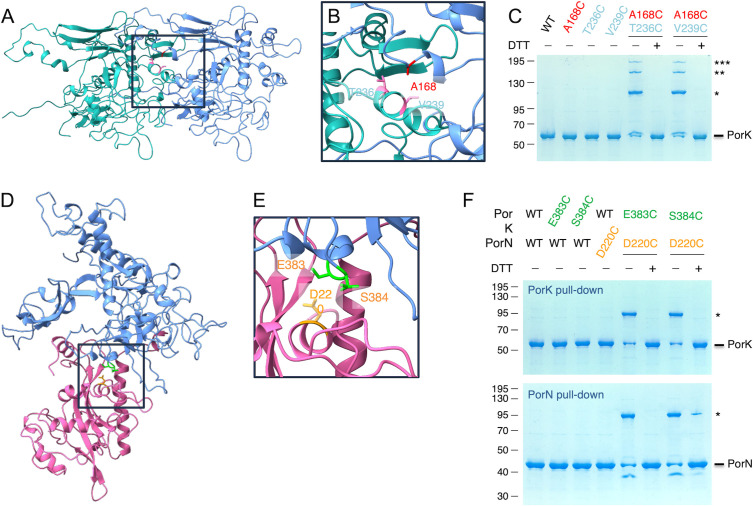
The type IX secretion system (T9SS) is a protein secretion machinery unique to the Bacteroidetes-Chlorobi-Fibrobacteres superphylum, which plays crucial roles in bacterial pathogenesis and gliding motility. It is composed of >15 proteins, including the proton-motive force-dependent PorLM motor, the PorKN ring anchored to the outer membrane, and the Sov translocon. Here, we present the cryo-electron microscopy (EM) structure of the PorKN ring complex from Porphyromonas gingivalis at 3.2 Å resolution. Our structural analysis reveals that PorK contains a repurposed formylglycine-generating enzyme-like fold, which serves as a structural hub for complex assembly rather than enzymatic activity. The complex exhibits a 33-fold symmetry with PorK and PorN assembling two tightly packed and wedged subrings. The structure reveals previously uncharacterized N- and C-terminal helices in PorN that are crucial for PorK binding and complex stability. By combining our high-resolution structure with in situ cryo-electron tomography data, we propose a mechanism whereby PorKN undergoes conformational changes during substrate transport, transitioning between 50° and 90° states relative to the membrane plane. Finally, structural predictions coupled to site-directed disulfide cross-linking identified contacts between PorM and the PorKN ring. Collectively, these findings provide crucial insights into the molecular architecture and dynamic behavior of the T9SS machinery, advancing our understanding of bacterial protein secretion mechanisms.IMPORTANCEThe bacterial type IX secretion system (T9SS) is essential for processes such as gliding motility and secretion of virulence factors. In Porphyromonas gingivalis, a major periodontal pathogen, the T9SS transports over 30 virulence-associated proteins, making it central to disease development. The T9SS core is composed of PorLM motors that are thought to energize the PorKN outer membrane-associated ring. However, the molecular architecture of the PorKN ring has remained unresolved. Here, we present its atomic-resolution cryo-EM structure, revealing a formylglycine-generating enzyme-like fold in PorK that mediates PorK-PorN interactions through specific insertion motifs. Our results show that the ring exhibits intrinsic structural plasticity, including dynamic flexibility and variable stoichiometry. AlphaFold models and disulfide cross-linking experiments further provide information on how PorLM motors are connected to the PorKN ring. These insights redefine our understanding of the T9SS mechanism of action and offer a structural framework for the development of targeted antimicrobial strategies.

Keywords: cryo-EM; surface structures; type 9 protein secretion.


Conflict of interest statement

The authors declare no conflict of interest.

Figures

Fig 1
Fig 1
PorKN rings: single and double forms with varying symmetries. (A) Schematic of the structure and function of the T9SS of P. gingivalis and representative image of PorKN ring particles visualized by cryo-EM. The 2D class averages show single and double rings in various orientations. (B) Top views of PorKN rings in 32-, 33-, and 34-fold symmetries. Corresponding cutaway views are shown in the second row. (C) Tilted views of single PorKN ring (left) and double ring (right) with 33-fold symmetry. Geometric measurements are displayed in the top-middle section.
Fig 2
Fig 2
Cryo-EM structure of PorKN ring. (A) Upper: focus-refined PorKN ring segment at 3.2 Å resolution, with three dissected subunits highlighted in different colors. Lower: corresponding atomic models of three consecutive PorK and PorN subunits. (B) Side view of a single unit of PorK and PorN, along with the corresponding atomic models. The outer membrane’s relative position is indicated.
Fig 3
Fig 3
In vivo validation of PorK-PorK and PorK-PorN interactions. (A) Cryo-EM structure of two contiguous PorK subunits (green and blue). (B) Magnification of a portion of the PorK-PorK interface, highlighting the side chains of the substituted residues: T236 and V239 from the “green” monomer (pink), and A168 from the “blue” monomer (red). (C) Solubilized membrane extracts of E. coli cells producing VSV-G-tagged PorL, FLAG-tagged PorM, Strep-tagged PorN, and 6× His-tagged wild type (WT) or indicated cysteine PorK variants were subjected to pull-down on Ni-NTA resin. The eluate proteins were treated (+) or not (−) with DTT and subjected to 10% acrylamide SDS-PAGE and Coomassie Blue staining. The positions of PorK and PorK multimers (*, **, and ***) are indicated on the right. Molecular weight markers (kDa) are indicated on the left. (D) Cryo-EM structure of a PorK-PorN heterodimer (blue and purple, respectively). (E) Magnification of a portion of the PorK-PorN interface, highlighting the side chains of the substituted residues: PorK E383 and S384 (green) and PorN D220 (orange). (F) Solubilized membrane extracts of E. coli cells producing VSV-G-tagged PorL, FLAG-tagged PorM, Strep-tagged WT or cysteine PorN variants, and 6× His-tagged WT or indicated cysteine PorK variants were subjected to pull-down on Ni-NTA (top panel) or StrepTactin (bottom panel) resins. The eluate proteins were treated (+) or not (−) with DTT and subjected to 10% acrylamide SDS-PAGE and Coomassie Blue staining. The positions of PorK, PorN, and PorK-N complex (*) are indicated on the right. Molecular weight markers (kDa) are indicated on the left.
Fig 4
Fig 4
Interactions between PorK subunits. (A) Structure of three consecutive PorK subunits, with FGE-like folding domains shown in gray and cyan, the major insertion domain in green and pink, and the minor insertion region and variable regions in orange and purple, respectively. (B) The middle PorK subunit is displayed in surface representation. (C) Three inter-subunit interaction sites are highlighted within boxed regions.
Fig 5
Fig 5
Interactions between PorN-PorN and PorK-PorN. (A, B, and C) PorN-PorN interactions. (A and B) Superimposition of the cryo-EM structure of PorN (this study, shown in rainbow) with the crystallographic structure (PDB ID: 7PVH [28]; colored gray) from different perspectives. Variable regions are labeled 1−10. (C) The interaction interface between two PorN subunits occurs between regions 9 and 5 of one unit and regions 2, 3, and 8 of the adjacent unit. (D and E) PorK-PorN interactions. (D) The extended N-terminal helix of PorN fits into a groove of PorK. (E) The extended C-terminal helix of PorN is stabilized by interaction with the VR3 variable region of PorK, shown in purple.
Fig 6
Fig 6
Structural model and in vivo validation of the PorM-PorN interaction. (A and B) Surface (A) and ribbon (B) representations of the AlphaFold2 structural model of PorN (red) with the PorM D4 domain (green). A magnification of a portion of the PorM-PorN interface, highlighting the side chains of the substituted residues: PorM S460 (green) and PorN A241 (orange) are shown as inset (i) in panel B. (C) Solubilized membrane extracts of E. coli cells producing VSV-G-tagged PorL, FLAG-tagged wild type (WT) or cysteine PorM variants, Strep-tagged WT or cysteine PorN variants, and 6× His-tagged PorK were subjected to pull-down on DYKD4K (top panel) or StrepTactin (bottom panel) resins. The eluate proteins were treated (+) or not (–) with DTT and subjected to 10% acrylamide SDS-PAGE and Coomassie Blue staining. The positions of PorM, PorN, and PorM-N complex (*) are indicated on the right. Molecular weight markers (in kDa) are indicated on the left. (D) AlphaFold2 model of the PorM D2-D4 dimer (blue and green) with one copy of PorN (red). (E) AlphaFold2 model of the PorM D2-D4 dimer/PorN plugged into the AlphaFold2 model of PorK2N2. PorM, PorN, and PorK are shown in light green, purple, and green, respectively.
Fig 7
Fig 7
FGE-like fold in PorK-like proteins functioning as a docking hub. Three key insertion sites are highlighted: H-helix (red), V-helix (blue), and B-loop (dark green).

Similar articles

References

    1. McBride MJ, Zhu Y. 2013. Gliding motility and por secretion system genes are widespread among members of the phylum bacteroidetes. J Bacteriol 195:270–278. doi: 10.1128/JB.01962-12 - DOI - PMC - PubMed
    1. Abby SS, Cury J, Guglielmini J, Néron B, Touchon M, Rocha EPC. 2016. Identification of protein secretion systems in bacterial genomes. Sci Rep 6:23080. doi: 10.1038/srep23080 - DOI - PMC - PubMed
    1. Lien YW, Amendola D, Lee KS, Bartlau N, Xu J, Furusawa G, Polz MF, Stocker R, Weiss GL, Pilhofer M. 2024. Mechanism of bacterial predation via ixotrophy. Science 386:eadp0614. doi: 10.1126/science.adp0614 - DOI - PubMed
    1. McBride MJ, Nakane D. 2015. Flavobacterium gliding motility and the type IX secretion system. Curr Opin Microbiol 28:72–77. doi: 10.1016/j.mib.2015.07.016 - DOI - PubMed
    1. Shrivastava A, Patel VK, Tang Y, Yost SC, Dewhirst FE, Berg HC. 2018. Cargo transport shapes the spatial organization of a microbial community. Proc Natl Acad Sci USA 115:8633–8638. doi: 10.1073/pnas.1808966115 - DOI - PMC - PubMed

MeSH terms

Substances

LinkOut - more resources