

[Anatomical variants of blood vessels and their surgical challenges related to oncological right hemicolectomy]
- PMID: 40773097
- PMCID: PMC12457557
- DOI: 10.1007/s00104-025-02342-8
[Anatomical variants of blood vessels and their surgical challenges related to oncological right hemicolectomy]
Abstract
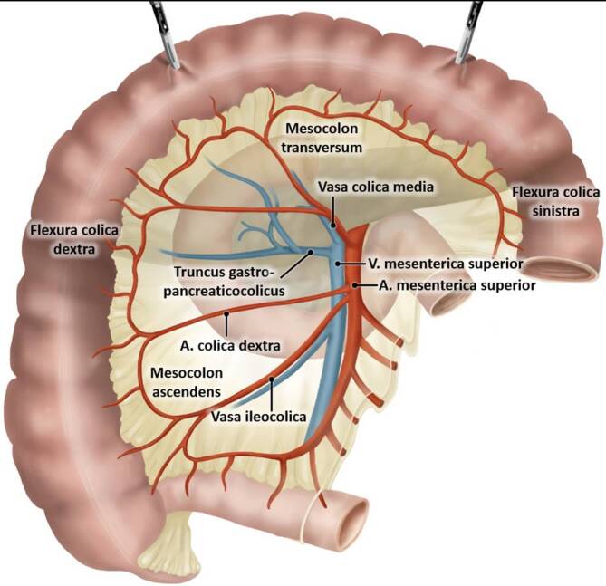
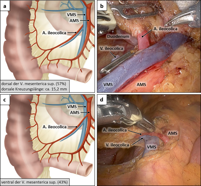
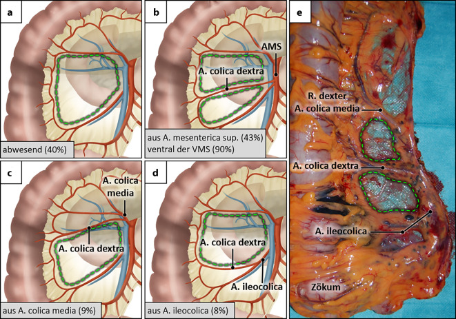
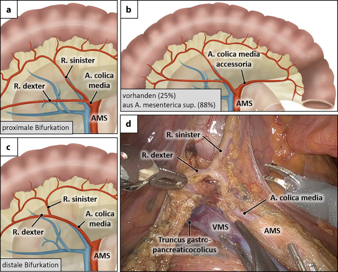
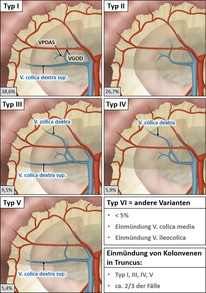
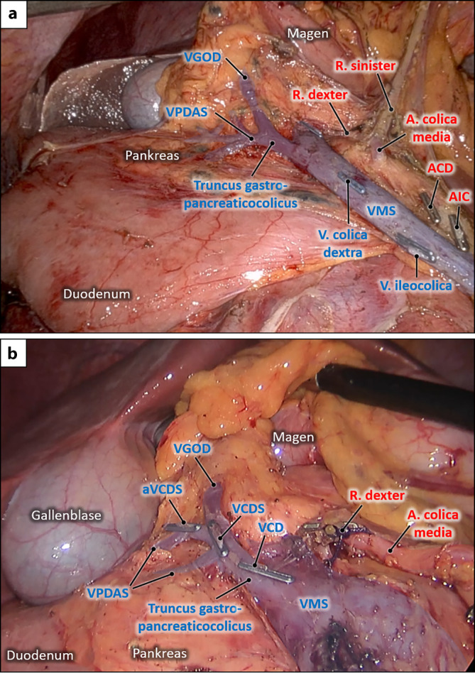
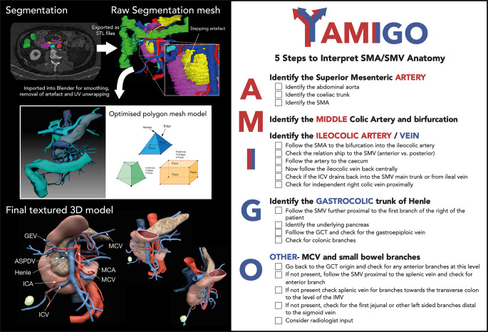
Oncological right hemicolectomy requires central ligation of all blood vessels supplying the right colon. This includes the ileocolic, right and middle colic vessels. The vascular variants comprise the prevalence, number, origin as well as the trajectory and involve both the arterial and venous systems. The course of the ileocolic artery ventral or dorsal to the superior mesenteric vein, the frequent absence of the right colic artery, the variable bifurcation of the middle colic artery and the presence of an accessory middle colic artery are of particular relevance. Venous drainage of the ascending colon and right colic flexure is provided by the right/right superior colic veins which frequently drain together with veins from the greater gastric curvature and the pancreatic head into the gastropancreaticocolic trunk (trunk of Henle). The surgical vascular management requires detailed knowledge of these vascular variants. Prevention of vascular complications is best accomplished by a preoperative vascular mapping, e.g., computed tomography (CT) angiography, AMIGO system, 3D reconstruction and the application of the critical view/open book concept.
Die onkologische Hemikolektomie rechts erfordert eine zentrale Absetzung aller Blutgefäße, die das rechte Kolon versorgen. Hierzu gehören die ileokolischen, rechten und mittleren Kolongefäße. Die vaskulären Varianten umfassen die Prävalenz, die Anzahl, den Ursprung sowie den Verlauf und betreffen sowohl das arterielle als auch das venöse System. Hervorzuheben sind der Verlauf der A. ileocolica vor oder hinter der V. mesenterica superior, das häufige Fehlen der A. colica dextra, die variable Aufzweigung der A. colica media sowie eine teilweise vorhandene A. colica media accessoria. Die venöse Drainage des aufsteigenden Kolons sowie der rechten Kolonflexur erfolgt über die V. colica dextra/dextra superior, die häufig gemeinsam mit Venen der großen Magenkurvatur und dem Pankreaskopf in den Truncus gastropancreaticocolicus (Henle-Truncus) münden. Das chirurgische Gefäßmanagement erfordert eine detaillierte Kenntnis dieser Blutgefäßvarianten. Dabei werden gefäßbedingte Komplikationen am besten vermieden durch eine präoperative Gefäßbeurteilung (CT[Computertomographie]-Angiographie, AMIGO-Methode, 3D-Rekonstruktion) und die Anwendung des Critical-View/Open-Book-Konzeptes.
Keywords: Gastropancreaticocolic trunk (Henle’s trunk); Ileocolic vessels; Middle colic vessels; Right colic vessels; Superior mesenteric vessels.
© 2025. The Author(s).
Conflict of interest statement
Einhaltung ethischer Richtlinien. Interessenkonflikt: T. Heinze, M. Heimke, T. Wedel und S.R. Benz geben an, dass kein Interessenkonflikt besteht. Für diesen Beitrag wurden von den Autor/-innen keine Studien an Menschen oder Tieren durchgeführt. Für die aufgeführten Studien gelten die jeweils dort angegebenen ethischen Richtlinien.
Figures
References
-
- Andersen BT, Stimec BV, Edwin B et al (2022) Re-interpreting mesenteric vascular anatomy on 3D virtual and/or physical models: positioning the middle colic artery bifurcation and its relevance to surgeons operating colon cancer. Surg Endosc 36:100–108. 10.1007/s00464-020-08242-8 - DOI - PMC - PubMed
-
- Benz S, Grützmann R, Stinner B (2021) Chirurgie des Kolonkarzinoms 10.1007/978-3-662-60453-3
Publication types
MeSH terms
LinkOut - more resources
Full Text Sources