. 2025 Aug 7;23(8):e3003312.
doi: 10.1371/journal.pbio.3003312. eCollection 2025 Aug.

An arginine switch drives the stepwise activation of β-arrestin by CXCR7

Affiliations

An arginine switch drives the stepwise activation of β-arrestin by CXCR7

Jeong Seok Ji et al. PLoS Biol. .

Abstract

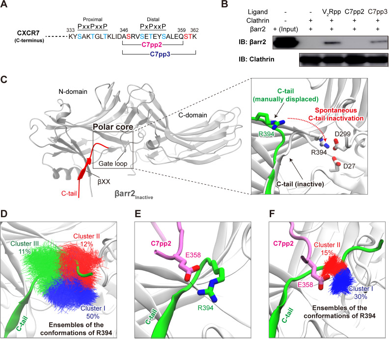
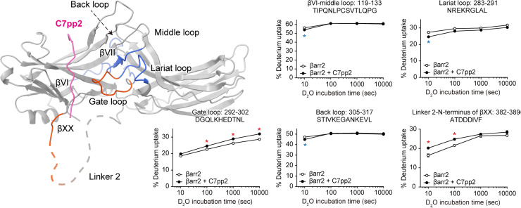
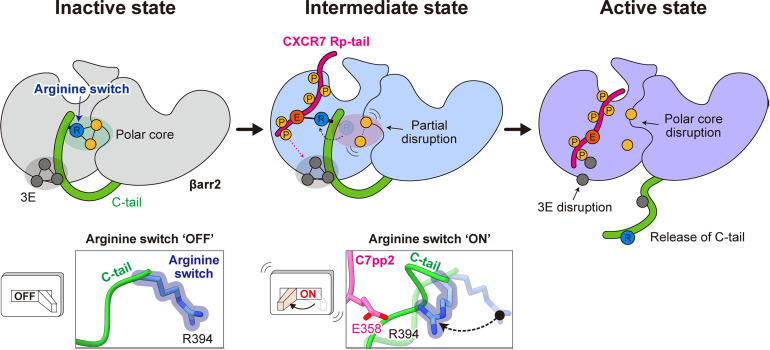
β-arrestins (βarrs) play a crucial role in regulating G protein-coupled receptor (GPCR) signaling and trafficking. Canonically, interactions of βarr with the phosphorylated intracellular GPCR-tail induce a multi-step conformational transition that results in the activation of βarr. Depending on the specific interaction pattern with the receptor, βarrs adopt multiple conformational states, each tightly linked to a specific functional outcome of βarr recruitment. Despite its physiological relevance, the structural determinants of βarr activation remain poorly understood. Using a combination of molecular dynamics simulations, biochemical and cell-based experiments, we reveal how specific interactions with a chemokine receptor 7 (CXCR7) promote the unbinding of the βarr2 C-tail-a crucial step in arrestin activation. Importantly, we observe that the expulsion of the C-tail is promoted by the displacement of a conserved arginine residue (Arg394) within the βarr polar core, which we dub "the arginine switch." Our study uncovers a role for the arginine switch that, upon engagement, destabilizes the polar core as a crucial step in the CXCR7-induced βarr activation.


Conflict of interest statement

The authors have declared that no competing interests exist.

Figures

Fig 1
Fig 1. Generating a model of the intermediate state of βarr2.
(A) The C-terminal sequence (residues 333-362) of CXCR7. The C7pp2 (residues 346-359) and C7pp3 (residues 346-362) peptides are indicated. Phosphorylation sites in the proximal and distal clusters (PxxPxxP and PxPxxP, where P represents pSer/pThr and x represents any other amino acid) are indicated in blue, while added phosphorylation sites on C7pp3 are denoted in red. All phosphorylated residues have been experimentally validated [–55]. (B) In vitro clathrin binding assay for βarr2. The βarr2 binding and input are shown. Clathrin binding to βarr2 was enhanced upon binding of V2Rpp and C7pp3. (C) Ribbon diagram of inactive βarr2. The C-tail of inactive βarr2 is highlighted in red. The polar core region, gate loop, and βXX motif are indicated. A zoomed-in view of the polar core region (right panel) shows an initial step of the simulation. The C-tail of βarr2 (residues 385–397) was manually displaced (shown in green) from the initial inactive conformation. The observed spontaneous inactivation of the C-tail during the simulation is indicated by a dotted arrow. (D) Representative conformations of R394 during spontaneous inactivation of the βarr2 C-tail. The frames obtained from simulations of the generated model (35 × 200 ns, restraints applied to the backbone of βarr2) were clustered based on the conformation of R394. The ensembles of the hit conformations of R394 are shown in blue (cluster I), red (cluster II), and green (cluster III), respectively. (E) The βarr2-C7pp2 complex model obtained from docking analysis. The most populated conformation of βarr2 obtained from clustering analysis was used to dock the C7pp2 peptide. (F) The representative conformations of R394 in each cluster from the simulation results. The ensembles of the hit conformations of R394 are shown in blue (cluster I) and red (cluster II). The βarr2-C7pp2 complex model obtained from docking result was used for simulations (35 × 300 ns, last 50 ns taken for analysis).
Fig 2
Fig 2. Model of the intermediate βarr2-C7pp2 state.
(A) Structural model of the intermediate βarr2-C7pp2 complex. Interactions between C7pp2 and clusters of positive residues in βarr2 have been categorized into site I and site II, respectively. (B) ITC analysis of the binding of C7pp2 to βarr2. Purified βarr2 proteins were incubated with increasing concentrations of C7pp2 peptide, and the binding parameters were calculated based on the dose-response curve. βarr2 induces biphasic binding by C7pp2. The indicated KD1 and KD2 values correspond to site I and site II, respectively. (C) RMSF analysis of the Cα atoms of the simulated C7pp2 peptide. Residues of C7pp2 belonging to sites I or II are indicated with arrows. The data underlying the graphs shown in the figure can be found in S1 Data.
Fig 3
Fig 3. Destabilization of the polar core by C7pp2.
(A) Comparison of polar core and three-element interactions between the inactive and intermediate conformations of βarr2. The ribbon diagram of βarr2 is colored dark gray. C7pp2 and the C-tail of βarr2IM are shown in pink and green, respectively. Hydrogen bonds stabilizing the polar core interactions are shown as dashed lines. (B) Boxplots comparing the distance between D299 and R394 (forming polar interactions in the inactive βarr2) monitored within MD simulations (5 × 300 ns). Outliers are depicted as points. The significance of differences between the groups was measured using a two-sided Mann–Whitney U test. (C) CXCL12-induced trafficking of βarr2 as monitored using confocal microscopy in HEK293 cells expressing CXCR7WT and CXCR7E358A. The “Surface” images depict βarr2 localization at the plasma membrane shortly after agonist stimulation, representing initial recruitment of βarr2 to CXCR7. The “Endosomal” images show intracellular βarr2 localization at later time points, indicating receptor internalization. Scale bar is 10 μm. (D) The whole-cell ELISA-based assay to measure the surface expression of CXCR7. Data are presented as mean ± SEM of three independent experiments, normalized to CXCR7WT. (E) CXCL12-induced βarr2 recruitment to CXCR7WT and CXCR7E358A in the Tango assay. Data are presented as mean ± SEM. CXCR7E358A is significantly compromised in inducing βarr2 trafficking upon CXCL12 stimulation. The data underlying the graphs shown in the figure can be found in S1 Data.
Fig 4
Fig 4. HDX profile changes of βarr2 upon co-incubation with C7pp2.
Regions with increased or decreased HDX upon co-incubation with C7pp2 are colored red or blue, respectively, on the structure of the βarr2IM-C7pp2 complex and the HDX profiles of the corresponding peptides are shown as graphs. Data represent the mean ± standard error of three independent experiments. Statistical analysis was performed using Student t test (*p < 0.05 compared with βarr2 alone). Differences smaller than 0.2 Da were not considered significant. The data underlying the graphs shown in the figure can be found in S1 Data.
Fig 5
Fig 5. Proposed arginine switch model for βarr2 activation by the CXCR7 Rp-tail.
In the inactive state, Arg394, which we name as an “arginine switch,” is an integral component of the polar core, facilitating interaction. Simultaneously, the βarr2 C-tail engages with the N-domain through a 3E interaction. Upon C7pp2 binding to βarr2, the arginine switch undergoes rotation and forms a direct interaction with Glu358 of C7pp2, leading to partial disruption of the polar core. This stage represents an intermediate state preceding βarr2 activation, where the 3E interaction remains intact. Phosphorylation of Ser360/Thr361 further contributes to the disruption of the 3E interaction, triggering the release of the βarr2 C-tail, ultimately leading to the activation of βarr2.

References

    1. Laporte SA, Oakley RH, Zhang J, Holt JA, Ferguson SS, Caron MG, et al. The beta2-adrenergic receptor/betaarrestin complex recruits the clathrin adaptor AP-2 during endocytosis. Proc Natl Acad Sci U S A. 1999;96(7):3712–7. doi: 10.1073/pnas.96.7.3712 - DOI - PMC - PubMed
    1. Shenoy SK, Lefkowitz RJ. Multifaceted roles of beta-arrestins in the regulation of seven-membrane-spanning receptor trafficking and signalling. Biochem J. 2003;375(3):503–15. doi: 10.1042/BJ20031076 - DOI - PMC - PubMed
    1. Lefkowitz RJ, Shenoy SK. Transduction of receptor signals by beta-arrestins. Science. 2005;308(5721):512–7. doi: 10.1126/science.1109237 - DOI - PubMed
    1. Ahn S, Shenoy SK, Luttrell LM, Lefkowitz RJ. SnapShot: β-arrestin functions. Cell. 2020;182(5):1362-1362.e1. doi: 10.1016/j.cell.2020.07.034 - DOI - PubMed
    1. Smith JS, Lefkowitz RJ, Rajagopal S. Biased signalling: from simple switches to allosteric microprocessors. Nat Rev Drug Discov. 2018;17(4):243–60. doi: 10.1038/nrd.2017.229 - DOI - PMC - PubMed

LinkOut - more resources