. 2025 Oct:60:102491.
doi: 10.1016/j.tranon.2025.102491. Epub 2025 Aug 6.

Triptonide stabilizes BIM to enhance oxaliplatin-induced ferroptosis and apoptosis in colorectal cancer

Affiliations

Triptonide stabilizes BIM to enhance oxaliplatin-induced ferroptosis and apoptosis in colorectal cancer

Ji Ma et al. Transl Oncol. 2025 Oct.

Abstract

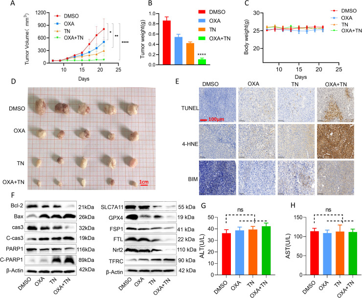
Oxaliplatin (OXA) is a common chemotherapeutic agent for advanced colorectal cancer. However, its effectiveness is limited by drug resistance, highlighting the need for combination therapies. In this study, Triptonide (TN), a diterpenoid compound is used to enhance the sensitivity of OXA, and the underlying mechanisms are investigated. Our findings indicated the combination of TN and OXA demonstrated strong synergistic anti-tumor effects across a broad concentration range in both HCT116 and LoVo cell lines, particularly at ratios ranging from 1:312 to 1:156. The combination of TN and OXA at low doses effectively inhibits growth and induces cell death in HCT116 and LoVo cells. TN and OXA cotreatment causes severe mitochondrial damage in colorectal cancer cells, leading to intracellular reactive oxygen species (ROS) accumulation, which subsequently triggers apoptosis and ferroptosis. Mechanistically, TN directly binds to BIM, a pro-apoptotic and ferroptotic protein, and stabilizes it. TN treatment led to increased expression of BIM and knockdown of BIM alleviated the growth inhibition of OXA in colorectal cancer cells. Finally, TN and OXA cotreatment significantly reduced the tumor weight and volume of LoVo-bearing nude mice in vivo. Taken together, our findings indicate that TN may serve as a novel therapeutic agent to enhance the efficacy OXA in the treatment of colorectal cancer.

Keywords: BIM; Colorectal cancer; Mitochondrial damage; Oxaliplatin; Oxidative stress; Triptonide.


Conflict of interest statement

Declaration of competing interest The authors declare that they have no known competing financial interests or personal relationships that could have appeared to influence the work reported in this paper.

Figures

Fig 1
Fig. 1
(A and B) CRC cells (LoVo, HCT116) and normal epithelial cells (NCM460) cells were treated with varying concentrations of OXA and TN for 48 h. Cell viability was assessed utilizing the CCK8 assay. (C and D) Subsequently, the cells were treated with specific concentrations of TN (5, 10, 20, 40, and 80 nM) and OXA (1.56, 3.125, 6.25, 12.5, and 25 μM) either individually or in combination. (E and F) Furthermore, CRC cells were treated with different ratios of TN to OXA (1:78, 1:156, 1:312) for 48 h, followed by cell viability assessment and CI calculation. (G) Additionally, the effects of TN and OXA (at concentrations of 5 nM or 1.56 μM) on colony formation of CRC cells were examined. (J)Quantitative analysis of G . (H, I and K) The proliferation of CRC cells following treatment with either the drug alone or the combination of OXA and TN was assessed using the EDU incorporation assay. The CCK8 assay was used to assess cell viability in HCT116, LoVo, and NCM460 cells, with the control group receiving an equivalent amount of DMSO.Scale bar = 10 μm. The experiment using the CCK8 assay was performed with at least three repetitions, and a total of 3 separate trials were executed. Results are expressed as mean ± standard deviation. Significance levels were indicated as ****P < 0.0001.
Fig 2
Fig. 2
(A and B) The MMP of LoVo and HCT116 cells was treated with 5 nM TN or 1.56 μM OXA alone or in combination. (C and E) After treatment with different groups, intra-mitochondrial ROS were analyzed using confocal microscopy following the staining of cells with 5 μM MitoSOX Red. (D)Intracellular ROS accumulation was analyzed using flow cytometry assay with 10 μM DCFH-DA dye. (F)Quantitative analysis of D. The results provided originate from three separate trials and are depicted as mean ± standard deviation.The control group received equal amounts of DMSO. Scale bar = 10 μm. The results are from three independent experiments. Data are presented as mean ± standard deviation. Significance levels were indicated as *P < 0.05,**P < 0.01, ***P < 0.001, ****P < 0.0001.
Fig 3
Fig. 3
(A) At a concentration of 5 nM, TN significantly enhanced the apoptosis induced by 1.56 µM OXA in HCT116 and LoVo cells, as demonstrated through Annexin V/PI staining. Furthermore, HCT116 and LoVo cells that were pre-incubated with or without NAC (5 mM) for 2 h prior to co-administration of OXA and TN exhibited alterations in apoptotic activity. (B)Quantitative analysis of A. (C)The impact of NAC pre-treatment on the combination of TN with OXA treatment on colony formation. (D)Western Blot was used to assess mitochondrial apoptosis-related proteins. (E)The cells were treated with 1.56 μM OXA and/or 5 nM TN, followed by immunoblotting to assess Cleaved Caspase3 and Cleaved PARP protein.This treatment effect was notably mitigated by NAC pre-treatment. (F)HCT116 and LoVo cells were pre-incubated with different concentrations (μM) of Caspase inhibitor Z-VAD-FMK for 2 h before co-administration of TN and OXA drugs. CCK8 method was used to detect cell activity, which was repeated at least 3 times. The control group was given the same amount of DMSO. The results provided originate from three separate trials and are depicted as mean ± standard deviation. Significance levels were indicated as *P < 0.05,**P < 0.01, ***P < 0.001, and ****P < 0.0001.
Fig 4
Fig. 4
(A and B) Flow cytometry analysis was performed on 10 μM BODIPY 581 / 591 C11 dye-filtered cells to quantify cellular lipid peroxidation levels. (C)Mitochondrial electron microscopy (TEM) imaging was conducted on HCT116 cells following 5 nM TN and 1.56 μM OXA treatment for 24 h. (D)Western blotting assessment of promoter or resist ferroptosis-related proteins was performed on HCT116 cells and LoVo cells treated with OXA and/or TN. (E)The GSH level of LoVo and HCT116 cells was treated with 5 nM TN or 1.56 μM OXA alone or in combination, the combination treatment effect was notably mitigated by fer-1 (20 μM) pre-treatment. (F)The cells were treated with OXA and/or TN, followed by Western blotting to assess cleaved SCL7A11 and GPX4 protein.This treatment effect was notably mitigated by Fer-1 pre-treatment. Controls were treated with equal amounts of DMSO. The data presented are from three independent experiments, expressed as mean ± standard deviation, and were assessed by Student's t-test.Significance levels were indicated as **P < 0.01, ***P < 0.001, ****P < 0.0001.
Fig 5
Fig. 5
Venn diagram showing the number of overlapping genes between TN and CRC(A), and the display of overlapping targets(B). Molecular docking results of TN and BIM(C and D). (E) TN was administered to LoVo and HCT116 cells at a designated time point, and the expression levels of BIM were subsequently assessed using Western blotting. (F)HCT116 and LoVo cells were treated with TN or an equivalent volume of DMSO for 12 h, followed by treatment with CHX (50µg/mL) for durations of 6, 12, and 24 h. The levels of BIM protein were assessed using western blot analysis. (G)Quantitative analysis of the BIM protein bands was performed using ImageJ software, and the percentage of remaining BIM was subsequently calculated using GraphPad Prism software. (H)The expression of BIM was detected by Western blotting in LoVo and HCT116 cells with BIM knockdown (si-BIM) or the corresponding negative control (si-nc). (I)LoVo and HCT116 cells transfected with si-nc or si-BIM-1# were stimulated with TN or equal volume of DMSO, and the expression of SLC7A11 and GPX4 was detected by Western blotting. (J)LoVo cells transfected with si-nc or si-BIM-1# were subjected to stimulation with OXA or an equivalent volume of DMSO. Subsequently, an EdU incorporation assay was conducted to assess cell proliferation. (K)LoVo and HCT116 cells transfected with si-nc or si-BIM-1# were stimulated with OXA or equal volume of DMSO, and the expression of GPX4, SLC7A11 was analyzed by Western blotting.Scale bar = 25 μm. The date were assessed by Student's t-test. Significance levels were indicated as*P < 0.05,**P < 0.01, ***P < 0.001, ****P < 0.0001.
Fig 6
Fig. 6
LoVo cells were injected into nude mice, followed by the administration of 5 mg/kg OXA and/or 2.5 mg/kg TN. (A and B)The combination treatment resulted in a reduction in tumor volume and tumor weight. (C) Effects of different treatments on body weight of nude mice. (D)The morphology of the tumor was analyzed. (E)Tumor samples were immunohistochemically stained for 4-HNE, TUNEL and BIM. (F)Tumor samples being lysed and subjected to Western blotting using specific antibodies and β-Actin as a reference control. (G and H)Liver function indicators, such as AST and ALT, were assessed in nude mice bearing LoVo cells xenografts following either single or combined treatment. Data were collected from three independent experiments and are presented as mean ± standard deviation. Significance levels were indicated as*P < 0.05,**P < 0.01, ***P < 0.001,****P < 0.0001.ns represents no significant difference.
Fig 7
Fig. 7
Proposed schematic diagram. TN targets BIM and amplifies OXA-induced ROS overload in CRC cells. The accumulation of ROS leads to oxidative stress and mitochondrial dysfunction, subsequently triggering apoptosis and ferroptosis.

References

    1. Shi J.-F., Wang L., Ran J.-C., Wang H., Liu C.-C., Zhang H.-Z., Yang L., Shi S.-S., Jiang L.-M., Fan J.-H., Zhang Y.-M., Wang W.-H., Ren J.-S., Zhu L., Zheng Z.-X., Sun Y.-K., Zou S.-M., Jiang J., Chen B., Chen H.-D., Liu G.-X., Yang L., Huang Y.-C., Guo L.-W., Wang D.-B., Zhang Y.-Z., Mao A.Y., Wang J.-L., Gong J.-Y., Wei D.-H., Qiu W.-Q., Song B.-B., Zhang K., Li N., Feletto E., Lew J.-B., Qiao Y.-L., Chen W.-Q., Dai M., He J. Clinical characteristics, medical service utilization, and expenditure for colorectal cancer in China, 2005 to 2014: overall design and results from a multicenter retrospective epidemiologic survey. Cancer. 2021;127:1880–1893. doi: 10.1002/cncr.33445. - DOI - PubMed
    1. McQuade R.M., Stojanovska V., Bornstein J.C., Nurgali K. Colorectal cancer chemotherapy: the evolution of treatment and new approaches. Curr. Med. Chem. 2017;24:1537–1557. doi: 10.2174/0929867324666170111152436. - DOI - PubMed
    1. Rottenberg S., Disler C., Perego P. The rediscovery of platinum-based cancer therapy. Nat. Rev. Cancer. 2021;21:37–50. doi: 10.1038/s41568-020-00308-y. - DOI - PubMed
    1. Ibrahim A., Hirschfeld S., Cohen M.H., Griebel D.J., Williams G.A., Pazdur R. FDA drug approval summaries: oxaliplatin. Oncologist. 2004;9:8–12. doi: 10.1634/theoncologist.9-1-8. - DOI - PubMed
    1. Martinez-Balibrea E., Martínez-Cardús A., Ginés A., Ruiz de Porras V., Moutinho C., Layos L., Manzano J.L., Bugés C., Bystrup S., Esteller M., Abad A. Tumor-related molecular mechanisms of oxaliplatin resistance. Mol. Cancer Ther. 2015;14:1767–1776. doi: 10.1158/1535-7163.MCT-14-0636. - DOI - PubMed

LinkOut - more resources