. 2025 Aug 5:35:e2504039.
doi: 10.4014/jmb.2504.04039.

Protective Effect of N-Acetylcysteine (NAC) on oxLDL-Induced Endothelial Dysfunction

Affiliations

Protective Effect of N-Acetylcysteine (NAC) on oxLDL-Induced Endothelial Dysfunction

Chathuri K Marasinghe et al. J Microbiol Biotechnol. .

Abstract

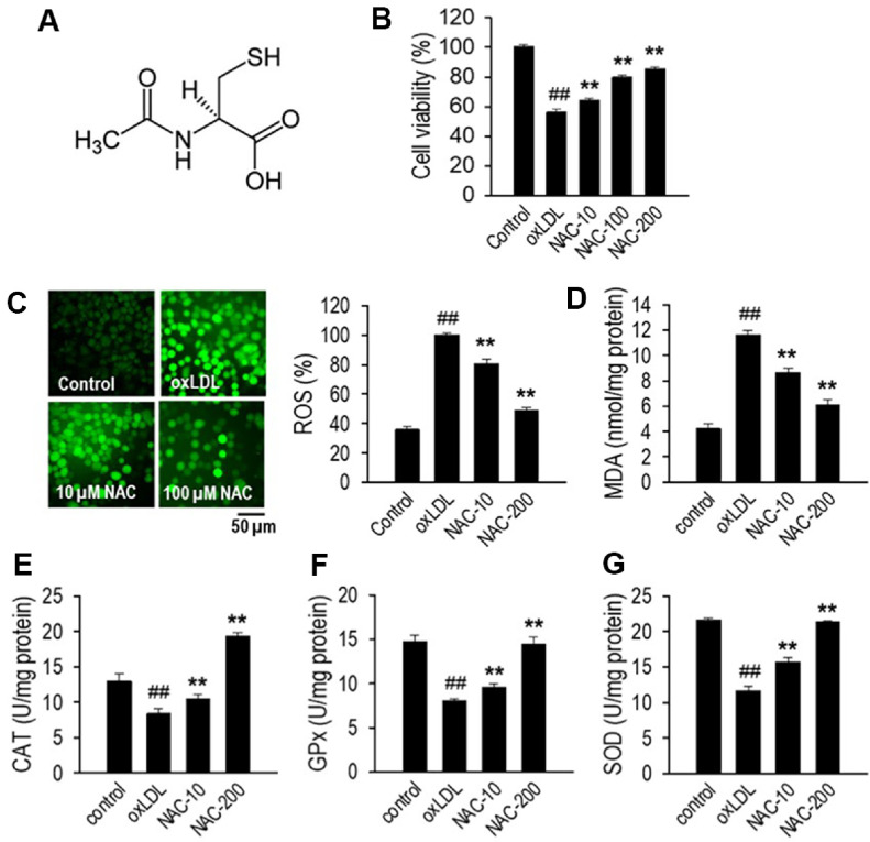
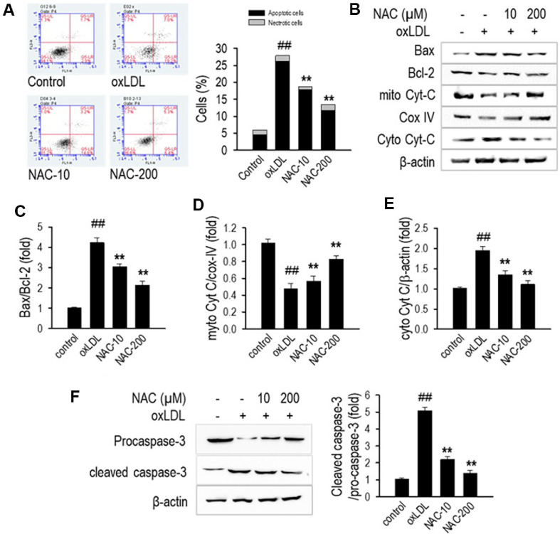
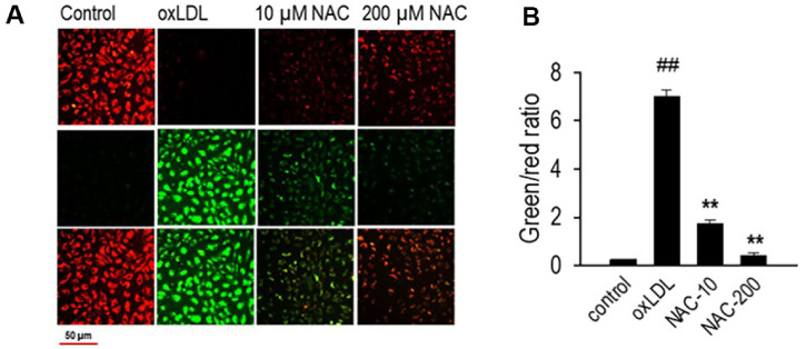
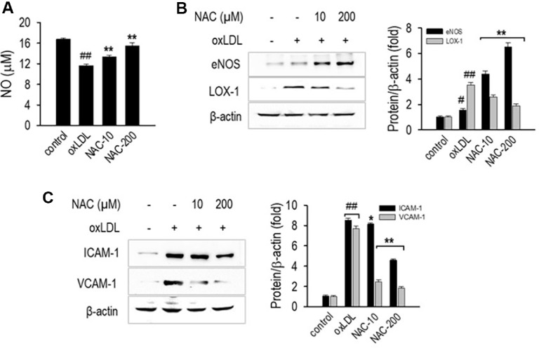
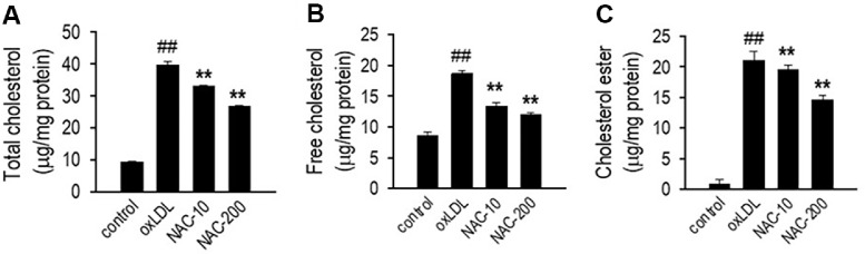
N-acetylcysteine (NAC), a well-known antioxidant and glutathione precursor, has been extensively studied for its free radical-scavenging properties, anti-inflammatory effects, and ability to enhance cellular redox balance. NAC has also been shown to mitigate oxidative damage in various disease models, yet its role in endothelial dysfunction remains underexplored. In this study, we evaluated the ability of NAC to counteract oxLDL-induced endothelial dysfunction in human umbilical vein endothelial cells (HUVECs). NAC treatment significantly reduced ROS levels, lipid peroxidation, and apoptotic markers while restoring mitochondrial membrane potential (MMP) and NO bioavailability. Additionally, NAC regulated the expression of eNOS, LOX-1, ICAM-1, and VCAM-1, demonstrating its role in reducing endothelial inflammation and improving vascular homeostasis. Furthermore, NAC prevented excessive cholesterol accumulation, suggesting its potential to regulate lipid metabolism in endothelial cells. These findings highlight the therapeutic potential of NAC in protecting against oxLDL-induced endothelial dysfunction and preventing vascular complications associated with cardiovascular diseases.

Keywords: Endothelial dysfunction; N-acetylcysteine; cardiovascular diseases; oxLDL; oxidative stress.


Conflict of interest statement

Conflict of Interest

The authors have no financial conflicts of interest to declare.

Figures

Fig. 1
Fig. 1. NAC modulates cell viability and oxidative stress in HUVECs exposed to oxLDL.
(A) Chemical structure of NAC; (B) Cell viability assessment: HUVECs were exposed to NAC (10, 100, or 200 μM) for 24 h, or pretreated with NAC for 1 h prior to 24 h oxLDL (100 μg/ml) exposure. Viability was measured using the MTT assay. (C) Representative fluorescence images showing ROS levels and corresponding quantification, normalized with protein concentration. (D) Quantification of malondialdehyde (MDA) as a marker of lipid peroxidation, assessed using an ELISA kit. (E-G) Evaluation of catalase (CAT), glutathione peroxidase (GPx), and superoxide dismutase (SOD) activity in HUVECs. Data are presented as the mean ± SD (n = 3). **p < 0.01 compared to the oxLDL-treated group; ##p < 0.01 compared to the non-treated control group.
Fig. 2
Fig. 2. NAC protects HUVECs from oxLDL-induced apoptosis.
(A) Apoptosis analysis by flow cytometry in HUVECs pretreated with NAC (10 or 200 μM) for 1 h, followed by 24-h exposure to oxLDL. Representative dot plots and quantification of apoptotic cells using Annexin V/PI staining. (B) Western blot (WB) analysis of apoptosis-related proteins. (C) Quantification of Bax/Bcl-2 ratio. (D) Quantification of mitochondrial Cyt-C/Cox-IV. (E) Quantification of cytosolic Cyt-C/β-actin. (F) Western blot analysis and densitometric quantification of the cleaved caspase-3 to pro-caspase-3 ratio. Proteins were isolated using RIPA buffer containing protease inhibitors, with β-actin serving as the loading control for normalization. Data are presented as the mean ± SD (n = 3). **p < 0.01 compared to the oxLDL-treated group; ##p < 0.01 compared to the non-treated control group.
Fig. 3
Fig. 3. NAC maintains mitochondrial membrane potential integrity.
(A) JC-1 staining analysis of MMP in HUVECs treated with NAC (10 and 200 μM) for 1 h, followed by oxLDL exposure for 24 h. Red fluorescence reflects functional mitochondria with preserved membrane potential, whereas green fluorescence signals mitochondrial depolarization. (B) Quantification of the red/green fluorescence ratio, reflecting MMP integrity. Following the treatments, HUVECs were stained with JC-1 dye following the manufacturer’s protocol, and fluorescence was measured using a fluorescence microscope. Values are expressed as mean ± SD (n = 3). **p < 0.01 compared to the oxLDL-treated group; ##p < 0.01 compared to the non-treated control group.
Fig. 4
Fig. 4. NAC restores NO production and reduces endothelial inflammation.
(A) NO levels measured using the Griess reagent assay. (B-C) Western blot (WB) images of eNOS, LOX-1, ICAM-1, and VCAM-1, with corresponding quantification of fold changes. HUVECs were treated with NAC (10 and 200 μM) for 1 h, followed by oxLDL treatment for 24 h. NO levels in the culture medium were quantified using the Griess reagent assay, following the previously outlined procedure. Western blot analysis was performed to assess the expression levels of eNOS, LOX-1, ICAM-1, and VCAM-1, using β-actin as the internal loading control. Data are presented as mean ± SD (n = 3). *p < 0.05, **p < 0.01 versus the oxLDL-treated group; ##p < 0.05, ##p < 0.01 versus the untreated control group.
Fig. 5
Fig. 5. NAC improves lipid profile in oxLDL-treated HUVECs.
(A) Total cholesterol, (B) Free cholesterol, and (C) Cholesterol ester levels in HUVECs treated with NAC (10–200 μM) for 1 h, followed by oxLDL exposure for 24 h. HUVECs were plated in 12-well culture plates and incubated for 24 h prior to NAC treatment. Total and free cholesterol levels were quantified using the Total Cholesterol and Cholesteryl Ester Colorimetric Assay Kit II (BioVision Inc., Mountain View, CA) according to the manufacturer’s protocol. Data are expressed as mean ± SD (n = 3). **p < 0.01 versus the oxLDL-treated group; ##p < 0.01 versus the untreated control.

Similar articles

References

    1. Little PJ, Askew CD, Xu S, Kamato D. Endothelial dysfunction and cardiovascular disease: history and analysis of the clinical utility of the relationship. Biomedicines. 2021;9:699. doi: 10.3390/biomedicines9060699. - DOI - PMC - PubMed
    1. Allbritton-King JD, García-Cardeña G. Endothelial cell dysfunction in cardiac disease: driver or consequence? Front. Cell Dev. Biol. 2023;11:1278166. doi: 10.3389/fcell.2023.1278166. - DOI - PMC - PubMed
    1. van Hinsbergh VW. 2012. Presented at the Seminars in immunopathology. - PMC - PubMed
    1. Sheikh AM, Yano S, Tabassum S, Nagai A. The role of the vascular system in degenerative diseases: mechanisms and implications. Int. J. Mol. Sci. 2024;25:2169. doi: 10.3390/ijms25042169. - DOI - PMC - PubMed
    1. Filippini A, Tamagnone L, D'Alessio A. Endothelial cell metabolism in vascular functions. Cancers. 2022;14:1929. doi: 10.3390/cancers14081929. - DOI - PMC - PubMed

MeSH terms