. 2025 Aug 7;15(1):28987.
doi: 10.1038/s41598-025-13764-z.

Metabolic dynamics of human external urethral sphincter myoblast differentiation and the effects of tricarboxylic acid cycle inhibition

Affiliations

Metabolic dynamics of human external urethral sphincter myoblast differentiation and the effects of tricarboxylic acid cycle inhibition

Hironori Kai et al. Sci Rep. .

Abstract

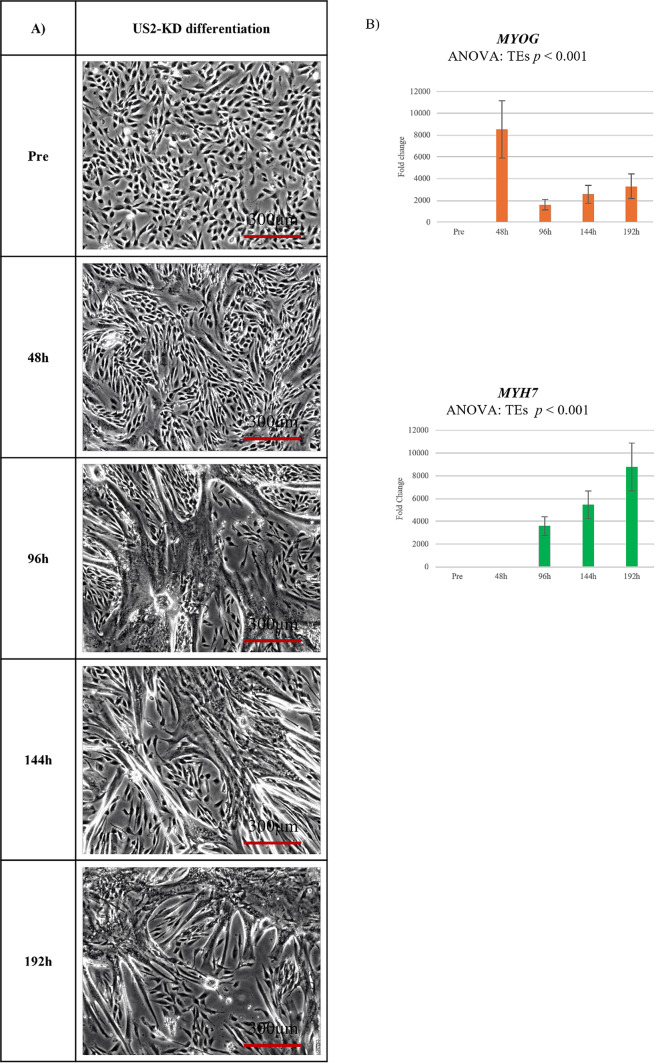
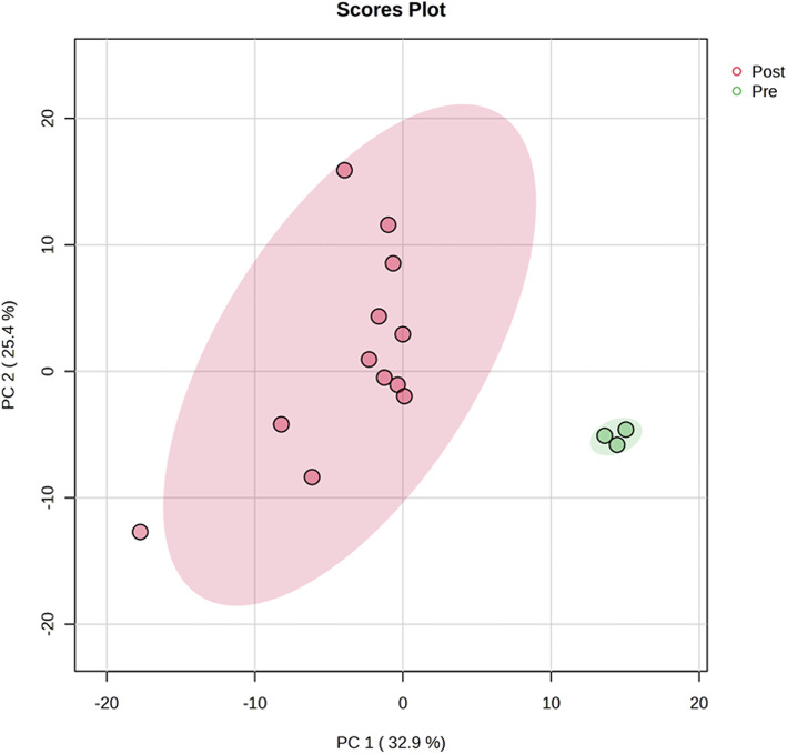
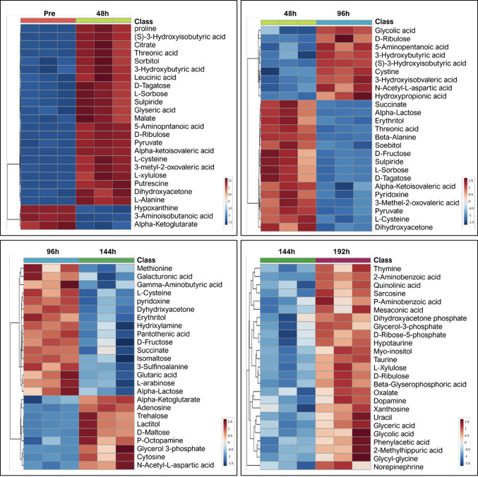
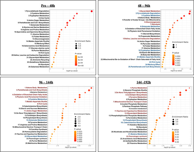
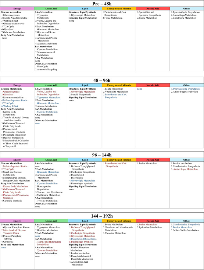
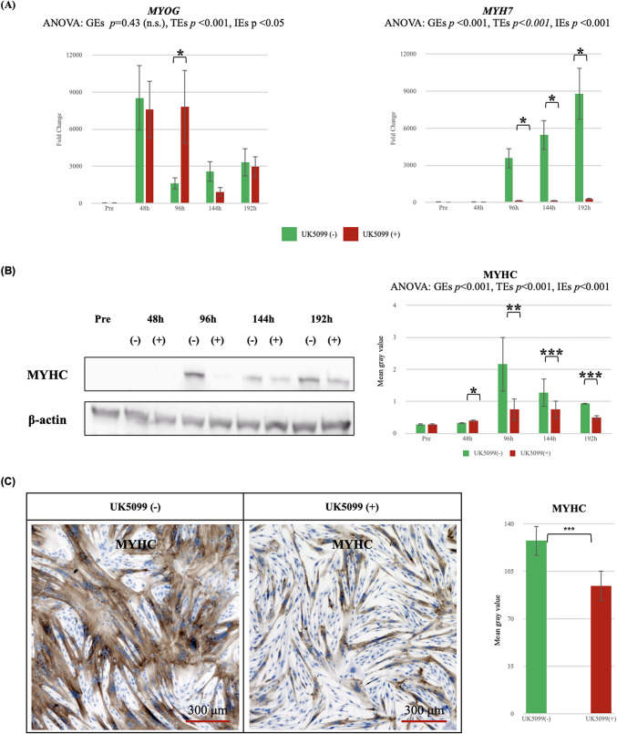
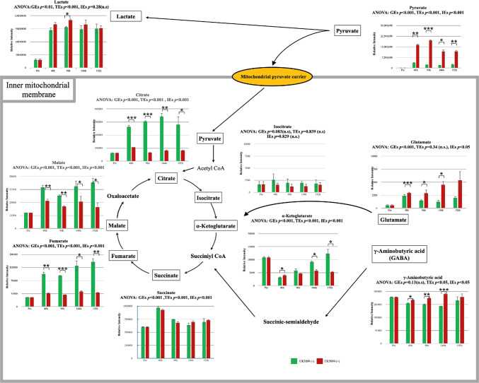
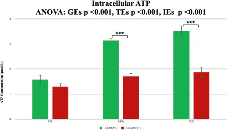
Stress urinary incontinence commonly arises with aging or following prostatectomy, yet its underlying mechanisms remain unclear. To address this, we investigated the role of metabolic pathways-particularly the tricarboxylic acid (TCA) cycle-in the differentiation of human external urethral sphincter myoblasts. Immortalized sphincter cells (US2-KD) were induced to differentiate over 192 h. Metabolomic profiling using gas chromatography-mass spectrometry, along with pathway enrichment analysis, identified key metabolic changes. Inhibition of mitochondrial pyruvate transport with UK5099 markedly suppressed TCA cycle metabolites, including citrate, α-ketoglutarate, fumarate, and malate. This inhibition also significantly reduced MYH7 expression and intracellular adenosine triphosphate levels throughout the differentiation period. These results demonstrate that the TCA cycle plays a critical role in both energy metabolism and the differentiation of urethral sphincter myoblasts. This study is the first to suggest that impaired TCA cycle activity may contribute to the pathogenesis of Stress urinary incontinence and represents a potential therapeutic target. Our findings offer new insight into age-related metabolic decline associated with Stress urinary incontinence and support the development of therapies that combine metabolic modulation with regenerative approaches.

Keywords: External urethral sphincter; Metabolomic pathways; Myoblast differentiation; Stress urinary incontinence; Tricarboxylic acid cycle.


Conflict of interest statement

Declarations. Competing interests: The authors declare no competing interests.

Figures

Fig. 1
Fig. 1
Differentiation of US2-KD myoblasts: Morphological changes and gene expression. (A) Phase-contrast images of US2-KD myoblasts during differentiation at 0 (Pre), 48, 96, 144, and 192 h. Scale bars = 300 μm. (B) Relative expression of MYOG (orange) and MYH7 (green) mRNA by qRT-PCR. Data: mean ± SD; n = 3 biological replicates. Significant time effects (TEs): p < 0.001 (repeated measures ANOVA). h, hours.
Fig. 2
Fig. 2
The results of principal component analysis (PCA) performed on metabolic profiles measured by GC-MS during the differentiation of human external urethral sphincter myoblasts. Pink points indicate samples before differentiation (Pre), while green points represent samples after differentiation (Post), including all four time points (48 h, 96 h, 144 h, and 192 h). Each point corresponds to an independent biological replicate (n = 3). GC-MS, gas chromatography-mass spectrometry. h, hours.
Fig. 3
Fig. 3
Dynamic metabolomic shifts during external urethral sphincter myoblast differentiation. The heatmap depicts the top 25 differentially abundant metabolites in the culture supernatant of differentiating external urethral sphincter myoblasts at 48-h intervals (0 [Pre]-48 h, 48–96 h, 96–144 h, and 144–192 h). Metabolomic profiling was performed in triplicate (n = 3) at each time point. h, hours.
Fig. 4
Fig. 4
Overview of enriched metabolic pathways during differentiation of external urethral sphincter myoblasts. During the differentiation of external urethral sphincter myoblasts, culture supernatants were collected every 48 h and analyzed using GC–MS for metabolomic profiling. Based on the resulting data, pathway enrichment analysis was performed, and the top 25 statistically significant pathways for each time interval (pre–48 h, 48–96 h, 96–144 h, and 144–192 h) are presented.
Fig. 5
Fig. 5
Dynamic classification of significant pathways during the differentiation of external urethral sphincter myoblasts. This figure presents the classification of the statistically significant metabolic pathways identified during the differentiation of external urethral sphincter myoblasts. Data were obtained from the culture supernatant collected every 48 h and analyzed using GC–MS. Pathways were grouped into six categories: energy, amino acid, lipid, coenzyme and vitamin, nucleic acid, and others. Black text indicates newly detected pathways, red text indicates pathways with increased rank compared with the previous interval, and blue text indicates pathways with decreased rank compared with the previous interval. TCA, tricarboxylic acid. h, hours. GC-MS, gas chromatography-mass spectrometry. EAA, essential amino acid. NEAA, non-essential amino acid. SAA, sulfur-containing amino acid. AA, amino acid.
Fig. 6
Fig. 6
Inhibition of muscle differentiation by UK5099 in US2-KD myoblasts. (A) qRT-PCR analysis of MYOG and MYH7 mRNA expression during differentiation (Pre, 48, 96, 144, 192 h) in US2-KD myoblasts treated with (red) or without (green) UK5099. n = 3. *p < 0.05, **p < 0.01. (B) Western blotting of MYHC protein at the same time points. β-actin was the loading control. (−): without UK5099; (+): with UK5099. n = 2; each blot quantified three times using ImageJ. Bar graph shows band intensities relative to β-actin (mean ± SD). Representative blots shown. Brightness and contrast were adjusted equally using Adobe Photoshop. Uncropped images are shown in Supplementary Figure S2-1 and S2-2. *p < 0.05, **p < 0.01, ***p < 0.001. (C) Phase-contrast and immunohistochemical images of MYHC at 96 h with or without UK5099. Representative images shown. Brightness and contrast were adjusted equally using Adobe Photoshop. Raw images are shown in Supplementary Figure S3-1 to S3-3. Quantification by ImageJ (mean ± SD, n = 3). ***p < 0.001. h, hours; GEs, group effects; TEs, time effects; IEs, interaction effects.
Fig. 7
Fig. 7
Effect of UK5099 on TCA cycle metabolite levels in culture supernatant during muscle differentiation. This figure presents the relative intensities of TCA cycle metabolites and related pathways in the culture supernatant of US2-KD cells during muscle differentiation, as analyzed by GC–MS. Green bars denote untreated US2-KD cells, whereas red bars indicate cells treated with UK5099. Statistical significance was determined using ANOVA, with GEs and TEs indicated. Significant differences are indicated by *p < 0.05, **p < 0.01, and ***p < 0.001. Error bars represent the standard deviation. (−): without UK5099 (+): with UK5099. TCA, tricarboxylic acid. h, hours. GC-MS, gas chromatography-mass spectrometry. GEs, group effects. TEs, time effects. IEs, interaction effects.
Fig. 8
Fig. 8
Intracellular ATP concentrations during muscle differentiation in US2-KD cells treated with UK5099. The intracellular ATP concentration (µmol/L) was measured in US2-KD cells during muscle differentiation at 96, 144, and 192 h. Green bars represent untreated cells, whereas red bars represent cells treated with UK5099. Statistical significance is indicated by *p < 0.05 and ***p < 0.001. Error bars represent the standard deviation. This process was performed in triplicate for each condition (with or without UK5099). h, hours. GEs, group effects. TEs, time effects. IEs, interaction effects.

Similar articles

References

    1. Patel, U. J., Godecker, A. L., Giles, D. L. & Brown, H. W. Updated prevalence of urinary incontinence in women: 2015–2018 National population-based survey data. Female Pelvic Med. Reconstr. Surg.28 (4), 181–187 (2022). - PubMed
    1. Gacci, M. et al. Latest evidence on post-prostatectomy urinary incontinence. J. Clin. Med.12 (3), 1190 (2023). - PMC - PubMed
    1. Wang, X. X., Zhang, L. & Lu, Y. Advances in the molecular pathogenesis and cell therapy of stress urinary incontinence. Front. Cell. Dev. Biol.11, 1090386 (2023). - PMC - PubMed
    1. Chiang, C-H. & Kuo, H-C. The efficacy and mid-term durability of urethral sphincter injections of platelet-rich plasma in treatment of female stress urinary incontinence. Front. Pharmacol.13, 847520. 10.3389/fphar.2022.847520 (2022). - PMC - PubMed
    1. Schmid, F. A. et al. Treatment of stress urinary incontinence with muscle stem cells and stem cell components: Chances, challenges and future prospects. Int. J. Mol. Sci.22 (8), 3981. 10.3390/ijms22083981 (2021). - PMC - PubMed

LinkOut - more resources