. 2025 Aug 7;16(1):7311.
doi: 10.1038/s41467-025-62380-y.

Anti-CV2/CRMP5 autoantibodies as drivers of sensory neuron excitability and pain in rats

Affiliations

Anti-CV2/CRMP5 autoantibodies as drivers of sensory neuron excitability and pain in rats

Laurent Martin et al. Nat Commun. .

Abstract

Paraneoplastic neurological syndromes arise from autoimmune reactions against nervous system antigens due to a maladaptive immune response to a peripheral cancer. Patients with small cell lung carcinoma or malignant thymoma can develop an autoimmune response against the CV2/collapsin response mediator protein 5 (CRMP5) antigen, with approximately 80% of these patients experiencing painful neuropathies. Here we investigate the mechanisms underlying anti-CV2/CRMP5 autoantibodies (CV2/CRMP5-Abs)-related pain and find that patient-derived CV2/CRMP5-Abs bind to their target on rat dorsal root ganglia (DRG) and superficial laminae of the spinal cord, to induce DRG neuron hyperexcitability and mechanical hypersensitivity. These effects from patient-derived Abs are recapitulated in rats immunized with a DNA vaccine for CRMP5, in which therapeutic treatment with anti-CD20 depleting B cells ameliorates autoimmunity and neuropathy. Our data thus reveal a mechanism of neuropathic pain in patients with paraneoplastic neurological syndromes and implicates CV2/CRMP5-Abs as a potential target for treating paraneoplastic neurological syndromes.


Conflict of interest statement

Competing interests: The authors declare no competing interests.

Figures
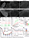
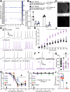
Fig. 1
Fig. 1. CV2/CRMP5-Abs labels nociceptive structures and induces mechanical hypersensitivity and DRG neuron hyperexcitability.
A Micrographs of rat dorsal root ganglia (DRG) and spinal cord immunolabelled with anti-CV2 sera from three patients. The right panels show a magnification of boxed regions of spinal cord sections. The experiment was repeated across five rats with consistent results. B Representative recordings of evoked action potentials recorded from small-diameter DRG neurons in response to depolarizing current injection of 30, 60, and 90 picoamperes (pA). Female rat DRG neurons were treated overnight with serum from patient #4 or #6 (1/100 dilution). C Quantification of the number of evoked action potentials in response to 0–100 pA of injected current. *p < 0.05, multiple Mann–Whitney tests. PBS n = 11 cells, Patient #4 n = 11 cells, Patient #6 n = 11 cells, obtained from at least three rats. D Representative traces of rheobase recordings from cells treated with PBS, serum from patient #4, or #6. E Bar graph with scatter plot showing a decreased rheobase in neurons treated with serum from patient #4 or #6. PBS n = 14 cells, Patient #4 n = 10 cells, Patient #6 n = 12 cells; error bars indicate mean ± SEM, *p < 0.05, Kruskal–Wallis test. F Graph showing the paw withdrawal threshold of male rats injected intrathecally (i.th.) with 10 µL of the indicated positive CV2/CRMP5-Abs or (G) depleted (cross-adsorbed with purified CRMP5) sera. n = 12; 3 rats per CV2/CRMP5-Abs serum; 4 different CV2/CRMP5-Abs sera. The black line shows the average of all patients tested. two-way ANOVA. H Bar graph with scatter plot showing the area under the curve for the data in (F, G), *p < 0.05, two-tailed, Mann–Whitney test. All data are shown with error bars that indicate mean ± SEM. See Supplementary Data 1 for additional statistical details. Source data are provided as a Source Data file.
Fig. 2
Fig. 2. Blocking CV2/CRMP5-Abs with epitope peptides prevents the sensitization of sensory neurons and mechanical hypersensitivity in rats.
A Heatmap of the immunoreactivity of CV2/CRMP5-Abs sera hybridized on a CRMP5 peptide array mapping the entire sequence of the protein in 15-mer peptides with three amino acid increments. Four main epitopes on CRMP5 are targeted by the CV2/CRMP5-Abs. B Bar graph with scatter plot showing that peptides 53, 142, and 146 can block the binding of CV2/CRMP5-Abs from patients #8 and #4 to purified CRMP5. 0.03% DMSO is the vehicle, purified CRMP5 was used as a positive control to achieve maximal displacement of the CV2/CRMP5-Abs. n = 3 independent measures from an average of three repeats. *p < 0.05, one-way ANOVA. C Micrograph of a rat DRG immunolabelled with CV2/CRMP5-Abs positive serum (1/100) from patient #1 and then with blocking peptides 53, 142, 146. Blocking peptides abolished the immunoreactivity of CV2/CRMP5-Abs for their protein target CRMP5. The experiment was repeated across five rats with consistent results. D Representative recordings of evoked action potentials recorded from small-diameter DRG neurons treated with serum from patient #1 (1/100 dilution) in combination with 100 ng/ml of peptides 53, 142, and 146 as indicated, overnight in response to depolarizing current injection of 30, 60, and 90 pA. E Quantification of the number of evoked action potentials in response to 0–100 pA of injected current. *p < 0.05, multiple Mann–Whitney tests. DMSO n = 15 cells, Patient #1 n = 15 cells, DMSO + peptides n = 14 cells, Patient #1 + peptides n = 16 cells. F Representative traces of rheobase recordings from cells treated with the serum from patient #1 and blocking peptides as indicated. G Bar graph with scatter plot showing unchanged rheobase in cells treated with the serum from patient #1 and with blocking peptides as indicated. *p < 0.05, Kruskal–Wallis test. DMSO n = 15 cells, Patient #1 n = 14 cells, Patient #1 + peptides n = 16 cells from at least three rats. H Graph showing the paw withdrawal threshold of rats injected with 15 µl of the indicated CV2/CRMP5-Abs positive sera (1/10 dilution). I Graph showing the paw withdrawal threshold of rats injected with CV2/CRMP5-Abs positive sera with blocking peptides 53, 142, and 146 (300 ng/ml). *p < 0.05, two-way ANOVA. J Bar graph with scatter plot showing the area under the curve of the data in (H, I) and color coded per treatment groups (n = 3 rats each), *p < 0.05, Mann–Whitney. All data are shown with error bars that indicate mean ± SEM. Experimenters were blind to the treatment groups. See Supplementary Data 1 for additional statistical details. Source data are provided as a Source Data file.
Fig. 3
Fig. 3. CRMP5 autoimmunity induces mechanical allodynia and hyperexcitability in both male and female rats.
Rats were immunized by three injections (indicated by arrows) of 50 µg of pCMV2 plasmid, allowing for the expression of CRMP5 or control (empty) in the spinodeltoidus muscle. Rats received an intramuscular injection of a plasmid (pCMV2-CRMP5) carrying the coding sequence for CRMP5 on day 0 and then two booster shots at weeks 2 and 4 after the first injection. Graph showing the paw withdrawal threshold of A male and E female rats injected with the CRMP5 coding plasmid compared to the control empty plasmid (n = 8 animals per group; *p < 0.05, two-way ANOVA). Bar graph with scatter plot showing the area under the curve for the mechanical thresholds in B male and F female rats injected as described above (n = 8 animals per group; *p < 0.05, two-tailed, Mann–Whitney test). Rats were tested for their thermal thresholds using Hargreave’s test, and no difference was found in C male and G female rats injected with the CRMP5 coding plasmid compared to the control empty plasmid (n = 8 animals per group; two-way ANOVA). Bar graph with scatter plot showing the area under the curve for the thermal thresholds in D male and H female rats injected as described above (n = 8 animals per group; two-tailed, Mann–Whitney test. I Representative recordings in response to a depolarizing current step to evoke action potentials (APs) in sensory neurons from male and female rats injected with a plasmid expressing CRMP5 or a control plasmid. J Summary of the number of APs in the indicated conditions. *p < 0.05, multiple Mann–Whitney tests. Male control n = 10 cells, Male CRMP5 n = 7 cells, Female control n = 11 cells, Female control n = 10 cells from at least three rats. K Representative recordings in response to various steps of depolarizing current to measure rheobase in sensory neurons prepared from rats with CRMP5 autoimmunity or control. L Summary of the measured rheobase in indicated conditions. Asterisks indicate significance compared with control *p < 0.05, two-tailed, Mann–Whitney test. Male control n = 10 cells, Male CRMP5 n = 7 cells, Female control n = 11 cells, Female control n = 10 cells from at least three rats. All data are shown with error bars that indicate mean ± SEM. See Supplementary Data 1 for additional statistical details. Source data are provided as a Source Data file.
Fig. 4
Fig. 4. The anti-CD20 monoclonal antibody reverses CRMP5 autoimmunity-induced mechanical hypersensitivity and hyperexcitability.
Male rats were immunized against CRMP5 and developed stable mechanical hypersensitivity up to day 86 after the first intramuscular injection. A Graph showing the paw withdrawal threshold of rats over time. Black arrows show the three plasmid injections necessary for the induction of the model. Anti-CD20 injection (4 mg/kg, i.p.) is indicated at days 56 and 63 by an antibody symbol. Control + Anti-CD20 n = 6, CRMP5 autoimmunity + Vehicle n = 8, CRMP5 autoimmunity + anti-CD20 n = 6, *p < 0.05 two-way ANOVA. B Bar graph with scatter plot showing the area under the curve for each individual from the indicated treatment groups. CRMP5 autoimmunity + Vehicle n = 8, CRMP5 autoimmunity + anti-CD20 n = 6. *p < 0.05, two-tailed, Mann–Whitney test. C Representative recordings of evoked action potentials recorded from rat small-diameter DRG neurons cultured from the indicated treatment groups in response to depolarizing current injection of 30, 60, and 90 pA. D Quantification of the number of current-evoked action potentials in response to 0–100 pA injected current. CRMP5 autoimmunity increased action potential firing compared to control DRG neurons. Anti-CD20 reversed this phenotype back to the level of control sensory neurons. *p < 0.05, multiple Mann–Whitney tests. Control + Anti-CD20 n = 9 cells, CRMP5 autoimmunity + Vehicle n = 9 cells, CRMP5 autoimmunity + anti-CD20 n = 8 cells obtained from at least three rats. All data are shown with error bars that indicate mean ± SEM. See Supplementary Data 1 for additional statistical details. Source data are provided as a Source Data file.

Similar articles

References

    1. Graus, F. et al. A clinical approach to diagnosis of autoimmune encephalitis. Lancet Neurol.15, 391–404 (2016). - PMC - PubMed
    1. Dubey, D. et al. Autoimmune CRMP5 neuropathy phenotype and outcome defined from 105 cases. Neurology90, e103–e110 (2018). - PubMed
    1. Dubey, D. et al. Amphiphysin-IgG autoimmune neuropathy: a recognizable clinicopathologic syndrome. Neurology93, e1873–e1880 (2019). - PubMed
    1. Antoine, J. C. et al. Paraneoplastic anti-CV2 antibodies react with peripheral nerve and are associated with a mixed axonal and demyelinating peripheral neuropathy. Ann. Neurol.49, 214–221 (2001). - PubMed
    1. Honnorat, J. et al. Ulip/CRMP proteins are recognized by autoantibodies in paraneoplastic neurological syndromes. Eur. J. Neurosci.11, 4226–4232 (1999). - PubMed

LinkOut - more resources