. 2025 Aug 7;16(1):7306.
doi: 10.1038/s41467-025-62681-2.

Efficacy of modified-vaccinia Ankara vaccine as pre- and post-exposure prophylaxis against monkeypox sexual transmission in non-human primate model

Affiliations

Efficacy of modified-vaccinia Ankara vaccine as pre- and post-exposure prophylaxis against monkeypox sexual transmission in non-human primate model

Cécile Herate et al. Nat Commun. .

Abstract

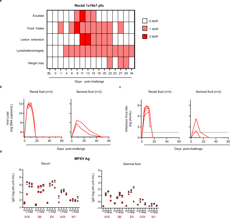
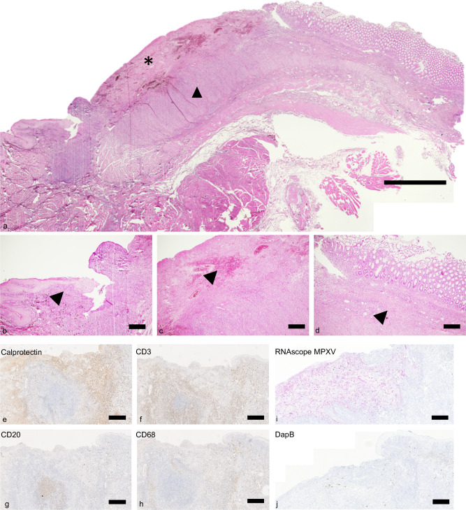
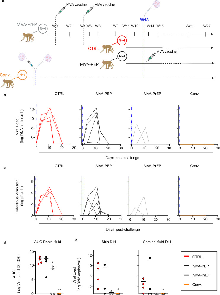
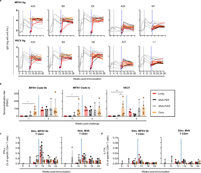
The monkeypox virus (MPXV) outbreak of 2022 caused a human disease with unusual epidemiological and clinical features, notably an increase in human-to-human transmission through sexual contact, predominantly among men who have sex with men (MSM). This evolution underscores the need to reassess prevention and control strategies in the context of a sexually transmitted disease. Here, we show that rectal challenge of male cynomolgus macaques with a 2022 clade IIb MPXV isolate mimics sexual transmission, leading to rectal infection, with systemic and male genital tract dissemination and seminal fluid shedding. Vaccination with modified-vaccinia Ankara (MVA) protected the macaques from subsequent rectal MPXV challenge. However, MVA failed to prevent the disease when administered four days post-exposure to MPXV. These findings have a critical impact on outbreak management and highlight the importance of reevaluating MVA post-exposure prophylaxis protocols.


Conflict of interest statement

Competing interests: Nathalie Silvestre and Catherine Brua are Transgene employees and may hold shares and/or stock options in the company. The remaining authors declare no competing interests.

Figures

Fig. 1
Fig. 1. Rectal challenge model of infection in cynomolgus macaques.
Four cynomolgus macaques were challenged with MPXV IIb virus on day 0 with 1 × 107 pfu of MPXV by the rectal route. Two animals were euthanized on day 11 and two were followed for 83 days for clinical and immunological monitoring (a). Five clinical signs were monitored for 34 days (n = 2): the presence of rectal exudate, loss of appetite, presence of lesions, lymphadenomegaly, and weight loss >10% were recorded for each animal. b Viral load assessment in rectal and seminal fluids (n = 4 for rectal fluid and n = 2 for seminal plasma). MPXV DNA was quantified by qPCR and is expressed as viral DNA copies/mL. The lower limit of detection (LLOD) was 90,000 copies/mL, and the lower limit of quantification (LLOQ) was 100,000 copies/mL. c The rectal and seminal fluid samples containing viral DNA were tested for virus infectivity in Vero cells. Viral infectivity is quantified in pfu/mL. The LLOD was established at 10 PFU/mL, indicated by the dotted line. All samples below this LLOD were considered negative and are represented by a value of 1. d Orthopoxvirus-binding IgG concentrations are reported in arbitrary units (arb. unit./mL). Serum (left) and seminal plasma (right) from two NHPs (n = 2) were evaluated for the total anti-MPXV protein IgG concentration before challenge and at various time points up to days 62 and 83 post-challenge for serum and seminal plasma, respectively. Each symbol represents one animal, and the different shapes represent different antigens. The dashes represent the mean. Ag: antigen. Source data are provided as a Source Data file.
Fig. 2
Fig. 2. Histological analysis of MPXV-infected rectal mucosa.
Representative Hematoxylin-Eosin staining of the rectal mucosa on day 11 post-challenge from a cynomolgus macaque infected with MPXV. The analysis was performed on 2 animals. a Ulcerated rectal mucosa at the margin of the anus showing ulcerative, necrotizing rectitis (*) characterized by b hyperplastic mucosa (arrowhead) and c fibrin and extravasated erythrocytes (hemorrhage) (arrow head). d rectal submucosa, with numerous inflammatory cells (arrow head), mostly e neutrophils (calprotectin staining), f T cells (CD3 staining), g follicular-like B-cell aggregates (CD20 staining), and h minimal diffuse macrophage infiltration (CD68 staining). i RNA scope using a specific MPXV probe in the submucosal ulcerated tissue and adjacent epithelium (purple staining localized mostly in the ulcerated area). j RNAscope with the negative control probe DapB. Scale-bars: a = 500 µm, bj = 100 µm.
Fig. 3
Fig. 3. MVA protection against MPXV challenge.
a Study design on NHP immunization and challenge periods in weeks, and a description of the four groups of cynomolgus macaques. One group was vaccinated before the challenge with two doses of MVA administered 4 weeks apart (MVA-PrEP, n = 4; gray). Another group was vaccinated with a single dose four days after the MPXV IIb challenge (MVA-PEP, n = 4; black). In addition, we challenged one group of convalescent NHPs previously exposed via the rectal or ID route alone or in combination 37–46 weeks before the challenge and described previously (Conv., n = 6; orange) and one control group (CTRL, n = 4; red). The four groups were challenged with 1 × 107 pfu of MPXV by the rectal route as indicated in blue. MVA-PrEP animals were monitored during the MVA immunization phase, with blood samplings for immunogenicity analysis. Following the MPXV challenge, blood, rectal fluid, seminal plasma, and skin swab samples were collected. The blue vertical dotted line represents the challenge. The image was created in BioRender. Herate, C. (2025) https://BioRender.com/lkzvios. b Rectal fluids collected during the challenge phase were evaluated for viral DNA content by qPCR. Viral load is expressed as viral DNA copies/mL. Each graph represents the viral kinetics of one group during 27 days. The LLOD was 36,000 copies/mL and the LLOQ 100,000 copies/mL. c Rectal fluid containing viral DNA was tested for virus infectivity in Vero cells and measured in pfu/mL. The LLOD was established at 10 PFU/mL, indicated by the dotted line. All samples below the LLOD were considered negative and are represented by a value of 1. When qPCR was negative, samples were considered negative and are represented by a value of 1. d Area under the curve (AUC) of viral DNA in rectal fluids during the 27 days of follow-up and statistical analysis. (CTRL, n = 4; red), (MVA-PEP, n = 4; black) (MVA-PrEP, n = 4; gray) (Conv., n = 6; orange). **:p = 0.0079. Each circle represents one animal. e Viral load measured by qPCR in skin swabs (left) and seminal plasma (right) sampled at day 11 and statistical analysis. The mean value is represented by the horizontal bar. (CTRL, n = 4 for skin and seminal fluid; red), (MVA-PEP, n = 3 for skin and n = 4 for seminal fluid; black) (MVA-PrEP, n = 3 for skin and n = 4 for seminal fluid; gray) (Conv., n = 5 for skin and n = 6 for seminal fluid; orange). *:p = 0.0333. Each circle represents one animal. Statistical analyses were performed using the Kruskal-Wallis test followed by a two-tailed non-parametric Mann-Whitney test (*p < 0.05, **p < 0.01). Source data are provided as a Source Data file.
Fig. 4
Fig. 4. MVA vaccine and MPXV challenge immunogenicity.
a The concentrations of five MPXV (upper panel) and homologous VACV (lower panel) protein-binding IgG were quantified in the serum of NHPs that were either immunized with MVA and/or challenged with MPXV. The data are expressed in arbitrary units (arb. unit./mL). Total IgG concentrations in NHP serum were monitored during the immunization phase for the MVA-PrEP group and after the challenge phase. For the remaining three groups, IgG follow-up began two weeks prior to the challenge. b Seroneutralization titers were measured in the serum collected at week -1 and week 4 post-challenge using Vero cells and live MPXV or VACV. The mean PRNT50 with standard deviation were calculated using infectious MPXV IIb (left panel): *:p = 0.0159, MPXV Ia (middle panel): *: = 0.0159 or VACV-107 (right panel): *:p = 0.0190 and **:p = 0.0095. (CTRL, n = 4; red), (MVA-PEP, n = 4; black), (MVA-PrEP, n = 4; gray), (Conv., n = 6 at W-1 and n = 5 at W4; orange). c, d Cellular immunity conferred by MVA vaccination and/or MPXV challenge. (CTRL, n = 4; red), (MVA-PEP, n = 4; black) (MVA-PrEP, n = 4; gray) (Conv., n = 6 and n = 5 from W14; orange). Intracellular staining of PBMCs, collected during the MVA immunization phase and after MPXV challenge, after an overnight MPXV or MVA stimulation in vitro. Panels represent the mean percentage with standard deviation of CD4+ CD154+ cells expressing IFN-γ (c) and IL17A (d) upon MPXV 2b (left) or MVA (right) stimulation. For (bd), each circle represents one animal. When represented, statistical analyses were performed using the Kruskal-Wallis test followed by a two-tailed non-parametric Mann-Whitney test (*p < 0.05, **p < 0.01). Ag: antigen; Stim. Stimulation. Source data are provided as a Source Data file.
Fig. 5
Fig. 5. Polyfunctional analysis of TH1 responder cells and CD8+ cells.
CD4+ and CD8+ cells producing TNF-α, IL-2, and IFN-γ are represented for each animal (one pie chart per animal) at W12 post-immunization (W−1 post-challenge) and W15 post-immunization (W2 post-challenge). The polyfunctionality of CD4+ CD154+ (left) and CD8+ CD137+ (right) cells upon MPXV IIb stimulation is shown. Non-responder cells are not represented. Cells secreting only one of the three cytokines are in blue, two of them in green, and three in orange. Source data are provided as a Source Data file.
Fig. 6
Fig. 6. Correlation analysis between infection parameters and immune response parameters before the MPXV challenge.
Correlation matrix between virological parameters measured after the MPXV challenge and humoral and cellular parameters measured 1 week before the challenge. Spearman's r values from −0.7 to −1 and from 0.7 to 1 are shown. Fl.: fluid.

Similar articles

References

    1. Gessain, A., Nakoune, E. & Yazdanpanah, Y. Monkeypox. N. Engl. J. Med.387, 1783–1793 (2022). - PubMed
    1. World-Health-Organization. WHO Director-General declares the ongoing monkeypox outbreak a Public Health Emergency of International Concern. https://www.who.int/azerbaijan/news/item/23-07-2022-who-director-general..., (2022).
    1. Ward, T. et al. Understanding the infection severity and epidemiological characteristics of mpox in the UK. Nat. Commun.15, 2199 (2024). - PMC - PubMed
    1. Girometti, N. et al. Demographic and clinical characteristics of confirmed human monkeypox virus cases in individuals attending a sexual health centre in London, UK: an observational analysis. Lancet Infect. Dis.22, 1321–1328 (2022). - PMC - PubMed
    1. Thornhill, J. P., Antinori, A. & Orkin, C. M. Monkeypox virus infection across 16 countries - April-June 2022. Reply. N. Engl. J. Med.387, e69 (2022). - PubMed

LinkOut - more resources