. 2025 Aug 7;8(1):1175.
doi: 10.1038/s42003-025-08593-9.

Structural Insights into the Protein Mannosyltransferase from Mycobacterium tuberculosis reveal a WW-Domain-Like Protein Motif in Bacteria

Affiliations

Structural Insights into the Protein Mannosyltransferase from Mycobacterium tuberculosis reveal a WW-Domain-Like Protein Motif in Bacteria

Nicolas Géraud et al. Commun Biol. .

Abstract

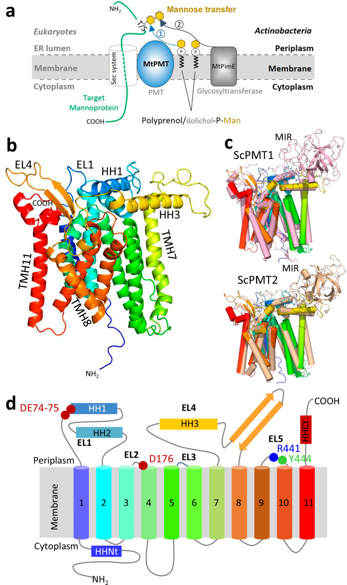
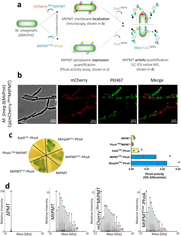
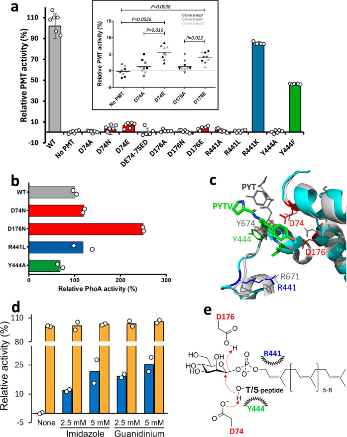
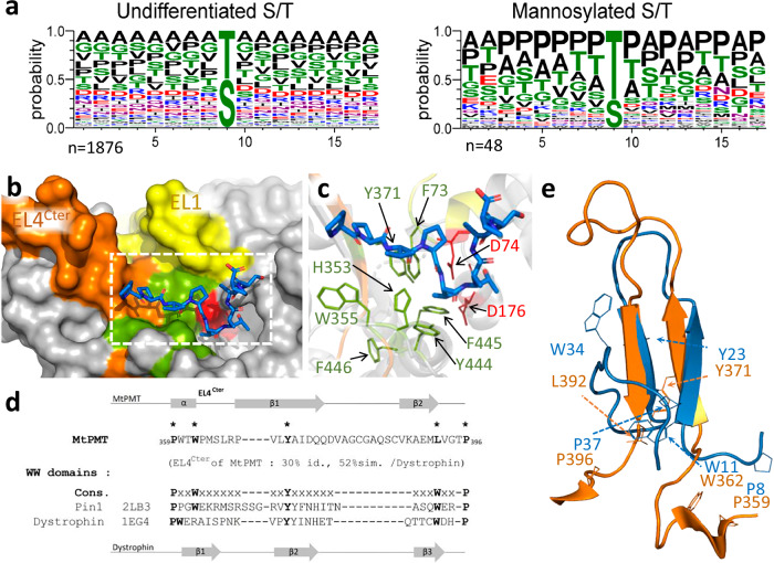
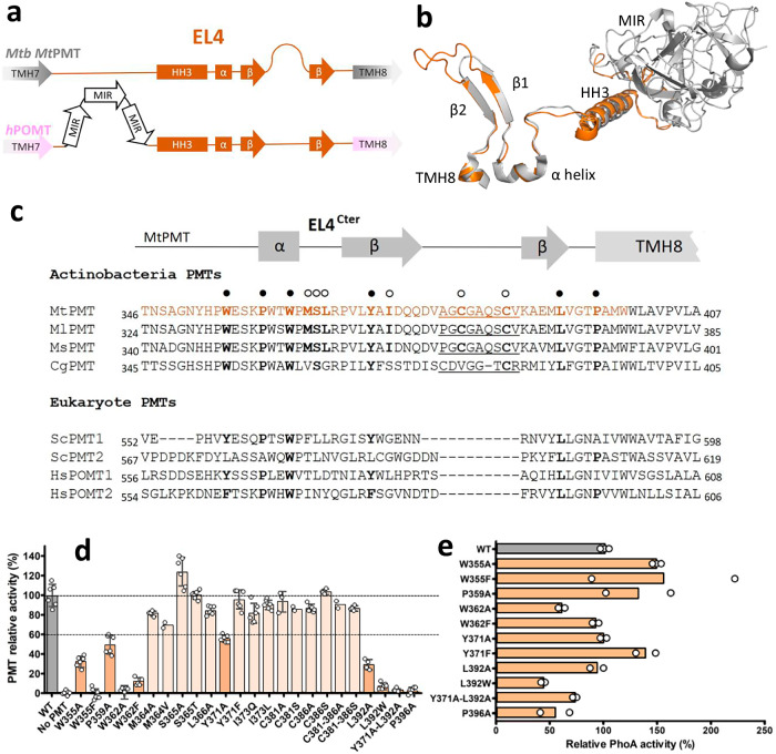
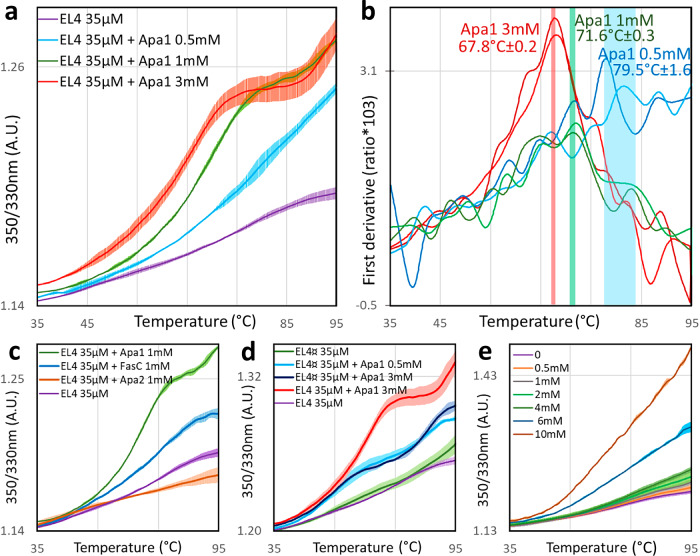
We have previously demonstrated that protein-O-mannosylation (POM), a widespread post-translational glycosyl modification of proteins, is a key virulence factor of Mycobacterium tuberculosis (Mtb), the world's deadliest infectious agent. Here, we report a detailed analysis of the structure-function relationship of MtPMT, the enzyme that catalyzes POM in Mtb. Using mutagenesis and in cellulo monitoring of POM activity, we demonstrate that, despite notable structural differences, MtPMT shares functional homologies with yeasts' PMTs in the mechanism of the sugar transfer from lipidic donors. Furthermore, we provide evidence that the selectivity for proline-rich target glycosylation sites that differentiates MtPMT from its eukaryotic homologues, relies on a WW-like domain, which preferentially interacts with proline-rich acceptor substrate analogues. This first identification of a functional WW-like domain in a prokaryotic protein raises questions about its potential evolutionary linkage with eukaryotic WW modules and provides new insights into PMT's acceptor-substrate recognition mechanism paving the way for the development selective inhibitors of MtPMT with potential therapeutic application against tuberculosis.


Conflict of interest statement

Competing interests: I declare the authors have no competing interests as defined by Nature Research or other interests that might be perceived to influence the interpretation of the article.

Figures

Fig. 1
Fig. 1. Role and conformational model of the Mycobacterium tuberculosis “dolichyl-phosphate-mannose-protein-O-mannosyltransferase” (#UniProt: P9WN05).
a: Similar to eukaryotes, M. tuberculosis protein-O-mannosylation is carried out at the membrane by a protein-O-mannosyltransferase, MtPMT, which transfers a mannosyl residue from a polyprenol-phosphate-mannose to a serine or a threonine on a nascent protein being translocated by the universal Sec protein secretion system. In Mtb, the primary mannose can be further elongated by sequential addition of mannosyl units by other glycosyltransferases, including the PimE mannosyl transferase. b: AlphaFold 3 ribbon conformational model of M. tuberculosis MtPMT (MtPMTAF3) colored in a rainbow sequence outlining the topological patterns addressed in the present paper (EL: external loop; HH: horizontal helix; TMH: transmembrane helix, see figure S1 for MtPMTAF3 model quality metrics). c: Superposition of the model and the structures of the Saccharomyces cerevisiae protein-O-mannosyl transferases ScPMT1 (top, in light pink, RMSD = 1.956) and ScPMT2 (bottom, in beige, RMSD = 1.721) resolved by Cryo-EM (PDB 6P2R), stressing the similarity between bacterial and eukaryotic PMTs. The MIR domain, specific to eukaryotes is indicated. d: Rainbow-colored topological sketch of MtPMT embedded in the bacterial membrane showing the eleven TMHs and the conserved amino-acids (circles, red: acidic, blue: basic and green: polar residue) presumed to be involved in the mannose transfer reaction. (Nt: N-terminus, Ct: C-terminus).
Fig. 2
Fig. 2. In-situ evidence for membrane insertion and functional topology of MtPMT.
a Diagram of experimental strategy: M. smegmatis Δ(MsPmt) was complemented with plasmids encoding chimeric subcellular compartment reporter proteins mCherry-NterMtPMT or MtPMTCter-PhoA. Ectopic expression and localization of the reporter tag were monitored by microscopy or quantification of PhoA activity. To test the functionality of MtPMT, an M. smegmatis Δ(MsPmt) strain expressing the wild type or hybrid MtPMT proteins was secondarily transformed with a plasmid encoding the MtPMT acceptor substrate protein FasCHis. MtPMT activity was monitored by LC-ESI-MS detection of the mannosylation patterns of native FasCHis. b Transmission and high-resolution fluorescence microscopy of M. smegmatis Δ(MsPmt) bacteria expressing the mCherry-NterMtPMT fusion protein (red) and stained with the membrane marker PKH67 (green), showing the peripheral location of the MtPMT fusion protein. (Scale bars: 2 µm). (Cp: complemented) c Periplasmic PhoA activity of M. smegmatis Δ(MsPmt) bacteria complemented with MmpS4Cter-PhoA (periplasmic control), MtPMTCter-PhoA, KatGCter-PhoA (cytoplasmic control), PhoA-NterMtPMT or untagged MtPMT. PhoA activity was detected on a solid support (left) using a chromogenic substrate and quantified in the periplasmic fractions by following pNPP hydrolysis at 420 nm (right). PhoA activity was quantified on three independent clones of each strain). d Intact protein high-resolution MS deconvoluted spectra showing the mannosylation patterns of native FasCHis purified by metal affinity column from the culture supernatants of M. smegmatis Δ(MsPmt) complemented with a mock plasmid (ΔPMT) or with plasmids encoding either the wild type MtPMT or the mCherry-NterMtPMT or MtPMTCter-PhoA fusion proteins (peaks relative intensity and symbol legend are reported in Table S1).
Fig. 3
Fig. 3. Identification of MtPMT active-site residues essential for enzymatic activity reveals its potential as a druggable Mtb virulence factor.
a Site-directed mutagenesis highlights the functional importance of residues D74, D176, R441, and Y444 within the MtPMT catalytic center. (Bars are color-coded according to the scheme shown in panel c; Data represent the mean of biological replicates from 2 clones of each mutant strains and the error bars represent the standard error of the mean (s.e.m.). Inset graph data were analyzed using an unpaired Student’s t-test (Pertinent and significant one-tailed p values p < 0.05 are indicated). b Periplasmic phosphatase activity assay confirms expression of inactive MtPMT mutant variants fused to a PhoA tag (as above, PhoA activity was quantified on two independent clones of each strain). c Close-up view of the AlphaFold 3 model of the PYTV peptide (shown as colored sticks) docked within the active site of MtPMT (cyan cartoon) (ipTM = 0.61, pTM = 0.92), superposed on the cryo-EM structure of ScPMT2 (gray cartoon) bound to a PYT peptide (gray lines; PDB: 6P2R, Bai et al.). The structural alignment (PyMOL RMSD_MtPMT–PYTV / ScPMT2–PYT = 2.39 Å) suggests a slightly altered peptide orientation in MtPMT, yet the threonine hydroxyl of PYTV remains within hydrogen-bonding distance of the catalytic D74, supporting the plausibility of the modeled interaction. d In vivo partial rescue of enzymatic activity in a genetically inactivated MtPMT mutant (R441A, blue bars) by supplementation with guanidinium or imidazole, mimicking the arginine side chain. Wild-type MtPMT is shown in orange. This supports the feasibility of chemically modulating enzyme activity in situ with small exogenous chemicals. e Proposed catalytic mechanism of MtPMT involving a general acid/base direct displacement reaction. D74 acts as a base to activate the hydroxyl group of the threonine or serine acceptor, while D176 serves as a proton donor. Residues R441 and Y444 stabilize both the phosphate group of the lipid-P-mannose donor and the acceptor substrate.
Fig. 4
Fig. 4. Evidence supporting the role of MtPMT EL4Cter in the selective recognition of [S/T]-containing proline-rich peptides for mannosylation.
a Sequence analysis reveals preferential mannosylation of [S/T]-containing proline-rich peptides. The amino acid environment of all serine/threonine (S/T) residues in a set of experimentally characterized Mtb mannoproteins (n = 44), (left panel; 17-mer peptides, n = 1876) was compared to that of experimentally validated glycosylation sites in the same proteins (right panel; 17-mer peptides, n = 48). Glycosylated S/T sites show significant enrichment in proline (P), alanine (A), and additional S/T residues surrounding the modification site (see Supplementary data 1 for full list). b Surface representation of the periplasmic face of the MtPMTAF3 complexed with the Nter truncated Apa1 decapeptide [PPVPTTAASP] (ipTM = 0.69, pTM = 0.88), illustrating the substrate-binding groove. The [PPVPTTAASP] peptide is represented with carbon and nitrogen atoms in blue and oxygen in red. Key structural elements are highlighted: EL1Nter (yellow), EL4Cter (orange), the aromatic cradle (green), and the catalytic residue D74 (red). c Cartoon representation of the putative acceptor peptide binding site in MtPMT, highlighting residues that form the aromatic cradle. Coloring is consistent with (b). d Sequence alignment of the MtPMT EL4Cter domain with the WW domains of human Pin1 and dystrophin, showing 52% sequence similarity. The WW domain consensus (Cons.) is shown with five conserved residues marked in bold and indicated by asterisks; four of these are conserved in MtPMT. Notably, the second tryptophan (W), typically located in the β3 strand of canonical WW domains, is replaced by leucine (L) in MtPMT EL4Cter. Secondary structural elements (α-helices and β-strands) are indicated by rectangles and arrows, respectively. e Alignment of the MtPMT EL4Cter domain (orange) from the AlphaFold 3 model with the WW domain of human Pin1 (PDB: 1PIN, blue), revealing conformational homology and suggesting a shared mode of peptide recognition mechanism.
Fig. 5
Fig. 5. Organization and essential function of MtPMT external loop 4 exposed at the bacterial periplasmic interface.
a Comparative organization scheme of the EL4 outer loop secondary structures of MtPMT and human hPOMTs depicting important divergences between bacterial and eukaryotic PMTs (segment size is not representative of the length of the corresponding sequences). b superposition of MtPMT AlphaFold 3 model (orange) and ScPMT1 (gray) EL4s showing the 3D structural similarity of the shared downstream domain of EL4s, except the eukaryotic MIR domain. c alignments of the MtPMT EL4 C terminal (EL4Cter) sequences (in orange) with representative corresponding domains of actinobacterial PMTs and eukaryotic PMTs. The amino acids (in bold) generally conserved in PMTs or, more specifically, in mycobacterial PMTs and addressed herein are respectively annotated with full or empty circles. The insertions of a 9 supplementary amino acid found in actinobacteria PMTs are underscored (Mt: M. tuberculosis, Ml: M. leprae, Ms: M. smegmatis, Cg: C. glutamicum, Sc: S. cerevisiae, Hs: H. sapiens). d In situ site-directed mutational evidence that EL4Cter is essential for MtPMT enzymatic activity. Effect of disruptive/rescuing mutagenesis of selected amino acids conserved in PMTs (dark orange) or specific of mycobacterial PMTs (light orange) on MtPMT relative activity. Dotted lines represent thresholds at 100% or 60% of activity, bars represent the mean, and the error bars figure the standard error of the mean (s.e.m.). MtPMT mutants with an activity less than or equal to 60% were selected for further study. e phosphatase activity of MtPMT-PhoA fusion proteins of MtPMT mutants with relative activity <60% supporting that the loss of activity is not due to an expression default of the mutated enzyme (PhoA activity was quantified on two to three independent clones of each strain).
Fig. 6
Fig. 6. NanoDSF thermal shift analysis showing dose-dependent specific alterations of the EL4Cter* peptide thermal stability in presence of ligands.
a Melting curves by nanoDSF of the EL4Cter* (35 µM) in the absence (purple curve) or in the presence of increasing concentrations of Apa1 peptide (Shown is the mean of three independent measurements (plain trace) ± SDM at each point (hatched trace)). b First derivative of the measured fluorescence ratio of the EL4Cter*(35 µM) in the presence of increasing concentrations of Apa1 (shown are two representative curves at each concentration), the mean ± SDM of the inflection points of the curves are indicated by vertical bars colored according the curves. c Melting curves of the EL4Cter* (35 µM) in presence of 1 mM of different peptide ligands (Apa1 in green, FasC P30–43 in blue and Apa2 in red). d Melting curve of the EL4Cter* Wild type (35 µM) in the presence of Apa1 (3 mM) (red curve) compared to that of the (W10A)EL4Cter* (EL4¤) in absence (green curve) or in presence of increasing concentration of Apa1 peptide (0.5 mM: light blue; 3 mM: marine blue). e Melting curves of the EL4Cter* (35 µM) in presence of increasing concentration of Zx95. Shown is the mean of three independent measurements (plain trace) ± SDM at each point (hatched trace). For clarity, the fluorescence ratio curves presented were fitted by linear translation adjustment to make their respective origin value to coincide.
Fig. 7
Fig. 7. Phylogenetic tree illustrating the potential evolutionary relationship between PRM-recognition WW domains (black branches) and the C-terminal domains of EL4 PMT loops (red branches).
The Muscle multiple sequence alignment analysis suggests that PMTs and the human dystrophin clade may share a common ancestor. For clarity, clades with an average branch length distance to their leaves below 0.35 have been collapsed, except for the PMT clade, for which leaves are grouped based on either “Mycobacteriales” genus or the respective homology of eukaryotic PMT orthologs. (Collapsed clades are represented by triangles annotated with the number of corresponding leaves; figure generated with iTOLv7). The AlphaFold-predicted conformations of WW domains from representative putative prokaryotic proteins are shown alongside the canonical WW motif of human dystrophin and the EL4Cter domains of MtPMT and ScPMTs (italicized annotations in brackets correspond to PDB accession codes).

Similar articles

References

    1. Hang, J. et al. Protein O-mannosylation across kingdoms and related diseases: from glycobiology to glycopathology. Biomed. Pharmacother.148, 112685 (2022). - PubMed
    1. Koff, M., Monagas-Valentin, P., Novikov, B., Chandel, I. & Panin, V. Protein O-mannosylation: one sugar, several pathways, many functions. Glycobiology33, 911–926 (2023). - PMC - PubMed
    1. Cowlishaw, D. A. & Smith, M. C. Glycosylation of a Streptomyces coelicolor A3(2) cell envelope protein is required for infection by bacteriophage phi C31. Mol. Microbiol.41, 601–610 (2001). - PubMed
    1. Mahne, M., Tauch, A., Puhler, A. & Kalinowski, J. The Corynebacterium glutamicum gene pmt encoding a glycosyltransferase related to eukaryotic protein-O-mannosyltransferases is essential for glycosylation of the resuscitation promoting factor (Rpf2) and other secreted proteins. FEMS Microbiol. Lett.259, 226–233 (2006). - PubMed
    1. VanderVen, B. C., Harder, J. D., Crick, D. C. & Belisle, J. T. Export-mediated assembly of mycobacterial glycoproteins parallels eukaryotic pathways. Science309, 941–943 (2005). - PubMed

MeSH terms

LinkOut - more resources