. 2025 Aug 7;23(1):885.
doi: 10.1186/s12967-025-06762-2.

SREBF1-mediated SND1 transcriptional activation promotes prostate cancer progression via MTDH interaction through the SESN2/AMPK/mTOR axis

Affiliations

SREBF1-mediated SND1 transcriptional activation promotes prostate cancer progression via MTDH interaction through the SESN2/AMPK/mTOR axis

Yijie Tang et al. J Transl Med. .

Abstract

Background: Prostate cancer (PCa) is a prevalent cancer and a major cause of cancer-related deaths in men worldwide. Growing evidence indicates that Staphylococcal nuclease and Tudor domain containing 1 (SND1) is a multifunctional protein extensively involved in transcriptional regulation, RNA maturation, post-transcriptional modifications, and other processes. However, previous studies have rarely investigated the function of SND1 as an RNA-binding protein in PCa tumorigenesis.

Methods: The Cancer Genome Atlas and NCBI Gene Expression Omnibus (GEO) databases were used to evaluate SND1 expression levels in PCa. We conducted a series of in vitro and in vivo functional experiments to assess the biological functions of SND1, including cell counting kit-8, colony formation, Transwell and wound-healing assays, and animal experiments in nude mice. Chromatin immunoprecipitation, dual-luciferase reporter assay, and DNA pull-down assay were performed to validate the association between the upstream transcription factor and SND1. Based on mass spectrometry, RNA-seq, and RNA immunoprecipitation (RIP)-seq, we identified the downstream targets of SND1- Sestrin 2 (SESN2), which were validated through qRT-PCR, Western blotting, RIP-qPCR, dual-luciferase reporter assay, and RNA pull-down assay. Finally, a series of functional assays and Western blotting analyses confirmed SESN2 as a downstream target of SND1.

Results: Our research identified that SND1 was significantly elevated in PCa, and knocking down SND1 repressed PCa multiplication and migration. Mechanistically, sterol regulatory element binding transcription factor 1 (SREBF1) bound to the promoter of the SND1 gene and activated its transcription, which subsequently formed a complex with metadherin (MTDH). This complex is directly bound to and degraded SESN2 mRNA, and disruption of this interaction with C26-A6 inhibited MTDH-SND1-mediated SESN2 degradation. Notably, SESN2 expression was inhibited in PCa and may exert tumor-suppressive effects by affecting the AMPK/mTOR signaling pathway. Rescue experiments indicated that knocking down SND1 or MTDH significantly inhibited PCa proliferation and migration, and knocking down SESN2 partially reversed this effect.

Conclusions: Our study reveals SND1 overexpression in PCa, which is transcriptionally activated by SREBF1. Mechanistically, SND1 interacts with MTDH and promotes SESN2 mRNA degradation, modulating PCa progression through the AMPK/mTOR pathway.

Keywords: MTDH; Prostate cancer; SESN2; SND1; mTOR.


Conflict of interest statement

Declarations. Ethics approval and consent to participate: All experiments conducted in this study were carried out in strict accordance with the Declaration of Helsinki. For studies involving human subjects, written informed consent was obtained from all participants, and their privacy and confidentiality were strictly maintained throughout the research process. Our study received approval from the Institutional Ethics Committee of the First Affiliated Hospital of Medical College, Zhejiang University (Hangzhou, China). The research adhered to the principles of transparency, honesty, and integrity in data collection, analysis, and reporting. Consent for publication: All authors agreed on the manuscript for publication. Conflict of interest: All authors declare no competing interests.

Figures
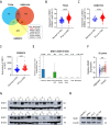
Fig. 1
Fig. 1
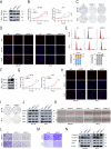
The expression of SND1 is elevated in PCa clinical samples and cell lines. A The Venn diagram shows the overlap of upregulated genes across TCGA, GSE21034, and GSE6919. B The expression mode of SND1 was analyzed between 497 PCa tissues and 53 adjacent normal prostate tissues (TCGA database). C, D The data from GSE21034 and GSE6919 revealed higher expression of SND1 in PCa tissues in comparison to benign tissues. E Single-cell sequencing data from GSE141445 showed that the relative expression level of SND1 in different types of PCa cells. E In our analysis of 32 clinical PCa samples, along with their paired normal tissues, SND1 levels were increased in 22 cases (68.75%). G Fourteen pairs of PCa samples were subject to western blot analysis of SND1. H Expression level of SND1 protein in PCa cell lines and RWPE-1 were analyzed. β-actin acted as internal reference
Fig. 2
Fig. 2
Knockdown of SND1 inhibits proliferation and migration of PCa cells in vitro. A Knockdown of SND1 was verified by western blot. B-D Knockdown of SND1 suppressed PCa proliferation evaluated by CCK-8, EdU and colony formation assay. E Cell cycle analysis indicated a notable increase of cells arrested in G1 phase in SND1 KD cells. F Overexpression of SND1 was verified by western blot. G-I Overexpression of SND1 promoted PCa proliferation and migration. J The expression of CDK2, CDK4, CCND1 was detected by western blot upon SND1 KD. K, L Knockdown of SND1 repressed PCa migration evaluated by trans-well assay and wound-healing assay (scale bar = 250 μm). M Overexpressing SND1 with plasmids promoted PCa migration evaluated by trans-well assay. N The expression of E-cadherin, N-cadherin and vimentin were detected by western blot upon SND1 KD. β-actin was the internal reference
Fig. 3
Fig. 3
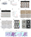
Knockdown of SND1 inhibits tumor growth and metastasis in vivo. A SND1 was stably knocked down using lentivirus-based shRNA technique in continuously expressing luciferase PC-3 cell lines (PC3-luci). B The growth curve of tumors was plotted (n = 5 each group) by measuring the tumor size with electronic vernier caliper every 4 days. C Subcutaneous xenografts were photographed and recorded at the end of observation. D The nude mice were first anesthetized and then subjected to dissection to obtain subcutaneous tumors, which were measured for size. E The dissected tumors were weighed in each group. F, G IHC staining of SND1 and Ki-67 in tumors were conducted (scale bar = 100 μm). H Intravenous tail vein tumor metastasis models in vivo were imaged at 6th weeks. I All imaged metastatic organs were anatomized and imaged again to confirm the metastatic sites. J H&E staining of metastasis (lung) were made to validate the metastatic tumor tissues. β-actin was the internal reference
Fig. 4
Fig. 4
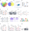
SREBF1 activates the transcription of SND1 in Pca A JASPAR, hTFtarget, TFDB and GeneCards were used to predict potential TFs of SND1. B Three TFs were screened out according to data from Linkedomics combined with online websites. C Correlation analysis was made between SND1 and SREBF1. D, E Expression level of SND1 mRNA was evaluated with qRT-PCR after knocking down SREBF1 or ZBTB7B with siRNAs. F Expression level of SND1 was evaluated with western blot after knocking down SREBF1 with siRNAs. G-I Knockdown of SREBF1 suppressed PCa proliferation and migration ability evaluated by CCK-8, colony formation assay and trans-well assay. J The motif of binding sites was downloaded from JASPAR. K ChIP-qPCR was performed and analyzed using SREBF1 antibody. L Dual luciferase reporter assay was conducted driven by SREBF1 plasmid and the SND1 promoters. M DNA pull-down assay was performed using biotin-labeled probe. β-actin was the internal reference
Fig. 5
Fig. 5
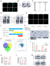
SND1 regulates the expression level of SESN2 through MTDH interaction A IF showed the subcellular localization of SND1. B Isolation of nuclear and cytoplasmic extract showed the localization of SND1 protein in PCa cells. C Proteins with the highest binding abundance to SND1 identified by LC/MS were listed. D Co-IP was performed to validate the interaction between MTDH and SND1. E IF was performed to validate the interaction between MTDH and SND1. F The diagram of truncated plasmids was as presented. G Co-IP was performed to validated the region of SND1 binding to MTDH. H Venn diagram suggested the number of overlapping genes preliminarily screened by eCLIP-seq, RNA-seq and RIP-seq (48 up). I Correlation analysis was made between SND1 and SESN2. J The expression of potential target mRNAs was detected by qRT-PCR upon SND1 KD. K, L The mRNA level of SESN2 was detected by qRT-PCR upon SND1 KD or MTDH KD. M The protein level of SESN2 was detected by western blot upon SND1 KD or MTDH KD. β-actin was the internal reference
Fig. 6
Fig. 6
MTDH-SND1 complex promotes the decay of SESN2 mRNA while C26-A6 suppresses this effect A, B C26-A6 inhibited the proliferation and migration of PCa in vitro. C-E C26-A6 inhibited the proliferation and migration of PCa in vivo.F, G C26-A6 significantly enhanced SESN2 mRNA and protein levels. H RIP-PCR assay clarified the association of MTDH, SND1 and SESN2 mRNA. I RIP-PCR assay clarified the regulation of MTDH to SESN2 depended on SND1. J RIP-PCR assay clarified the regulation of SND1 to SESN2 depended on MTDH. K RNA pull-down assay confirmed the interaction between SND1 and SESN2 mRNA. L Relative luciferase activities of SESN2-WT and SESN2-Mut in SND1 KD or MTDH KD cells were detected. M-P RNA stability assays revealed that SND1 KD, or MTDH KD, or C26-A6 prolonged SESN2 mRNA half-life. β-actin was the internal reference
Fig. 7
Fig. 7
SESN2 are responsible for SND1 and MTDH induced regulation of PCa progression A The expression pattern of SESN2 was plotted in 497 PCa tissues and 52 normal prostate tissues using data downloaded from TCGA database. B Three pairs of PCa samples were subject to western blot analysis of SND1. C-E Overexpression of SESN2 inhibited PCa tumor growth and metastasis accessed by colony formation assay and trans-well assay (scale bar = 250 μm). F-I Knockdown of SESN2 promoted PCa tumor growth and metastasis accessed by CCK-8, colony formation assay and trans-well assay (scale bar = 250 μm). J The expression of CDK2, CCND1, E-cadherin, N-cadherin and vimentin was detected by western blot after overexpressing SESN2. K The expression of AMPK, p-AMPK, mTOR and p-mTOR were detected by western blot after overexpressing SESN2. L, M Western blot showed knockdown of SESN2 partly reversed the increase of SESN2 expression induced by SND1 KD or MTDH KD. N, O CCK-8 and trans-well assay after SND1 knockdown alone or co-transfected with siSESN2 (scale bar = 250 μm). P The expression of p-AMPK after knocking down SND1 was detected. β-actin was the internal reference

References

    1. Siegel RL, Giaquinto AN, Jemal A. Cancer statistics, 2024. Cancer J Clin. 2024;74:12–49. - PubMed
    1. Chen W, Zheng R, Baade PD, et al. Cancer statistics in china, 2015. Cancer J Clin. 2016;66:115–32. - PubMed
    1. Galsky MD, Small AC, Tsao C, et al. Clinical development of novel therapeutics for castration-resistant prostate cancer: historic challenges and recent successes. Cancer J Clin. 2012;62:299–308. - PubMed
    1. Yang J, Aittomäki S, Pesu M, et al. Identification of p100 as a coactivator for STAT6 that bridges STAT6 with RNA polymerase II. EMBO J. 2002;21:4950–8. - PMC - PubMed
    1. Yang J, Välineva T, Hong J, et al. Transcriptional co-activator protein p100 interacts with SnRNP proteins and facilitates the assembly of the spliceosome. Nucleic Acids Res. 2007;35:4485–94. - PMC - PubMed

MeSH terms

LinkOut - more resources