

Cross-sectional fat fraction analysis of the gluteus medius and minimus muscle in asymptomatic vs. symptomatic hips using 2-point Dixon MRI
- PMID: 40775346
- PMCID: PMC12333093
- DOI: 10.1186/s12891-025-09043-7
Cross-sectional fat fraction analysis of the gluteus medius and minimus muscle in asymptomatic vs. symptomatic hips using 2-point Dixon MRI
Abstract
Background: To evaluate cross-sectional fat fractions (FF) of the hip abductors in asymptomatic compared to symptomatic hips in elderly patients using 2-point Dixon MRI, and compare them to the Goutallier classification.
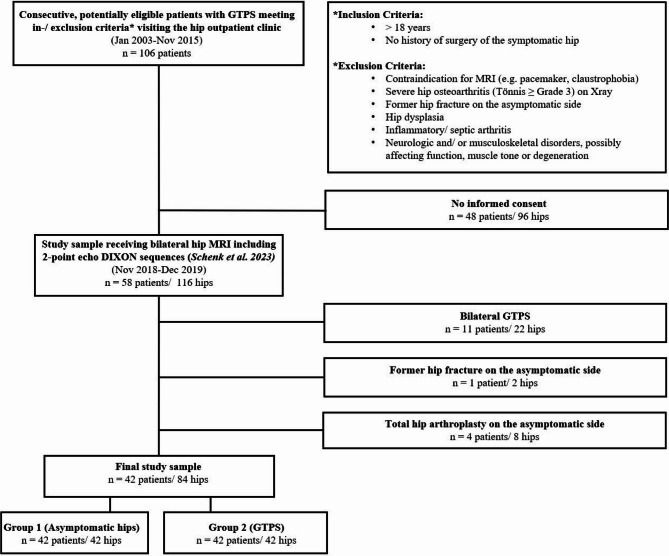
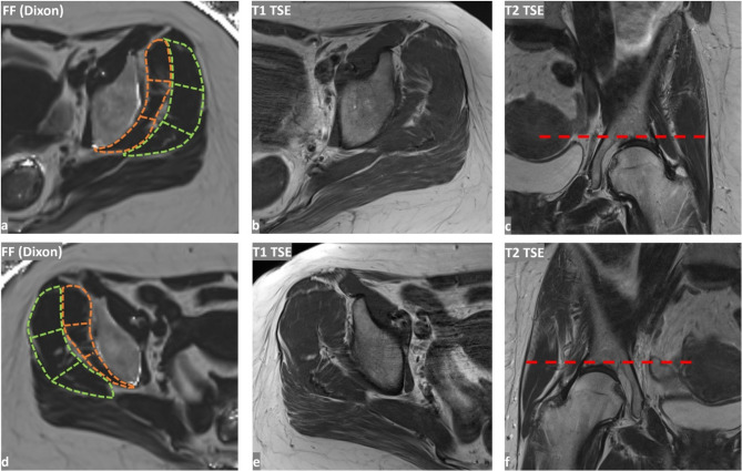
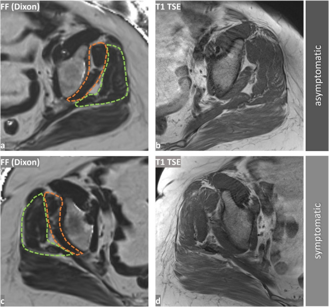
Methods: In this retrospective (clinicial trial number: not applicable) single-center study, two radiologists assessed cross-sectional fatty infiltration of the gluteus minimus (Gmin) and medius (Gmed) muscles in both hips of patients with unilateral greater trochanteric pain syndrome (GTPS) using 2-point Dixon MRI-derived fat fractions (FF). Additionally, fatty infiltration was assessed on T1w sequences using the Goutallier classification. Differences in fatty infiltration for both methods between the symptomatic and asymptomatic hips were assessed using the Wilcoxon signed-rank test.
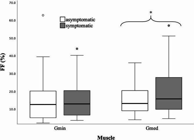
Results: 42 patients (mean age 65.1 ± 13.7 years, 28 females) were analyzed. Median FF in asymptomatic hips was 13.5 ± 15.2% for the Gmin and 14.2 ± 11.6% for the Gmed muscle (vs. 13.9.±14.1% and 16.2 ± 18.4% on the symptomatic side). FF of Gmed were significantly lower on the asymptomatic side than on the symptomatic side (p ≤ .043). No significant difference was observed neither for FF of the Gmin muscle (p ≥.30) nor for the Goutallier gradings of the Gmin (p ≥ .06) and Gmed (p ≥ .13) muscle between the asymptomatic vs. symptomatic sides.
Conclusion: Median FF were close to 14% for Gmin and Gmed in elderly asymptomatic hips, with a tendency for lower fat fractions in males. Significantly higher fat content of the Gmed was present in symptomatic hips, which was solely detectable by FF analysis and not by Goutallier grading. Reproducibility of FF analysis surpassed the Goutallier assessment.
Keywords: 2-point-Dixon; Fat fraction; Hip; Magnetic resonance imaging.
Conflict of interest statement
Declarations. Ethics approval and consent to participate: All procedures performed in studies involving human participants were in accordance with the ethical standards of the institutional and/or national research committee and with the 1964 Helsinki declaration and its later amendments or comparable ethical standards. The local ethic commission (Kantonale Ethikkommission Zürich) approved this study. Consent for publication: Not applicable. Competing interests: The authors declare no competing interests.
Figures

Similar articles
-
[Comparison of muscle injury between piriformis muscle release and preservation in total hip arthroplasty via supercapsular percutaneously-assisted total hip approach].Zhongguo Xiu Fu Chong Jian Wai Ke Za Zhi. 2025 Jun 15;39(6):715-722. doi: 10.7507/1002-1892.202502039. Zhongguo Xiu Fu Chong Jian Wai Ke Za Zhi. 2025. PMID: 40545460 Free PMC article. Clinical Trial. Chinese.
-
Prevalence of Gluteus Medius Pathology on Magnetic Resonance Imaging in Patients Undergoing Hip Arthroscopy for Femoroacetabular Impingement: Asymptomatic Tears Are Rare, Whereas Tendinosis Is Common.Am J Sports Med. 2020 Oct;48(12):2933-2938. doi: 10.1177/0363546520952766. Epub 2020 Sep 3. Am J Sports Med. 2020. PMID: 32881581
-
A Comparison between 6-point Dixon MRI and MR Spectroscopy to Quantify Muscle Fat in the Thigh of Subjects with Sarcopenia.J Frailty Aging. 2019;8(1):21-26. doi: 10.14283/jfa.2018.16. J Frailty Aging. 2019. PMID: 30734827 Free PMC article.
-
Muscle activation, strength, and volume in people with patellofemoral osteoarthritis: a systematic review and meta-analysis.Osteoarthritis Cartilage. 2022 Jul;30(7):935-944. doi: 10.1016/j.joca.2022.01.013. Epub 2022 Mar 5. Osteoarthritis Cartilage. 2022. PMID: 35257862
-
Gluteus medius muscle activity in patellofemoral pain syndrome during squats: A Systematic Review.J Bodyw Mov Ther. 2024 Oct;40:1536-1543. doi: 10.1016/j.jbmt.2024.03.007. Epub 2024 Apr 7. J Bodyw Mov Ther. 2024. PMID: 39593485
References
-
- Fischer MA, Pfirrmann CW, Espinosa N, Raptis DA, Buck FM. Dixon-based MRI for assessment of muscle-fat content in phantoms, healthy volunteers and patients with achillodynia: comparison to visual assessment of calf muscle quality. Eur Radiol. 2014;24(6):1366–75. - PubMed
-
- Hochreiter B, Germann C, Feuerriegel GC, Sutter R, Selman F, Gressl M, et al. Natural history of quantitative fatty infiltration and 3D muscle volume after nonoperative treatment of symptomatic rotator cuff tears: A prospective MRI study of 79 patients. J Bone Joint Surg Am. 2024;106(8):690–9. - PubMed
-
- Horiuchi S, Nozaki T, Tasaki A, Yamakawa A, Kaneko Y, Hara T, Yoshioka H. Reliability of MR quantification of rotator cuff muscle fatty degeneration using a 2-point Dixon technique in comparison with the goutallier classification: validation study by multiple readers. Acad Radiol. 2017;24(11):1343–51. - PubMed
-
- Wieser K, Joshy J, Filli L, Kriechling P, Sutter R, Furnstahl P, et al. Changes of supraspinatus muscle volume and fat fraction after successful or failed arthroscopic rotator cuff repair. Am J Sports Med. 2019;47(13):3080–8. - PubMed
LinkOut - more resources
Full Text Sources
