

Clinical management and retrieval of foreign body inclusion in a primary tooth: a case report
- PMID: 40775656
- PMCID: PMC12330185
- DOI: 10.1186/s13256-025-05468-9
Clinical management and retrieval of foreign body inclusion in a primary tooth: a case report
Abstract
Background: While foreign body ingestion is a frequent pediatric emergency, instances of foreign objects becoming lodged in teeth are uncommon. These can lead to infections, pain, and abscesses if left untreated. Imaging techniques such as radiovisiography and cone beam computed tomography help in detection and diagnosis.
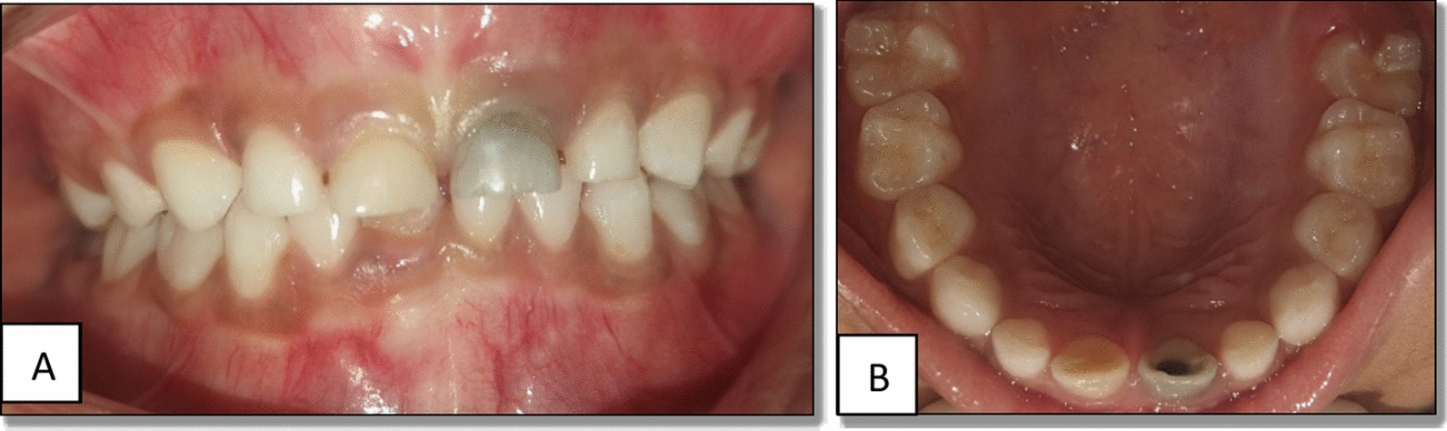
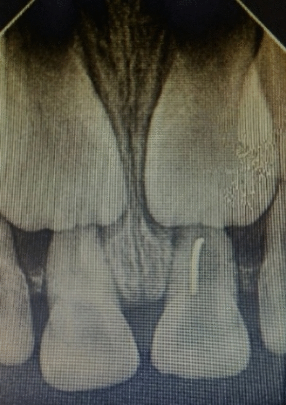
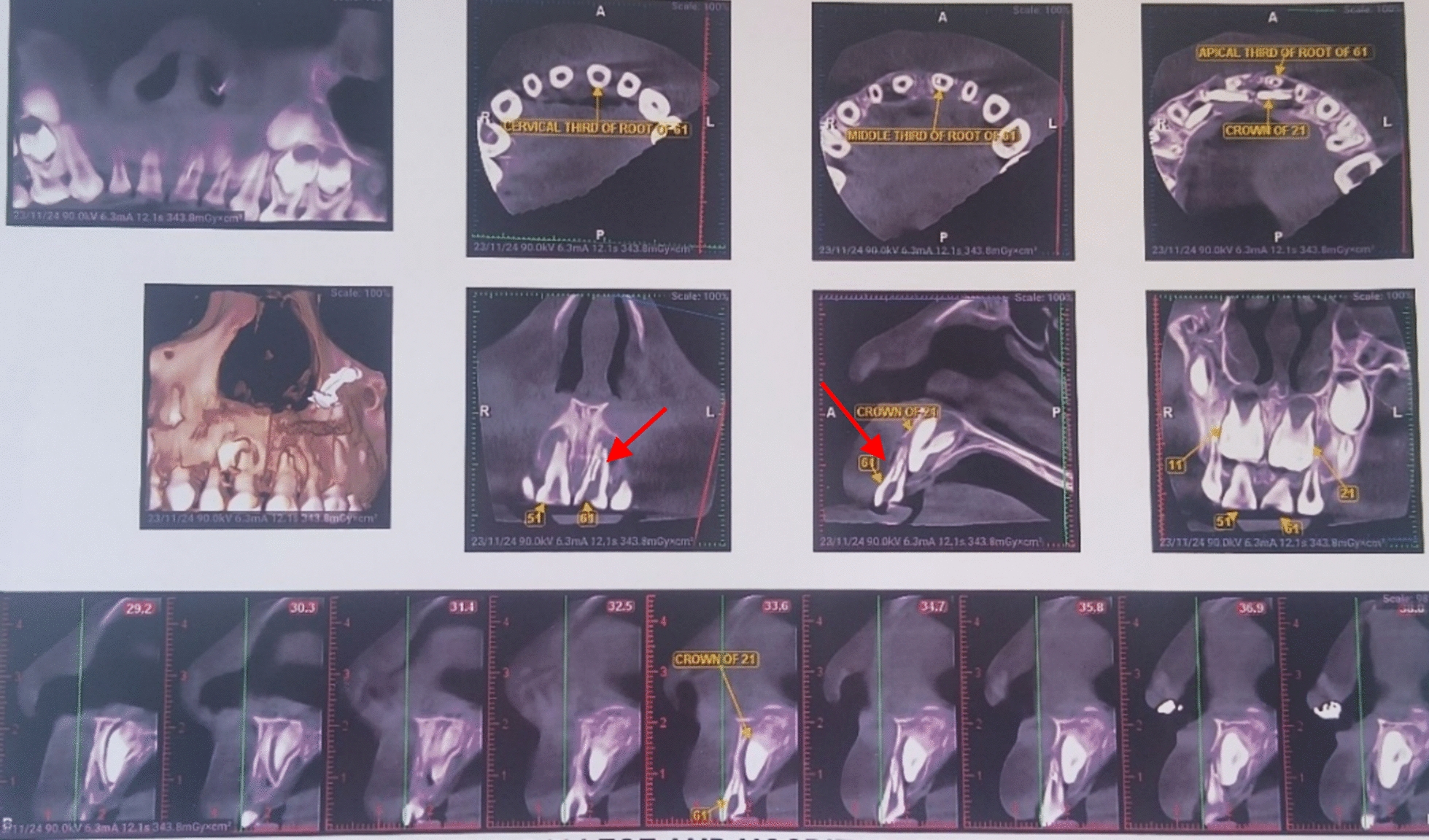
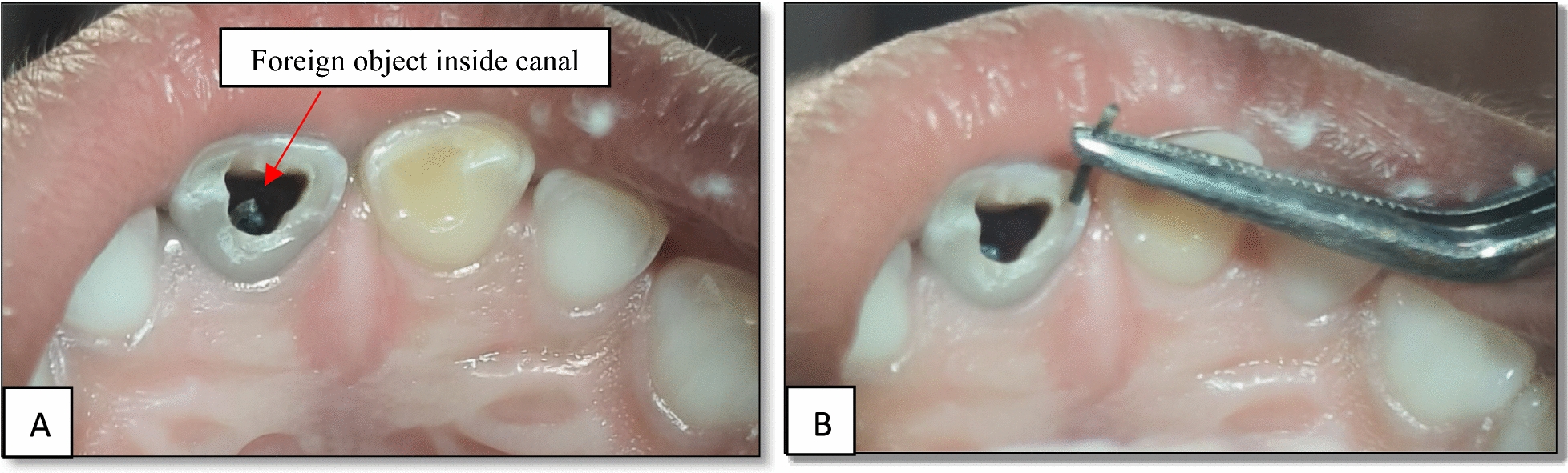
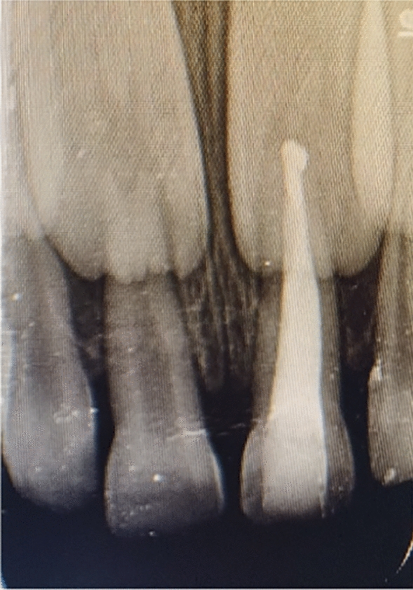
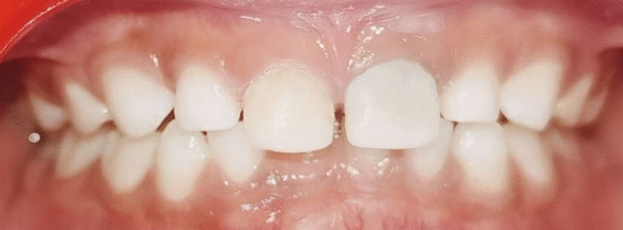
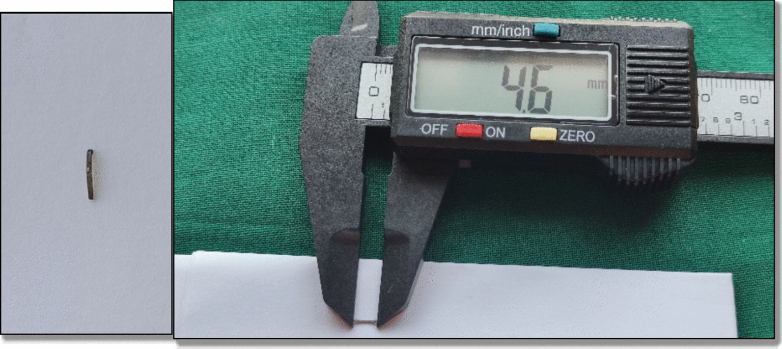
Case presentation: A 6-year-old Indian girl was brought in with black discoloration in her upper front tooth (61) for 6 months. The initial history of biting a stone was inconsistent with radiographic findings. Radiovisiography showed a radiopaque object, and cone beam computed tomography confirmed a metallic foreign body-later identified as a stapler pin-embedded in the root canal. Upon further questioning, the child disclosed self-insertion of the pin. The object was retrieved, and the tooth was successfully treated with pulpectomy and strip crown cementation.
Conclusion: Timely diagnosis and intervention are crucial in managing foreign body inclusions in teeth. Parents should be advised about the risks of children placing small objects in their mouths, and early treatment of carious lesions is essential.
Keywords: Foreign object; Pediatric patient; Traumatic injury.
© 2025. The Author(s).
Conflict of interest statement
Declarations. Ethics approval and consent to participate: Ethical approval was not required as per our institution’s guidelines, but informed written consent was obtained from the patient’s legal guardian for publication of his case along with radiographic images. Consent for publication: Written informed consent was obtained from the patient’s legal guardian for publication of this case report and any accompanying images. A copy of the written consent is available for review by the Editor-in-Chief of this journal. Competing interests: There were no conflicts of interest.
Figures
References
-
- Van As AB, du Toit N, Wallis L, et al. The South African experience with ingestion injury in children. Int J Pediatr Otorhinolaryngol. 2003;67:S175–8. - PubMed
-
- Aduri R, Reddy RE, Kiran K. Foreign objects in teeth: retrieval and management. J Indian Soc Pedodontics Prevent Dent. 2009;27(3):179–83. - PubMed
-
- McAuliffe N, Drage NA, Hunter B. Staple diet: a foreign body in a tooth. Int J Pediatr Dent. 2005;15(6):468–71. - PubMed
-
- Palit MC, Agrawal J, Saha S. Radiographic revelation of foreign body in primary tooth: a rare case report. J Dent App. 2015;2:251–3.
Publication types
MeSH terms
LinkOut - more resources
Full Text Sources
Medical
