. 2025 Aug;55(8):e70004.
doi: 10.1002/eji.70004.

Granulysin Antimicrobial Activity Promotes Dormancy in Mycobacterium tuberculosis

Affiliations

Granulysin Antimicrobial Activity Promotes Dormancy in Mycobacterium tuberculosis

Sarah Schmidiger et al. Eur J Immunol. 2025 Aug.

Abstract

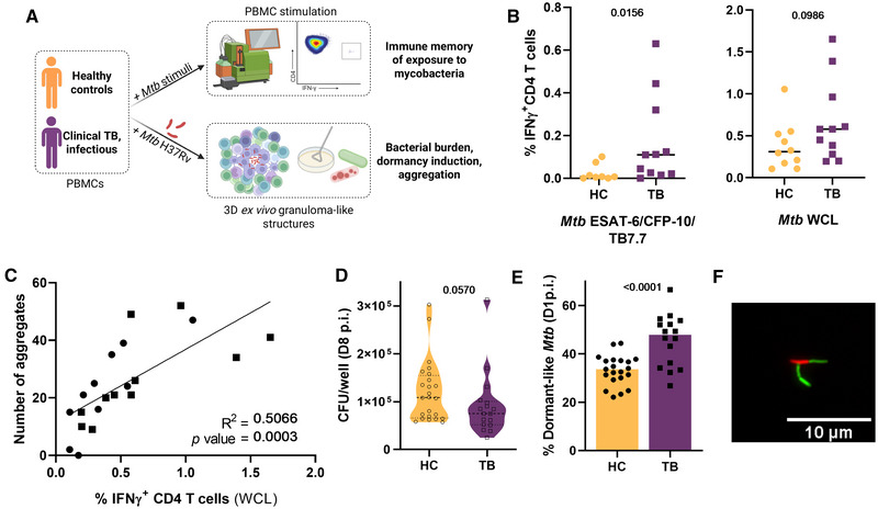
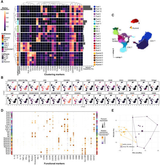
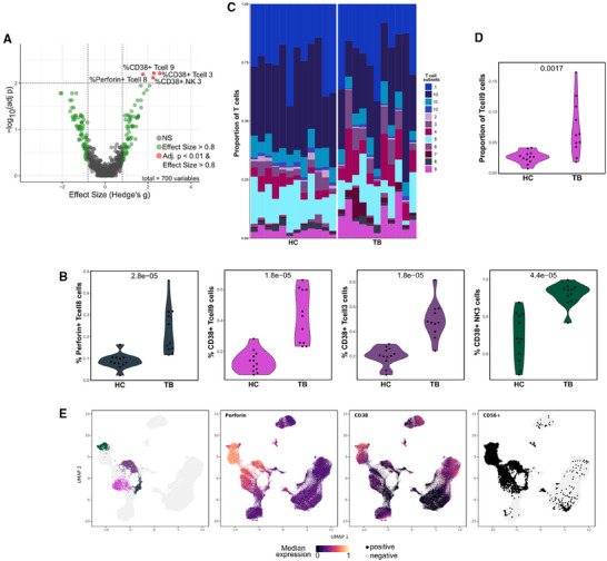
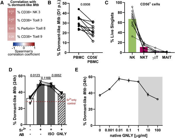
Human tuberculosis (TB) caused by Mycobacterium tuberculosis (Mtb) remains a global public health threat. Granulomas constitute a hallmark of TB pathogenesis that can clear, contain, or exacerbate an infection. Containment is exploited by Mtb as a hideout to persist in a dormant, antibiotic-tolerant state, only to resuscitate upon immunosuppression. The immune determinants of a granulomatous response driving Mtb persistence remain elusive. We here generated ex vivo granuloma-like structures from peripheral blood mononuclear cell specimens of TB patients and applied high-dimensional mass cytometry to elucidate immune factors prompting Mtb dormancy. Compared with healthy controls, patient-derived specimens rapidly forced Mtb to become dormant-like ex vivo. This observation correlated with an enrichment in activated, innate (-like) cytotoxic lymphocytes and required the presence of CD56+ lymphocytes or, more specifically, the content of their granules. Finally, we demonstrated that direct exposure to granulysin induces Mtb dormancy, thereby unravelling an immune escape mechanism to cytotoxic lymphocyte activity.

Keywords: CyTOF; Mycobacterium tuberculosis; NK cells; bacterial infections; cellular immunology; cytotoxicity; dormancy; granuloma; granulysin; immune evasion.


Conflict of interest statement

The authors declare no conflicts of interest.

Figures

FIGURE 1
FIGURE 1
TB patient‐derived specimens promote a dormant‐like Mtb phenotype ex vivo. (A) Schematic overview of the study design. Peripheral blood mononuclear cells (PBMC) from healthy controls (HC) or patients diagnosed with infectious clinical TB (TB) were either exposed to Mycobacterium tuberculosis (Mtb)‐derived stimuli prior to flow cytometry analysis or infected with Mtb H37Rv, prior to embedding within an extracellular matrix for formation of 3D ex vivo granuloma‐like structures and assessment of the indicated microbiological read‐outs. (B) Dot plot displaying background‐subtracted frequency of IFNγ‐producing CD4 T cells measured by flow cytometry after overnight stimulation with ESAT‐6/CFP‐10/TB7.7 peptide pool or Mtb whole‐cell lysate (WCL); lines indicate median. Each dot represents an independent study participant (HC: n = 10, TB: n = 11; panels with fewer dots due to limited cell availability). (C) Correlation between IFN‐γ+ CD4 T cells following overnight stimulation of PBMC samples with Mtb WCL and the number of aggregate‐like structures assessed by microscopy examination of the respective PBMC samples eight days postinfection (p.i.). (D) Bacterial load in colony‐forming units (CFU), 8 days p.i. Each dot represents an independent study participant (HC: n = 24, TB: n = 17). (E) Frequency of dormant‐like Mtb based on auramine‐O/Nile red dual staining, one day p.i.; bars depict the median. Each dot represents an independent study participant (HC: n = 21, TB: n = 16). (F) Representative fluorescence microscopy picture of the dual staining phenotypes. Dormant‐like bacilli (Nile red+) in red and metabolically active ones (Auramine‐O+) in green; 63× magnification. All p‐values represent Mann–Whitney tests. Circle and square symbols systematically represent healthy control and patient study participants, respectively.
FIGURE 2
FIGURE 2
CyTOF profiling of immune cells exposed to Mtb ex vivo characterizes 25 cell subsets further subdivided into 640 host cell phenotypes that readily discriminate responses of TB patients from those of healthy controls. (A) Heatmap of the median expressions of FlowSOM clustering markers with a horizontal bar plot representing the median frequency of the respective cell subsets specified by adjacent colored circles in the legend. UMAP visualization colored by (B) median expression of clustering markers, and (C) FlowSOM cluster identity. (D) Percent expressed and median expression level of functional markers characterizing the 25 subsets (host cell phenotypes, n = 700). (E) Principal component analysis on the host cell phenotype (n = 640) frequencies common to all study participants’ ex vivo responses, colored by disease group. Small symbols represent independent study participants (HC: n = 12, TB: n = 11), bigger symbols are the centroids.
FIGURE 3
FIGURE 3
Cytotoxic, activated NK and (unconventional) T cells are significantly enriched in patient immune cells exposed to Mtb. (A) Volcano plot (x: Hedge's g effect size, y: false discovery rate (FDR) adjusted p values of pairwise Wilcoxon rank sum tests) of functional marker expression (patients vs healthy controls). (B) Frequency of host cell phenotype hits identified in (A). (C) T cell subset proportions among T cells, and (D) frequency of T cell 9 subset within all cells (irrespective of functional marker positivity), split by disease group (inset p‐values from Wilcoxon tests (unadjusted)). Each dot (panels B and D) and stacked bar (panel C) represents an independent study participant (HC: n = 12, TB: n = 11). (E) UMAP visualizations of host cell phenotype hits identified in (A) (left), and cells’ median expression of perforin (second left) and CD38 (second right) and CD56 positivity (right).
FIGURE 4
FIGURE 4
Exposure to submycobactericidal concentrations of granulysin drives Mtb to adopt a dormant‐like state. (A) Spearman's correlation analysis between the frequency of dormant‐like Mtb retrieved from ex vivo infection responses and the prevalence of indicated host cell phenotypes in the respective sample (asterisks for significant p‐values after Benjamini–Hochberg correction). (B) Percentage of dormant‐like (Nile red+, auramine‐O) bacilli observed one day postinfection (p.i.) within ex vivo infection responses from TB patients, depleted or not of CD56+ cells (n = 10 independent patients). Inset p‐value of a paired t‐test. (C) Composition of the CD56+ fraction (n = 10 independent patients) depleted in b) as percentage of total live singlet cells: NK (CD3CD56+), NKT (CD3+CD56+), γδ T (CD3+TCRγδ+CD56+), MAIT (CD3+TCRVa7.2+CD56+); gating strategy provided as Figure S5B. (D) Frequency of dormant‐like Mtb upon 24 h incubation with strontium‐degranulated (+Sr2+) or not (‐Sr2+) cell‐free supernatants of CD56+ cells, in the presence or absence of anti‐granulysin (GNLY) or isotype control (ISO) antibodies (AB) (n = 4 independent patients). The red dotted line indicates the Mtb dormancy level measured following incubation in cell‐free strontium‐containing medium. Inset p‐values from an ANOVA with Holm–Sidak's multiple comparisons test. (E) Dose‐response effect of native 9 kDa granulysin [46] on Mtb dormancy induction (based on auramine‐O/Nile red staining). Error bars indicate the range, and open circles the mean of two experimental replicates. The shaded area depicts the mycobactericidal concentrations based on the study by Stenger et al. [31].

Similar articles

References

    1. Global tuberculosis report 2023 . Geneva: World Health Organization; 2023. Licence: CC BY‐NC‐SA 3.0 IGO.
    1. Peyron P., Vaubourgeix J., Poquet Y., et al., “Foamy Macrophages From Tuberculous Patients' granulomas Constitute a Nutrient‐Rich Reservoir for M. tuberculosis Persistence,” Plos Pathogens 4 (2008): e1000204, 10.1371/journal.ppat.1000204. - DOI - PMC - PubMed
    1. Ehlers S. and Schaible U. E., “The Granuloma in Tuberculosis: Dynamics of a Host‐pathogen Collusion,” Frontiers in Immunology 3 (2012): 411, 10.3389/fimmu.2012.00411. - DOI - PMC - PubMed
    1. Pagán A. J. and Ramakrishnan L., “Immunity and Immunopathology in the Tuberculous Granuloma,” Cold Spring Harbor Perspectives in Medicine 5 (2014), 10.1101/cshperspect.a018499. - DOI - PMC - PubMed
    1. Ramakrishnan L., “Revisiting the Role of the Granuloma in Tuberculosis,” Nature Reviews Immunology 12 (2012): 352–366, 10.1038/nri3211. - DOI - PubMed

Substances

LinkOut - more resources