

Macular vascular density changes in different stages of chronic primary angle-closure glaucoma
- PMID: 40776918
- PMCID: PMC12328391
- DOI: 10.3389/fmed.2025.1620673
Macular vascular density changes in different stages of chronic primary angle-closure glaucoma
Abstract
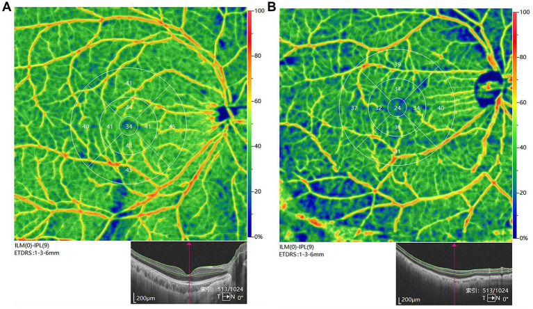
Objective: This study aims to investigate differences in macular vascular density (MVD) between individuals with chronic primary angle-closure glaucoma (CPACG) and healthy controls, as well as to evaluate cross-sectional changes in MVD at various stages of CPACG.
Method: This is a retrospective study based on the epidemiological survey of eye diseases in the local community, including 47 eyes of CPACG subjects (20 eyes at the early stage and 27 eyes at the middle-to-severe stages). All subjects underwent optical coherence tomography angiography (OCTA) imaging to detect MVD, as well as macular retinal nerve fiber layer (RNFL) and ganglion cell layer (GCL) thickness. Linear regression analysis was performed to evaluate other ophthalmic indicators related to vascular density loss.
Results: Compared to the control group, the MVD in CPACG eyes significantly declined by 11.5% in the superficial capillary plexus (p = 0.012) and 6.8% in the deep capillary plexus. Single correlation analysis showed that MVD in CPACG eyes was significantly correlated with axial length (r = 0.493, p = 0.036), RNFL thickness (r = 0.488, p = 0.047), and mean deviation of the visual field (r = -0.546, p = 0.010). In addition, multiple regression analysis also suggested that MVD was positively correlated with GCL/RNFL thickness and negatively correlated with the mean deviation of the visual field (p = 0.004).
Conclusion: Our study demonstrated that OCTA was a valuable tool for detecting vascular deterioration in CPACG eyes, with a stronger association between MVD and visual field damage. Further research is warranted to explore the potential of MVD as a biomarker for glaucoma progression.
Keywords: ganglion cell layer; macular retinal nerve fiber layer; macular vascular density; optical coherence tomography angiography; primary angle closure glaucoma; visual field defects.
Copyright © 2025 Zhang, Lu, Yu, Liu, Yang, Wang and Wang.
Conflict of interest statement
The authors declare that the research was conducted in the absence of any commercial or financial relationships that could be construed as a potential conflict of interest.
Figures

Similar articles
-
Ganglion Cell Layer Thickness as a Biomarker for Amyotrophic Lateral Sclerosis Functional Outcome: An OCT study.Rom J Ophthalmol. 2025 Apr-Jun;69(2):200-207. doi: 10.22336/rjo.2025.32. Rom J Ophthalmol. 2025. PMID: 40698100 Free PMC article.
-
Progressive Macular Vessel Density Loss Observed on Optical Coherence Tomography Angiography in Glaucoma Patients With Single-Hemifield Visual Field Defects.J Glaucoma. 2023 Aug 1;32(8):658-664. doi: 10.1097/IJG.0000000000002225. Epub 2023 Mar 30. J Glaucoma. 2023. PMID: 37054404
-
Assessment of Retinal Microcirculation in Primary Open-Angle Glaucoma Using Adaptive Optics and OCT Angiography: Correlation with Structural and Functional Damage.J Clin Med. 2025 Jul 14;14(14):4978. doi: 10.3390/jcm14144978. J Clin Med. 2025. PMID: 40725669 Free PMC article.
-
Peripheral iridotomy for pigmentary glaucoma.Cochrane Database Syst Rev. 2016 Feb 12;2(2):CD005655. doi: 10.1002/14651858.CD005655.pub2. Cochrane Database Syst Rev. 2016. PMID: 26871761 Free PMC article.
-
Optical coherence tomography (OCT) for detection of macular oedema in patients with diabetic retinopathy.Cochrane Database Syst Rev. 2015 Jan 7;1(1):CD008081. doi: 10.1002/14651858.CD008081.pub3. Cochrane Database Syst Rev. 2015. PMID: 25564068 Free PMC article.
References
LinkOut - more resources
Full Text Sources