Intraocular pressure and ocular biomechanical parameters vary between generations of the ocular response analyzer in healthy and ectatic eyes
- PMID: 40776931
- PMCID: PMC12328285
- DOI: 10.3389/fmed.2025.1605641
Intraocular pressure and ocular biomechanical parameters vary between generations of the ocular response analyzer in healthy and ectatic eyes
Abstract
Introduction: This study evaluated the agreement between a third-generation (G3) ocular response analyzer (ORA) and a first-generation (G1) ORA, and tested the ability of the keratoconus match index (KMI) to identify keratoconus.
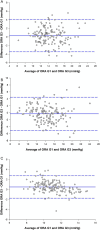
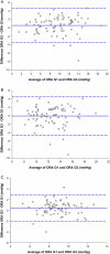
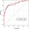
Methods: Healthy participants (n = 149 eyes) and participants with keratoconus (n = 78 eyes) were enrolled for this study. Four measurements were taken bilaterally using the G1 and G3 ORA. Goldmann-correlated intraocular pressure (IOPg), corneal-compensated IOP (IOPcc), corneal hysteresis (CH), waveform score, KMI, and waveform parameters area under the first applanation peak (p1area), area under the second applanation peak (p2area), width of the first applanation peak (w1), width of the second applanation peak (w2), height of the first applanation peak (h1), and height of the second applanation peak (h2) were recorded from the measurement with the highest waveform score in the left eye. Paired t-tests or Wilcoxon signed-rank tests were used to assess agreement between the devices, and receiver-operating characteristic curves determined the ability of KMI to identify eyes with keratoconus.
Results: There was no difference in IOPcc or IOPg between the devices in both cohorts. CH was significantly greater for the G3 than for the G1 in healthy participants but not in keratoconus participants. For both cohorts, measurements of waveform score, KMI, p1area, p2area, w2, h1, and h2 were greater for the G3 than for the G1. Only w1 was smaller for the G3 than for the G1. There was no difference in the ability of KMI to differentiate ectatic from healthy eyes between the devices.
Discussion: Although the G1 and G3 can identify keratoconus using KMI, there is meaningful variation between them in IOP and biomechanical outcome parameters. Thus, clinicians and researchers should compare results between the devices with caution and should state which generation produced the data.
Keywords: cornea; corneal hysteresis; intraocular pressure; keratoconus; tonometry; waveform parameters.
Copyright © 2025 Yuhas, Fortman, Nye, Mahmoud and Roberts.
Conflict of interest statement
PY has received an honorarium from CooperVision, Inc. for work unrelated to this manuscript. CR is a consultant for Ziemer Ophthalmic Systems AG and for Oculus Optikerate GmbH. Reichert provided technical expertise but did not influence the study in any way. The remaining authors declare that the research was conducted in the absence of any commercial or financial relationships that could be construed as a potential conflict of interest.
Figures


Similar articles
-
Waveform Score Influences the Outcome Metrics of the Ocular Response Analyzer in Patients with Keratoconus and in Healthy Controls.Curr Eye Res. 2025 Jul;50(7):700-709. doi: 10.1080/02713683.2025.2489607. Epub 2025 Apr 10. Curr Eye Res. 2025. PMID: 40210210
-
Reliability of Measurements Using Ocular Response Analyzer as a Screening Tonometer and Corneal Hysteresis Values in the Presence or Absence of Glaucomatous Changes in Fundus.J Glaucoma. 2024 Mar 1;33(3):183-188. doi: 10.1097/IJG.0000000000002312. Epub 2023 Sep 12. J Glaucoma. 2024. PMID: 37748090 Free PMC article.
-
Assessment of Tonometry Methods in Keratoconic Eyes Following Intracorneal Ring Segments Implantation: A Comparative Study.Korean J Ophthalmol. 2025 Jun;39(3):231-240. doi: 10.3341/kjo.2025.0016. Epub 2025 May 7. Korean J Ophthalmol. 2025. PMID: 40329769 Free PMC article.
-
Perioperative medications for preventing temporarily increased intraocular pressure after laser trabeculoplasty.Cochrane Database Syst Rev. 2017 Feb 23;2(2):CD010746. doi: 10.1002/14651858.CD010746.pub2. Cochrane Database Syst Rev. 2017. PMID: 28231380 Free PMC article.
-
Deep anterior lamellar keratoplasty versus penetrating keratoplasty for treating keratoconus.Cochrane Database Syst Rev. 2014 Jul 22;2014(7):CD009700. doi: 10.1002/14651858.CD009700.pub2. Cochrane Database Syst Rev. 2014. PMID: 25055058 Free PMC article.
References
-
- Ehrlich JR, Haseltine S, Shimmyo M, Radcliffe NM. Evaluation of agreement between intraocular pressure measurements using Goldmann applanation tonometry and Goldmann correlated intraocular pressure by Reichert's ocular response analyser. Eye (Lond). (2010) 24:1555–60. doi: 10.1038/eye.2010.83, PMID: - DOI - PubMed
-
- Kamalipour A, Moghimi S, Eslani M, Nishida T, Mohammadzadeh V, Micheletti E, et al. A prospective longitudinal study to investigate corneal hysteresis as a risk factor of central visual field progression in glaucoma. Am J Ophthalmol. (2022) 240:159–69. doi: 10.1016/j.ajo.2022.02.025, PMID: - DOI - PMC - PubMed
LinkOut - more resources
Full Text Sources

