. 2025 Jul 22;26(7):37399.
doi: 10.31083/RCM37399. eCollection 2025 Jul.

Accelerated Non-Contrast-Enhanced Three-Dimensional Cardiovascular Magnetic Resonance Deep Learning Reconstruction

Affiliations

Accelerated Non-Contrast-Enhanced Three-Dimensional Cardiovascular Magnetic Resonance Deep Learning Reconstruction

Sukran Erdem et al. Rev Cardiovasc Med. .

Abstract

Background: Cardiovascular magnetic resonance (CMR) is a time-consuming, yet critical imaging method. In contrast, while rapid techniques accelerate image acquisition, these methods can also compromise image quality. Meanwhile, the effectiveness of Adaptive CS-Net, a vendor-supported deep-learning magnetic resonance (MR) reconstruction algorithm, for non-contrast three-dimensional (3D) whole-heart imaging using relaxation-enhanced angiography without contrast and triggering (REACT) remains uncertain.

Methods: Thirty participants were prospectively recruited for this study. Each underwent non-contrast imaging that included a modified REACT sequence and a standard 3D balanced steady-state free precession (bSSFP) sequence. The REACT data were acquired through six-fold undersampling and reconstructed offline using both conventional compressed sensing (CS) and an Adaptive CS-Net algorithm. Subjective and objective image quality assessments, as well as cross-sectional area measurements of selected vessels, were conducted to compare the REACT images reconstructed using Adaptive CS-Net against those reconstructed using conventional CS, as well as the standard bSSFP sequence. For a statistical comparison of image quality across these three image sets, the nonparametric Friedman test was performed, followed by Dunn's post-hoc test.

Results: The Adaptive CS-Net and CS-reconstructed REACT images exhibited superior image quality for pulmonary veins, neck, and upper thoracic vessels compared to the standard 3D bSSFP sequence. Adaptive CS-Net and CS reconstructed REACT images displayed significantly higher contrast-to-noise ratio (CNR) compared to those reconstructed using the 3D bSSFP sequence (all p-values < 0.05) for the left upper (5.40, 5.53, 0.97), left lower (6.33, 5.84, 2.27), right upper (5.49, 6.74, 1.18), and right lower pulmonary veins (6.71, 6.41, 1.26). Additionally, REACT methods showed a statistically significant improvement in CNR for both the ascending aorta and superior vena cava compared to the 3D bSSFP sequence.

Conclusions: The Adaptive CS-Net reconstruction for the REACT images consistently delivered superior or comparable image quality compared to the CS technique. Notably, the Adaptive CS-Net reconstruction provides significantly enhanced image quality for pulmonary veins, neck, and upper thoracic vessels compared to 3D bSSFP.

Keywords: Adaptive CS-Net; artificial intelligence; cardiovascular magnetic resonance; congenital heart disease; deep learning.


Conflict of interest statement

The authors declare no conflict of interest.

Figures
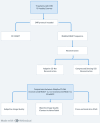
Fig. 1.
Fig. 1.
Study design and analysis workflow. CHD, congenital heart disease; CMR, cardiovascular magnetic resonance; REACT, relaxation-enhanced angiography without contrast and triggering; bSSFP, balanced steady-state free precession; 3D, three-dimensional.
Fig. 2.
Fig. 2.
Reviewer agreement heatmaps based on Cohen’s kappa analysis for interrater reliability across three imaging modalities. These heatmaps visualize interrater agreement between two reviewers for the qualitative assessment of cardiac image quality across three MRI reconstruction techniques: Adaptive CS-Net reconstructed REACT (left), CS reconstructed REACT (center), and 3D bSSFP (right). Each matrix cell represents the count of ratings given by Reviewer 1 (y-axis) and Reviewer 2 (x-axis), for image quality scores ranging from 0 to 4. Higher diagonal counts indicate stronger agreement. Cohen’s kappa values were computed for each modality to assess the strength of interrater agreement, with red hues denoting higher agreement counts. 3D bSSFP, three-dimensional balanced steady state free precession; CS, compressed sense; REACT, relaxation-enhanced angiography without contrast and triggering; MRI, magnetic resonance imaging.
Fig. 3.
Fig. 3.
Bland-Altman analysis of cross-sectional area comparisons between imaging techniques. The Blant-Altman analysis comparing cross-sectional areas (CSA) (millimeter square (mm2)) between 6-fold accelerated acquisition with deep learning (DL) (Adaptive CS-Net) reconstructed REACT (relaxation-enhanced angiography without contrast and triggering) and 6-fold accelerated acquisition with conventional CS (compressed sensing) reconstructed REACT (a–c) and between 6-fold accelerated acquisition with Adaptive CS-Net reconstructed REACT and 3D bSSFP (d–f). The black line indicates the mean difference of the diameter measurements, whereas the red lines represent the 95% confidence interval. The bias reflects the p-value of the regression coefficient when the mean difference is regressed against the average. (a) Coaxial right ventricular outflow tract (RVOT) at valvar level CSA measurements of Adaptive CS-Net and CS reconstructed REACT images demonstrate excellent agreement with a mean difference of 0.16 mm2 (95% confidence interval (CI) –36.70 to 37.03). (b) Coaxial right pulmonary artery (RPA) CSA measurements of Adaptive CS-Net and CS reconstructed REACT images demonstrate excellent agreement of a mean difference of 1.72 mm2 (95% CI –33.63 to 37.08). (c) Coaxial Transverse Arch CSA measurements of Adaptive CS-Net and CS reconstructed REACT demonstrate excellent agreement of a mean difference of 0.7 mm2 (95% CI –30.81 to 32.20). (d) Coaxial RVOT at valvar level CSA measurements of Adaptive CS-Net and 3D bSSFP images demonstrate good agreement with a mean difference of –20.6 mm2 (95% CI –134.87 to 93.67). (e) Coaxial RPA CSA measurements of Adaptive CS-Net and 3D bSSFP images demonstrate excellent agreement of a mean difference of –7.58 mm2 (95% CI –53.46 to 38.29). (f) Coaxial Transverse Arch CSA measurements of Adaptive CS-Net and 3D bSSFP demonstrate excellent agreement of a mean difference of –6.31 mm2 (95% CI –40.14 to 27.51). 3D bSSFP, three-dimensional balanced steady state free precession.
Fig. 4.
Fig. 4.
Comparison of thoracic vessel imaging using Adaptive CS-Net reconstructed REACT, CS reconstructed REACT, and 3D bSSFP. The upper thoracic and neck vessels in the coronal view (column a) and multiplanar reformats (columns b and c) of the ascending aorta (AAo, red arrow) and aortic arch (blue arrow) are displayed. The REACT (relaxation-enhanced angiography without contrast and triggering) technique produced uniform signals for the AAo and aortic arch, offering better border delineation compared to 3D bSSFP, which was maintained in both Adaptive CS-Net and CS (compressed sense) reconstructed images. Additionally, both REACT techniques provided higher-resolution images of the upper thoracic (yellow arrow) and neck vessels (green arrow) compared to 3D bSSFP. Multiplanar reformats of the right and left ventricles in the sagittal view (column d) show mild blurring in CS reconstructed REACT images which were reduced with Adaptive CS-Net reconstruction. The left ventricular endocardial border, papillary muscles, and right ventricular trabeculations and papillary muscles were sharply defined with 3D bSSFP compared to REACT techniques. Comparing the Adaptive CS-Net reconstructed REACT and CS reconstructed REACT, we can see significant noise reduction in the images, illustrating better image quality from Adaptive CS-Net reconstructed REACT. LV, left ventricle; RV, right ventricle; 3D bSSFP, three-dimensional balanced steady state free precession.
Fig. 5.
Fig. 5.
Performance comparisons of 3D whole-heart techniques in demonstrating pulmonary veins and pulmonary arteries. Acquisitions were performed with relaxation-enhanced angiography without contrast and triggering (REACT) and 3D bSSFP. REACT images were reconstructed with Adaptive CS-Net and compressed sensing (CS). Multiplanar reconstructions of pulmonary veins with close-up views show that REACT techniques provided significantly better image quality by suppressing flow and off-resonance artifacts. In column a, red arrows represent the upper branch of the right pulmonary artery, which was visualized with Adaptive CS-Net reconstructed REACT but not with CS reconstructed REACT and 3D bSSFP. Adaptive CS-Net reconstruction was not only a useful method in imaging pulmonary veins but also proved to be a significant aid in imaging pulmonary arteries. Column b shows left pulmonary veins and right upper pulmonary vein. Signal loss seen with 3Db SSFP is not seen with REACT images, significantly improving image quality. In column c, the blue arrows represent the left lower pulmonary veins. REACT images demonstrate the left lower pulmonary veins in full length and better resolution, while 3D bSSFP exhibited dephasing artifacts and reduced image quality. 3D bSSFP, three-dimensional balanced steady state free precession.
Fig. 6.
Fig. 6.
Coronary artery imaging: adaptive CS-Net vs. CS reconstructed REACT and 3D bSSFP. All methods successfully visualized the coronary arteries. Multiplanar reformats (with the right coronary artery labeled by a red arrow in columns a and c, and the left main coronary artery labeled by a blue arrow in columns b and d) demonstrated that 3D bSSFP provided superior image quality compared to relaxation-enhanced angiography without contrast and triggering (REACT) images reconstructed with conventional compressed sensing (CS), which showed blurred vessel borders. In contrast, Adaptive CS-Net reconstruction improved image sharpness, providing higher signal quality, reduced noise, and less blurring than standard CS reconstruction. 3D bSSFP, three-dimensional balanced steady state free precession.

Similar articles

References

    1. Seetharam K, Lerakis S. Cardiac magnetic resonance imaging: the future is bright. F1000Research . 2019;8:1636. doi: 10.12688/f1000research.19721.1. F. - DOI - PMC - PubMed
    1. Demirkiran A, Everaars H, Amier RP, Beijnink C, Bom MJ, Götte MJW, et al. Cardiovascular magnetic resonance techniques for tissue characterization after acute myocardial injury. European Heart Journal. Cardiovascular Imaging . 2019;20:723–734. doi: 10.1093/ehjci/jez094. - DOI - PubMed
    1. Scheffler K, Lehnhardt S. Principles and applications of balanced SSFP techniques. European Radiology . 2003;13:2409–2418. doi: 10.1007/s00330-003-1957-x. - DOI - PubMed
    1. Krishnam MS, Tomasian A, Deshpande V, Tran L, Laub G, Finn JP, et al. Noncontrast 3D steady-state free-precession magnetic resonance angiography of the whole chest using nonselective radiofrequency excitation over a large field of view: comparison with single-phase 3D contrast-enhanced magnetic resonance angiography. Investigative Radiology . 2008;43:411–420. doi: 10.1097/RLI.0b013e3181690179. - DOI - PubMed
    1. Weber OM, Martin AJ, Higgins CB. Whole-heart steady-state free precession coronary artery magnetic resonance angiography. Magnetic Resonance in Medicine . 2003;50:1223–1228. doi: 10.1002/mrm.10653. - DOI - PubMed

LinkOut - more resources