. 2025 Jul 24:16:1631530.
doi: 10.3389/fimmu.2025.1631530. eCollection 2025.

Non-invasive physical plasma activates stimulator of interferon genes pathway in triple negative breast cancer and is associated with increased host immune response

Affiliations

Non-invasive physical plasma activates stimulator of interferon genes pathway in triple negative breast cancer and is associated with increased host immune response

Guilin Wang et al. Front Immunol. .

Abstract

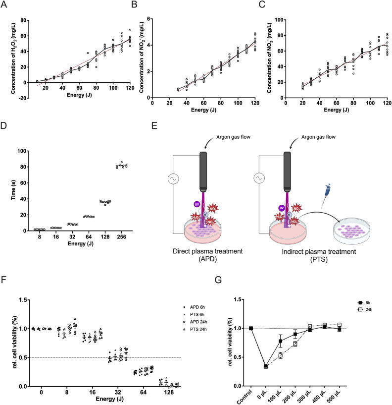
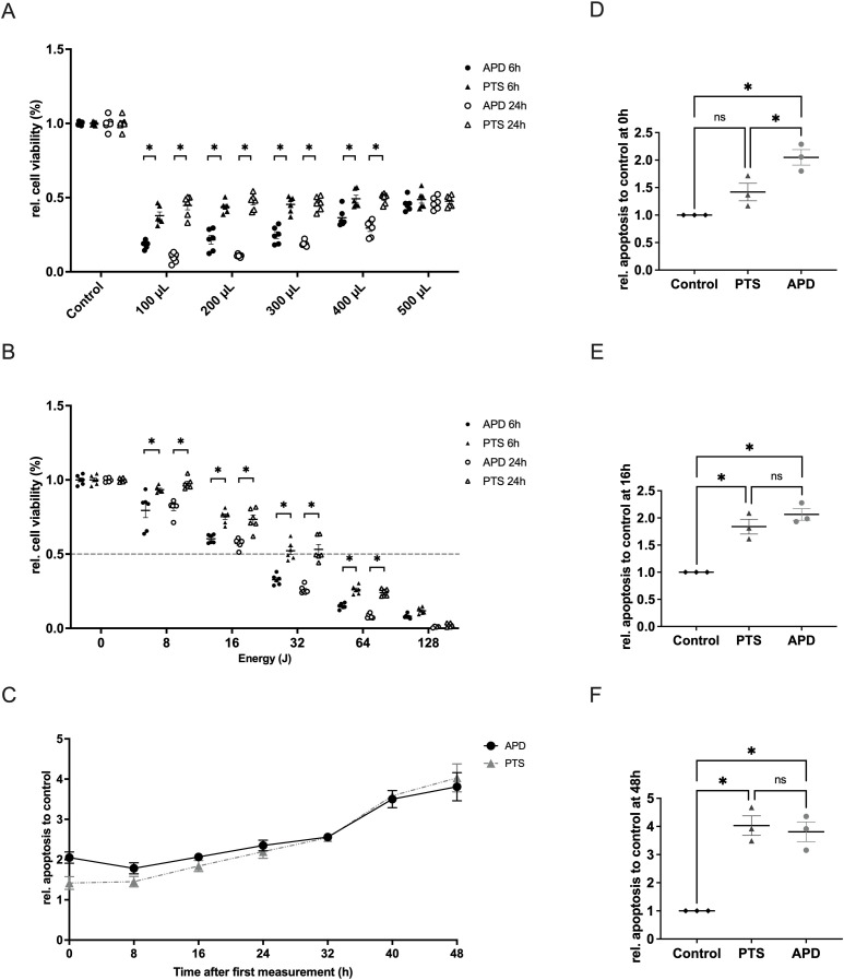
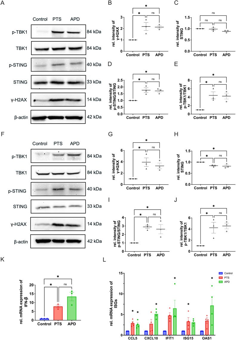
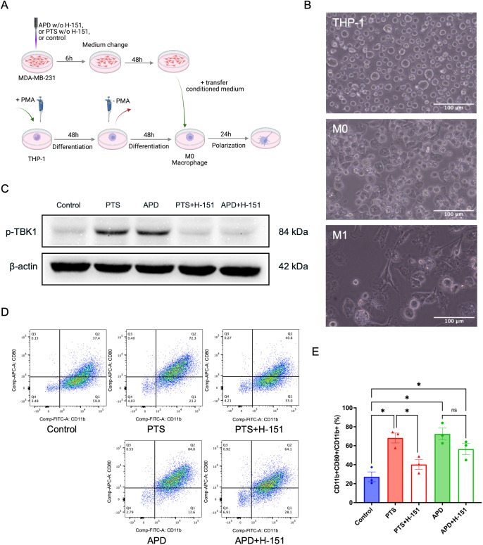
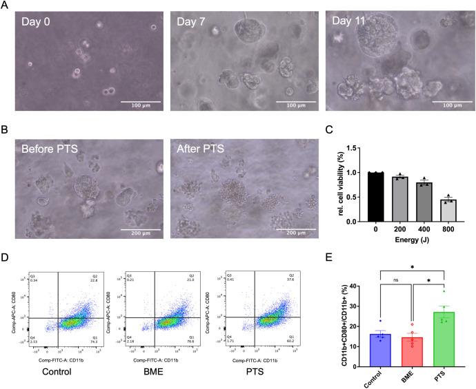
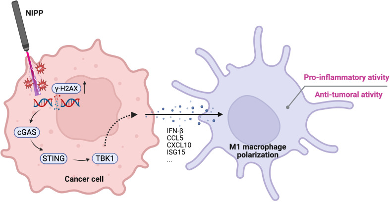
Triple-negative breast cancer (TNBC), characterized by the absence of ER, PR, and HER2 receptors, remains one of the most aggressive breast cancer subtypes, with limited therapeutic options and a high relapse rate. While immune checkpoint inhibitors (ICIs) have shown promise by leveraging TNBC's immunogenic profile, their use is often accompanied by significant toxicity, necessitating the development of safer immunomodulatory strategies. Non-invasive physical plasma (NIPP), a novel low thermal plasma technology that can be generated using various gases, including argon, and producing reactive oxygen and nitrogen species (RONS), has emerged as a potential alternative. This study investigates the capacity of direct (argon plasma devitalization, APD) and indirect (plasma-treated solution, PTS) plasma modalities to induce cytotoxicity and activate immune signaling via the stimulator of interferon genes (STING) pathway in TNBC. Dose-dependent RONS generation by APD and PTS correlated with reduced viability and apoptosis induction in MDA-MB-231 TNBC cells. Both plasma modalities caused DNA damage and upregulated key proteins in the STING pathway, including γ-H2AX, p-STING, and p-TBK1, with sustained activation observed up to 24 hours post-treatment. Furthermore, STING-dependent transcription of IFN-β and interferon-stimulated genes (ISGs) confirmed the immunogenic potential of NIPP. Conditioned media from plasma-treated TNBC cells induced M1 polarization in THP-1-derived macrophages, an effect significantly reduced upon specific STING inhibition with H-151. The immunomodulatory effects of NIPP were validated in patient-derived TNBC organoids, where plasma treatment disrupted organoid structure, reduced viability, and promoted M1 macrophage polarization. Collectively, these findings highlight the dual cytotoxic and immunostimulatory potential of NIPP in TNBC through STING pathway activation, claiming it as a promising, low-toxicity component in combination with conventional immunotherapy.

Keywords: argon plasma devitalization (APD); cold atmospheric plasma (CAP); non-invasive physical plasma (NIPP); plasma-treated solution (PTS); triple negative breast cancer.


Conflict of interest statement

ME is employee of Erbe Elektromedizin GmbH. The remaining authors declare that the research was conducted in the absence of any commercial or financial relationships that could be construed as a potential conflict of interest.

Figures

Figure 1
Figure 1
Characterization of NIPP and its cytotoxic effect in triple-negative breast cancer cells. (A–C) Correlation between plasma energy and the production of H2O2 (A), NO2 - (B), and NO3 - (C). Data points represent measured values. The regression line is shown as a red solid line. (D) Time required for the NIPP device to generate specific plasma energy. (E) Schematic representation of direct plasma treatment (APD) and indirect plasma treatment (PTS) using NIPP. (F) Relative cell viability of MDA-MB-231 cells after 6 hours or 24 hours of incubation following APD, PTS, and control treatments. Cells were covered with 500 µL of M2 medium during APD treatment. (G) Relative cell viability of MDA-MB-231 cells after 6 hours or 24 hours of incubation following 36 seconds of gas flow intervention and control treatment. Cells were covered with various volumes of M2 medium (0 µL, 100 µL, 200 µL, 300 µL, 400 µL, and 500 µL) and topped up to 500 µL during incubation. Statistical comparison was performed with paired Student’s t-tests. (mean ± SEM; *p < 0.05). Panel (E) was created in BioRender. Weiss, M. (2025) https://BioRender.com/9zkxpqs.
Figure 2
Figure 2
Characterization of the cellular effects of APD and PTS on MDA-MB-231 cells when the protective effect of culture medium was excluded. (A) Relative cell viability of MDA-MB-231 cells after 6 hours or 24 hours of incubation following 32 Joules of APD, PTS, or control treatments. Cells were covered with various volumes of M2 medium (0 µL, 100 µL, 200 µL, 300 µL, 400 µL, and 500 µL) and topped up to 500 µL during incubation. (B) Relative cell viability of MDA-MB-231 cells after 6 hours or 24 hours of incubation following APD, PTS, or control treatments. Cells were covered with 400 µL of M2 medium during treatment. (C) Relative apoptosis levels of MDA-MB-231 cells within 48 hours after 6 hours of incubation following APD, PTS, or control treatments. Time point 0 h refers to the time immediately after the 6 hours incubation following treatment. (D–F) Comparison of apoptosis levels among the APD, PTS, and control groups at the 0 h (D), 16 h (E), and 48 h (F) time points. Statistical comparisons were performed using the Student’s t-test for two-group comparisons or one-way ANOVA for comparisons across multiple groups. (mean ± SEM; *p < 0.05).
Figure 3
Figure 3
APD and PTS activate the STING pathway in MDA-MB-231 cells. (A) Representative immunoblots showing γ-H2AX, total STING, p-STING, TBK1, and p-TBK1 expression in MDA-MB-231 cells after 6 hours of incubation following APD, PTS, or control treatment. β-actin was used as a loading control. (B–E) Quantification of relative protein expression levels: γ-H2AX (B), total STING (C), p-STING/STING ratio (D), and p-TBK1/TBK1 ratio (E). (F) Representative immunoblots of γ-H2AX, total STING, p-STING, TBK1, and p-TBK1 expression in MDA-MB-231 cells after 24 hours of incubation following APD, PTS, or control treatment. β-actin was used as a loading control. (G–J) Quantification of relative protein expression levels: γ-H2AX (G), total STING (H), p-STING/STING ratio (I), and p-TBK1/TBK1 ratio (J). (K) RT-qPCR analysis of the gene expression of IFN-β in MDA-MB-231 cells treated with APD, PTS, or control and incubated for 6 hours. (L) RT-qPCR analysis of the gene expression of CCL5, CXCL10, IFIT1, ISG15, and OAS1 in MDA-MB-231 cells treated with APD, PTS, or control and incubated for 6 hours. GAPDH was used as a reference gene. Statistical comparisons were conducted using one-way ANOVA for parametric data and the Kruskal-Wallis test for non-parametric data. (mean ± SEM; *p < 0.05).
Figure 4
Figure 4
APD and PTS polarize THP-1 derived macrophages into an M1 phenotype in a STING-dependent manner. (A) Schematic of conditioned media experiments using macrophages derived from THP-1 cells. (B) Representative brightfield images of THP-1, M0, and M1 macrophages. Scale bar: 100 μm. (C) Representative immunoblots showing p-TBK1 expression in MDA-MB-231 cells following APD, PTS, or control treatment, with or without the STING inhibitor H-151. β-actin was used as a loading control. (D) Representative flow cytometry plots of macrophages exposed to various conditioned media derived from MDA-MB-231 cells treated with APD, PTS, or control, with or without the STING inhibitor H-151. Subpopulations were gated based on CD11b and CD80 expression, with M1 macrophages identified as CD11b+ CD80+. (E) The respective statistical analysis of the flowcytometry results. Statistical comparison was performed with one-way ANOVA. (mean ± SEM; *p < 0.05). Panel (A) was created in BioRender. Weiss, M. (2025) https://BioRender.com/a98tm0v.
Figure 5
Figure 5
PTS induces M1 polarization of THP-1 derived macrophages in a TNBC patient-derived organoid model. (A) Representative brightfield images showing the formation of TNBC patient-derived organoids on day 0, day 7, and day 11. Scale bar: 100 μm. (B) Representative brightfield images of TNBC patient-derived organoids before and after PTS treatment. Scale bar: 200 μm. (C) Relative cell viability of TNBC patient-derived organoids after 24 h of incubation following PTS treatment, assessed using the CellTiter-Glo® 3D Cell Viability Assay. (D) Representative flow cytometry plots of macrophages exposed to conditioned media derived from TNBC patient-derived organoids treated with PTS or control. ‘BME’ refers to a subgroup with the BME biomaterial scaffold but without cells. M1 macrophages were identified as CD11b+ CD80+. (E) The respective statistical analysis of the flowcytometry results. Statistical comparison was performed with one-way ANOVA. (mean ± SEM; *p < 0.05).
Figure 6
Figure 6
Proposed mechanism of NIPP-induced immune activation. NIPP treatment generates RONS, leading to DNA damage in cancer cells, as indicated by elevated γ-H2AX expression. The aberrant cytosolic DNA fragments are detected by cGAS, which subsequently activates the STING pathway. This signaling cascade promotes the polarization of THP-1 derived macrophages toward a pro-inflammatory M1 phenotype, contributing to the anti-tumor immune response. Created in BioRender. Weiss, M. (2025) https://BioRender.com/mxg10am.

Similar articles

References

    1. Bianchini G, De Angelis C, Licata L, Gianni L. Treatment landscape of triple-negative breast cancer - expanded options, evolving needs. Nat Rev Clin Oncol. (2022) 19:91–113. doi: 10.1038/s41571-021-00565-2, PMID: - DOI - PubMed
    1. Costa RLB, Gradishar WJ. Triple-negative breast cancer: current practice and future directions. JOP. (2017) 13:301–3. doi: 10.1200/JOP.2017.023333, PMID: - DOI - PubMed
    1. Nolan E, Lindeman GJ, Visvader JE. Deciphering breast cancer: from biology to the clinic. Cell. (2023) 186:1708–28. doi: 10.1016/j.cell.2023.01.040, PMID: - DOI - PubMed
    1. Onkar SS, Carleton NM, Lucas PC, Bruno TC, Lee AV, Vignali DAA, et al. The great immune escape: understanding the divergent immune response in breast cancer subtypes. Cancer Discov. (2023) 13:23–40. doi: 10.1158/2159-8290.CD-22-0475, PMID: - DOI - PMC - PubMed
    1. Schmid P, Cortes J, Dent R, McArthur H, Pusztai L, Kümmel S, et al. Overall survival with pembrolizumab in early-stage triple-negative breast cancer. N Engl J Med. (2024) 391:1981–91. doi: 10.1056/NEJMoa2409932, PMID: - DOI - PubMed

MeSH terms

LinkOut - more resources