[Preprint]. 2025 Jul 23:2025.07.20.665823.
doi: 10.1101/2025.07.20.665823.

Comparative Proteomic Profiling of Receptor Kinase Signaling Reveals Key Trafficking Components Enforcing Plant Stomatal Development

Affiliations

Comparative Proteomic Profiling of Receptor Kinase Signaling Reveals Key Trafficking Components Enforcing Plant Stomatal Development

Pengfei Bai et al. bioRxiv. .

Abstract

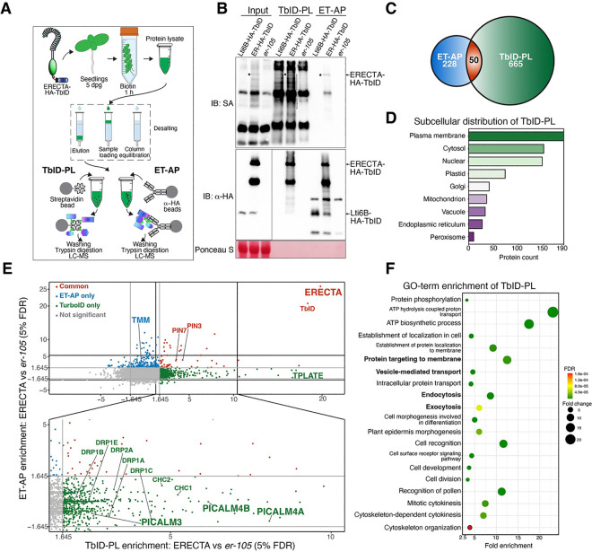
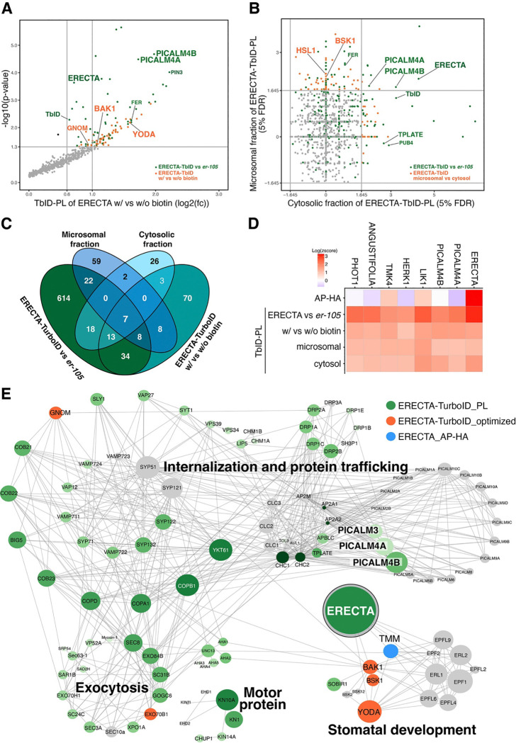
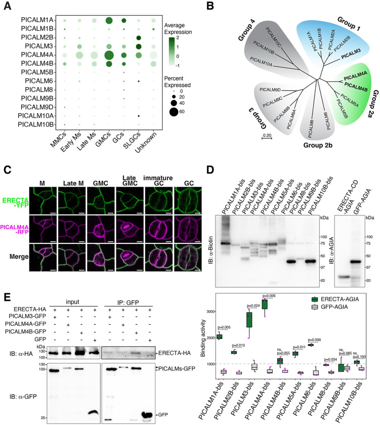
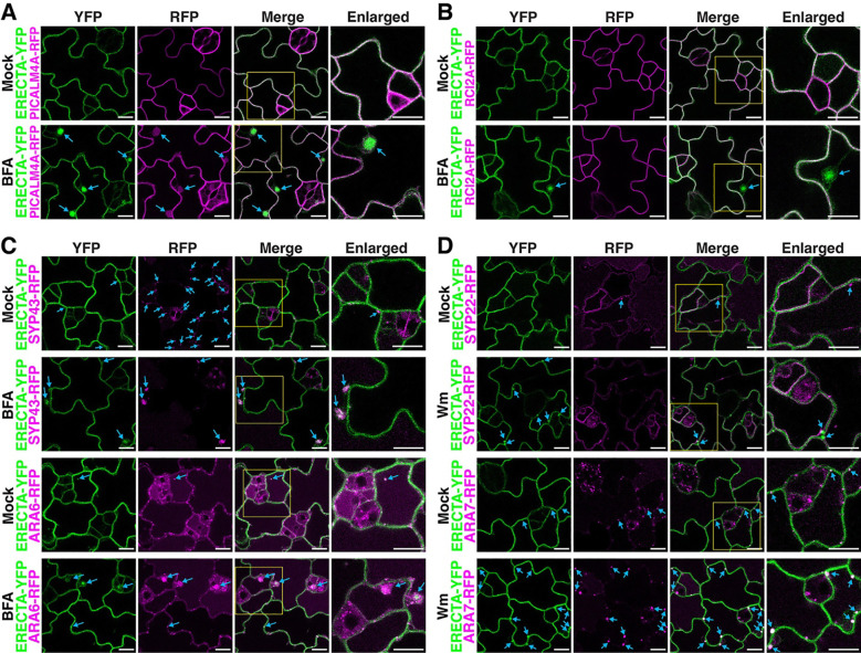
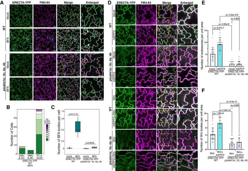
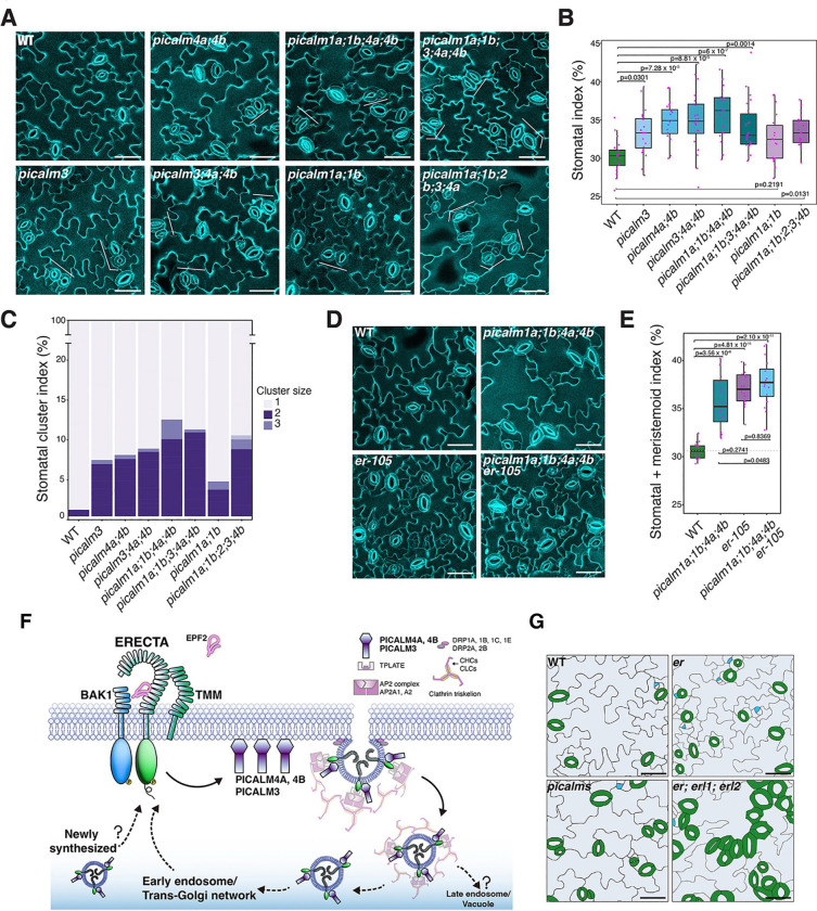
Receptor kinases are pivotal for growth, development, and environmental response of plants. Yet, their regulatory mechanisms and spatial dynamics are still underexplored. The ERECTA-family receptor kinases coordinate diverse developmental processes, including stomatal development. To understand the proteomic landscape of the ERECTA-mediated signaling pathways, we here report comparative analyses of the ERECTA interactome and proximitome by epitope-tagged affinity-purification (ET-AP) and TurboID-based proximity labeling (TbID-PL) mass-spectrometry, respectively. While ET-AP successfully recovered receptor complex components (e.g., TOO MANY MOUTHS), TbID-PL effectively captured transient associations with the components of endosomal trafficking, i.e. clathrin-mediated endocytosis (CME) machinery. We further identify that specific subfamily members of phosphatidylinositol-binding clathrin assembly proteins (PICALMs) interact with and synergistically regulate ERECTA internalization. Mutations of these PICALMs impair ERECTA endocytosis and lead to excessive stomatal clustering. Taken together, our work provides a proteomic atlas of the ERECTA signaling network and suggests that timely removal of receptor kinase by the endocytosis machinery is essential for active signal transduction enforcing stomatal patterning.


Conflict of interest statement

DECLARATION OF INTERESTS The authors declare no competing interests.

Figures

Figure 1.
Figure 1.. Comparative proteomic profiling of ERECTA-associated proteins.
(A) Schematic representation of the experimental workflow for profiling ERECTA-associated proteins using TbID-PL and ET-AP. Parallel enrichments of biotinylated proteins and HA-tagged protein complexes were subjected to mass spectrometry analysis. (B) Immunoblot validation of ERECTA and its associated proteins. Streptavidin-HRP (SA-HRP) (top) confirms successful biotinylation of proximal proteins via TbID-PL. α-HA immunoblot (middle) verifies the expression and enrichment of ERECTA-HA-TbID. Ponceau S staining (bottom) indicates loading control. (C) Venn diagram of identified proteins reveals 665 unique to TbID-PL, 228 unique to ET-AP, and 50 shared proteins. (D) Subcellular localization of TbID-PL enriched proteins, showing enrichment at the plasma membrane and cytosol, with additional representation in endomembrane compartments. (E) Scatterplot comparing protein enrichment between TbID-PL and ET-AP. Notable components of clathrin-mediated endocytosis (CME), such as PICALMs, DRPs, and TPLATE complex members, are preferentially captured by TbID-PL. In contrast, stable receptor partners like TMM are enriched via ET-AP. Some auxin transporters (PIN3, PIN7) are recovered in both methods. (F) GO-term enrichment of TbID-PL-identified proteins reveals overrepresentation of membrane trafficking, endocytosis, protein localization, and epidermal development terms. Circle size represents fold enrichment; color indicates false discovery rate (FDR) values.
Figure 2.
Figure 2.. Optimized TbID-PL enhances detection of ERECTA-associated signaling components and compartment-specific proteomes.
(A) Scatterplot showing differential enrichment of biotinylated proteins in ERECTA-TbID-PL with biotin versus without biotin treatments. Several known ERECTA signaling components, including BAK1, YODA (MAPKKK4) are identified (orange) alongside clathrin-mediated endocytosis proteins (e.g., PICALM4A, PICALM4B) identified (green) in Figure 1. (B) Scatterplot comparing enrichment between microsomal and cytosolic fractionation in TbID-PL proteomes. Membrane-attached receptor-like cytoplasmic kinase BSK1 is predominantly enriched in the microsomal fraction, while CME components (e.g., PICALM4A, 4B) and ERECTA are detected in both fractions. (C) Venn diagram showing overlap among TbID-PL datasets derived from comparisons of ERECTA-HA-TbID vs er-105, +biotin vs −biotin, and microsomal vs cytosolic fractions. Seven candidates are consistently detected across all comparisons. (D) Heatmap showing normalized enrichment (log2 z-scores) of the top 7 consistently identified ERECTA-proximal proteins across four TbID-PL conditions. These include ERECTA itself, PICALM4A/B, and kinases implicated in receptor signaling (LIK1, HERK1, TMK4), as well as ANGUSTIFOLIA and PHOT1. (E) STRING network analysis of ERECTA-associated proteins identified by TbID-PL (green), optimized TbID-PL (orange), or ET-AP (blue). Proteins are clustered into major functional categories: internalization/protein trafficking, exocytosis, motor proteins, and stomatal development. ERECTA, CME components (PICALMs, TPLATE, DRPs), and signaling partners (e.g., BAK1, YODA, TMM) are highlighted. Node size reflects interaction confidence.
Figure 3.
Figure 3.. ERECTA selectively interacts with PICALM family of clathrin-mediated endocytosis (CME) components.
(A) Dot plot showing transcriptional expression of Arabidopsis PICALM family genes across stomatal lineage cell types from re-analyzed single-cell RNA-seq data. PICALM1, PICALM3, PICALM4A, and PICALM4B are present across different stages of stomatal lineage cells. PICALM1, PICALM4A, and PICALM4B are highly enriched in guard mother cell (GMC), guard cell (GC) stages, while PICALM2B and PICALM3 are highly enriched in stomatal lineage ground cells (SLGCs). The distinct expression of PICALMs suggest their potential roles in different stage of stomatal development. Dot size indicates proportion of expressing cells; dot color reflects expression levels. (B) Phylogenetic tree of Arabidopsis PICALM family members. PICALM3 (Group 1, blue) and PICALM4A/B (Group 2a, green) cluster into distinct clades with high proximity to ERECTA based on TbID-PL. The phylogenetic tree illustrates evolutionary relationships and potential functional specialization within PICALM family. (C) Confocal microscopy showing subcellular colocalization of endogenous promoters-driven ERECTA-YFP and PICALM4A-RFP in cotyledon epidermal cells at multiple stomatal lineage stages. M, meristemoid; GMC, guard mother cell; GC, guard cell. Scale bars, 10 μm. ERECTA-YFP colocalizes with PICALM4A-RFP at the plasma membrane in stomatal lineage cells, except at cell plates for dividing cells. (D) In vitro protein-protein interacting assay between ERECTA and PICALM family proteins. ERECTA cytoplasmic domain and PICALMs were in vitro translated for the AlphaScreen assay. Quantification of normalized binding activity (right) confirms the preferential binding for PICALM3, PICALM4A, and PICALM1A. Boxplots represent mean with SD from three biological replicates and significance was assessed by unpaired t-test. (E) Co-immunoprecipitation assays validating interactions between ERECTA and PICALM proteins transiently co-expressed in Arabidopsis protoplasts. Immunoblot analysis confirmed in-vivo binding of ERECTA-HA to PICALM3-GFP, PICALM4A-GFP, and PICALM4B-GFP, as shown in the red-boxed lanes.
Figure 4.
Figure 4.. ERECTA-YFP co-localizes with PICALM4A and endosomal markers in stomatal-lineage cells.
(A) Confocal microscopy showing colocalization of ERECTA-YFP with PICALM4A-RFP under mock and BFA-treated conditions. In untreated cells, both proteins localize to the plasma membrane. Brefeldin A (BFA) treatment results in BFA-induced endosomal compartments, where PICALM4A-RFP encapsulates and colocalizes with ERECTA-YFP (arrowheads). (B) Confocal microscopy showing that ERECTA-YFP does not colocalize with the plasma membrane marker RCI2A-mCherry in BFA bodies. Under BFA treatment, ERECTA-YFP is redistributed to endosomal compartments (arrowheads), while RCI2A-mCherry remains at the plasma membrane, indicating selective internalization of ERECTA. (C) Confocal microscopy imaging of ERECTA-YFP with the trans-Golgi network (TGN) marker SYP43-RFP (top) and the plasma membrane/early endosome marker ARA6-RFP (bottom). Under mock conditions, ERECTA-YFP localizes to the plasma membrane and partially colocalizes with SYP43 and ARA6 (arrowheads). Following BFA treatment, ERECTA-YFP colocalized with both SYP43-RFP and ARA6-RFP in BFA-induced endosomal compartments, confirming its trafficking through the TGN and early endosomes. (D) Confocal microscopy showing colocalization of ERECTA-YFP with the late endosome marker ARA7-RFP and the vacuolar membrane marker SYP22-RFP. Under mock conditions, limited colocalization is observed. Upon wortmannin treatment, ERECTA-YFP co-localizes with ARA7-RFP in these enlarged endosomes, indicating ERECTA’s progression toward late endosome and vacuoles.
Figure 5.
Figure 5.. PICALM proteins regulate ERECTA internalization.
(A) Confocal images of ERECTA-YFP internalization in WT and picalm1a;1b;4a;4b mutants under mock and BFA treatment. In WT, BFA induces the formation of ERECTA-YFP–positive endosomal compartments (BFA bodies, arrowheads). Whereas the number of BFA body is markedly reduced in the picalm1a;1b;4a;4b mutant, indicating impaired endocytic recycling. (B) Quantification of the number of cells with BFA bodies across stomatal lineage stages in WT. ERECTA-YFP endocytic activity is enriched in pavement cells (PCs) and stomatal lineage ground cells (SLGCs). (C) Box plot quantifying the number of BFA bodies per cell under mock and BFA treatments in WT and picalm1a;1b;4a;4b mutants. WT shows a significant increase in BFA bodies upon treatment, while picalm1a;1b;4a;4b mutant exhibits no significant response. (D) Confocal microscopy images showing ERECTA-YFP internalization in response to mEPF2 peptide, wortmannin, or their combination in WT and picalm1a;1b;4a;4b mutants. In WT, mEPF2 triggers endocytic accumulation of ERECTA-YFP that overlaps with FM4–64, and Wm enlarges late endosomes containing ERECTA-YFP. These responses are strongly reduced in the picalm1a;1b;4a;4b mutant (arrowheads). (E) Quantification of mEPF2-induced ERECTA-YFP puncta in WT and picalm1a;1b;4a;4b mutants shows impaired ligand-triggered ERECTA endocytosis in the mutant. (F) Quantification of ERECTA-YFP positive Wm bodies per cell in WT and picalm1a;1b;4a;4b mutant. Wm-induced accumulation is significantly reduced in the mutant, indicating defective trafficking to late endosomes.
Figure 6.
Figure 6.. PICALMs are required for proper stomatal patterning
(A) Confocal images of abaxial epidermis from wild-type (WT) and various picalm single, double, and higher-order mutants. WT plants exhibit normal stomatal development, whereas picalm mutants exhibit increased stomatal index with varying degrees of stomatal clustering. Scale bars, 40 μm. (B) Quantification of stomatal index in WT and picalm mutants. Box plots show a significant increase in stomatal index in picalm mutants compared to WT. Statistical analysis was determined using one-way ANOVA followed by Tukey’s HSD test. (C) Stomatal cluster index analysis in WT and picalm mutants. Stomatal clusters are categorized by size (e.g., two, three, or four stomata per cluster), highlighting a dramatic rise in clustering frequency in higher-order picalm mutants. (D) Confocal images of abaxial epidermis from WT, er-105, picalm1a;1b;4a;4b, and the quintuple mutant picalm1a;1b;4a;4b;er-105. The quintuple mutant shows similar stomatal numbering and clustering to er-105. Scale bars, 40 μm. (E) Quantification of combined stomatal and meristemoid index. The picalm1a;1b;4a;4b;er-105 quintuple mutant exhibits values comparable to er-105, indicating that PICALMs act in the same genetic pathway as ERECTA. (F) Proposed model of the ERECTA receptor internalization facilitated by PICALM proteins. ERECTA is internalized through CME involving adaptor proteins such as PICALM3, PICALM4A, and PICALM4B, along with the AP2 complex and TPLATE. Internalized receptors are sorted via early endosomes or the trans-Golgi network (TGN) for recycling, or trafficked to vacuoles for degradation. In picalm mutants, defective CME impairs receptor pool balance, disrupting stomatal patterning. The secretion of newly synthesized ERECTA and trafficking to late endosomes/vacuoles remain unresolved in picalm mutants. (G) Representative images comparing stomatal patterning in WT, picalms, er, and er; erl1; erl2 mutants. picalm mutants phenocopy er family mutants, displaying increased stomatal number and clustering, supporting a functional link between CME regulation and ERECTA-mediated signaling.

Similar articles

References

    1. Torii K.U. (2004). Leucine-rich repeat receptor kinases in plants: structure, function, and signal transduction pathways. Int Rev Cytol 234, 1–46. 10.1016/S0074-7696(04)34001-5. - DOI - PubMed
    1. Shiu S.H., and Bleecker A.B. (2001). Receptor-like kinases from Arabidopsis form a monophyletic gene family related to animal receptor kinases. Proc Natl Acad Sci U S A 98, 10763–10768. 10.1073/pnas.181141598. - DOI - PMC - PubMed
    1. Boutrot F., and Zipfel C. (2017). Function, Discovery, and Exploitation of Plant Pattern Recognition Receptors for Broad-Spectrum Disease Resistance. Annu Rev Phytopathol 55, 257–286. 10.1146/annurev-phyto-080614-120106. - DOI - PubMed
    1. Nolan T.M., Vukasinovic N., Liu D., Russinova E., and Yin Y. (2020). Brassinosteroids: multidimensional regulators of plant growth, development, and stress responses. Plant Cell 32, 295–318. 10.1105/tpc.19.00335. - DOI - PMC - PubMed
    1. Chen X., Ding Y., Yang Y., Song C., Wang B., Yang S., Guo Y., and Gong Z. (2021). Protein kinases in plant responses to drought, salt, and cold stress. J Integr Plant Biol 63, 53–78. 10.1111/jipb.13061. - DOI - PubMed

Publication types

LinkOut - more resources