[Preprint]. 2025 Jul 23:2025.07.19.665690.
doi: 10.1101/2025.07.19.665690.

An allosteric network governs Tom70 conformational dynamics to coordinate mitochondrial protein import

Affiliations

An allosteric network governs Tom70 conformational dynamics to coordinate mitochondrial protein import

Max J Bachochin et al. bioRxiv. .

Abstract

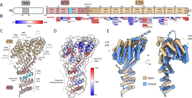
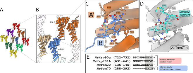
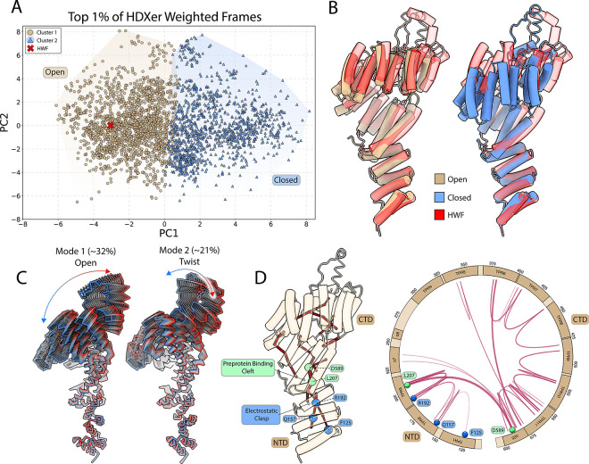
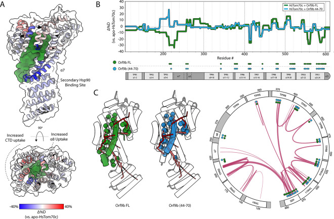
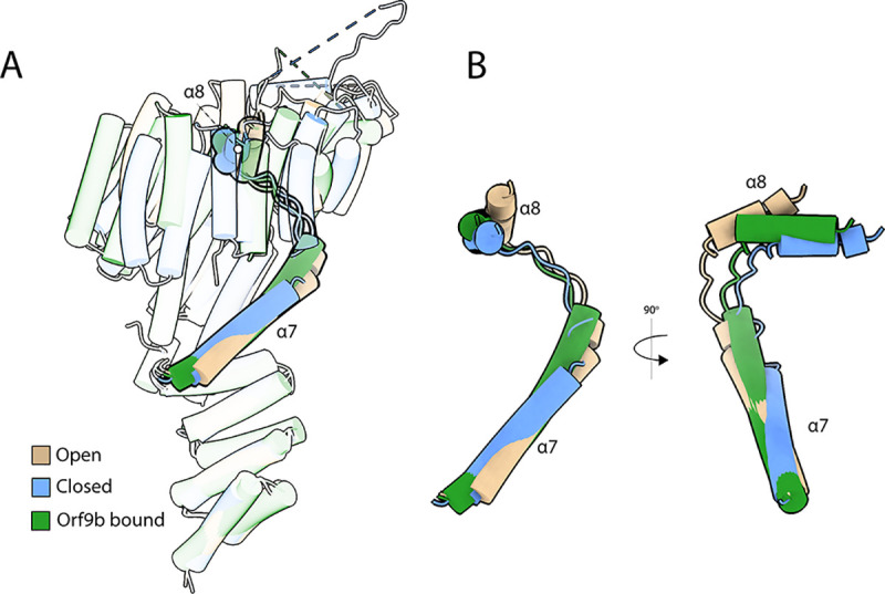
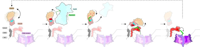
Tom70 mediates mitochondrial protein import by coordinating the transfer of cytosolic preproteins from Hsp70/Hsp90 to the translocase of the outer membrane (TOM) complex. In humans, the cytosolic domain of Tom70 (HsTom70c) is entirely α-helical and comprises modular TPR motifs divided into an N-terminal chaperone-binding domain (NTD) and a C-terminal preprotein-binding domain (CTD). However, the mechanisms linking these functional regions remain poorly understood. Here, we present the 2.04 Å crystal structure of unliganded HsTom70c, revealing two distinct conformations - open and closed - within the asymmetric unit. These states are stabilized in part by interdomain crystal contacts and are supported in solution by hydrogen-deuterium exchange mass spectrometry (HDX-MS) and molecular dynamics (MD) simulations. Principal component and dynamical network analyses reveal a continuum of motion linking the NTD and CTD via key structural elements, notably residues in helices α7, α8, and α25. Engagement of the CTD by the viral protein Orf9b interrupts this network, stabilizing a partially-closed intermediate conformation and dampening dynamics at distal NTD sites. Collectively, our findings lay the groundwork for understanding Tom70 allostery and provide a framework for dissecting its mechanistic roles in chaperone engagement, mitochondrial import, and viral subversion.


Conflict of interest statement

Competing Interests Statement The authors declare no competing interests.

Figures

Figure 1:
Figure 1:. Structure and dynamics of the cytosolic domain of HsTom70.
(A) Topology diagram of HsTom70 showing the transmembrane domain (TMD; grey), N-terminal domain (NTD; rose brown), and C-terminal domain (CTD; tan). Tetratricopeptide repeat (TPR) motifs and helices are also indicated. Dashed line at residue 108 indicates cytosolic domain of HsTom70 (residues 108–608; HsTom70c) used in this study. (B) HDX-MS peptide coverage map of HsTom70c aligned to the topology diagram. Peptides are colored by relative fractional deuterium uptake at the 2-minute exchange time point (0% uptake: blue; 66% uptake: red).(C) Ribbon representation of the 2.04 Å HsTom70c crystal structure (“open” conformation) annotated according to panel A. (D) HDX-MS relative fractional deuterium uptake heatmap at the 2-minute exchange time point overlaid on the open conformation ribbon structure. The chaperone-binding interface and NTD-CTD linker region are highlighted. (E) Structural superposition of open (tan) and closed (cornflower blue) HsTom70c conformations, aligned by the NTD. Conformational shifts in the C-terminal TPR motifs are emphasized.
Figure 2:
Figure 2:. Crystallographic contacts between asymmetric units mimic acidic peptide engagement of the electrostatic clasp.
(A) Arrangement of asymmetric units (ASUs) within the HsTom70c crystal lattice. (B) Interface between two neighboring ASUs, each containing one “open” (ASU1; chain A; tan) and one “closed” (ASU1; chain B; cornflower blue) conformation of HsTom70c. The disordered loop of the closed chain (chain B; cornflower blue) interacts with the electrostatic clasp of the adjacent open chain from a neighboring ASU (ASU2; chain A’; burnt orange). (C) Detailed molecular contacts mediating electrostatic clasp engagement. Residues 288–292 of the disordered loop from the closed chain (cornflower blue) form stabilizing interactions with clasp residues on the neighboring open chain (burnt orange). (D) Electrostatic clasp engagement of the acidic C-terminal tail of the yeast Hsp90 homolog ScHsp82 (cyan) with the yeast Tom70 homolog ScTom71c (light gray).
Figure 3:
Figure 3:. Molecular dynamics (MD) analysis of HsTom70c reveals coordinated motion and allosteric paths between the NTD and CTD.
(A) Top 1% of weighted frames from HDXer analysis plotted in principal component (PC) space and mapped onto the experimentally determined open and closed HsTom70c structures. Two major clusters were identified along the PC mode 1 (PC1) axis, corresponding to open (dark green circles) and closed (light green triangles) conformations. Cluster centroids (white crosses) and the highest-weighted frame (HWF; yellow circle) are indicated. (B) Structural alignment of the HWF (yellow) with the open and closed HsTom70c structures. (C) PC modes 1 and 2 illustrate the dominant opening/closing and twisting motions of HsTom70c, respectively. The percentage of total observed motion captured by each PC is indicated. (D) Representative dynamical network mapped onto the open state crystal structure (cartoon, tan) and a corresponding circular topology plot of HsTom70c. Nodes and edges persistent in ≥75% of the trajectory are shown as red lines. Line thickness indicates more frequently observed interactions. Key residues involved in acidic peptide binding at the NTD (F125, Q157, R192) are highlighted as blue circles; L207 and D589 are highlighted as green circles.
Figure 4:
Figure 4:. Orf9b tunes HsTom70c conformational dynamics via dual mechanisms of interaction.
(A) Heatmap of relative fractional deuterium uptake differences between the HsTom70c-Orf9b complex and apo-HsTom70c at the 2-minute exchange time point (−40% uptake: blue; +40% uptake: red; no sequence coverage: black). Data are mapped onto a ribbon representation of HsTom70c (transparent surface) bound to Orf9b (forest green surface). (B) Butterfly difference plot showing fractional deuterium uptake relative to apo-HsTom70c for two complexes: HsTom70c + full-length Orf9b (Orf9b FL, green) and HsTom70c + Orf9b (residues 44–70; blue). The plot is aligned with a topology diagram of HsTom70c annotated with contact regions derived from crystal structures.(C) Crystal structure of HsTom70c bound to either full-length Orf9b (PDB: 7DHG, green) or Orf9b(44–70, blue), shown as transparent cartoons. Contact residues between HsTom70c and Orf9b are depicted as solid-colored spheres (FL, green; 44–70, blue) and mapped onto both the representative dynamic network (red lines) and a circularized topology plot.
Figure 5:
Figure 5:. Orf9b locks HsTom70c into an intermediate conformation at a α7- α8 hinge point.
(A) Transparent overlay of the ”open” state (tan), “closed” state (cornflower blue), and Orf9b-bound (forest green; PDB: 7DHG) structures of HsTom70c, highlighting the positions of the α7 and α8 helices in each structure of the alignment. (B) Inset showing the overlay of α7 and α8 from the ”open”, ”closed”, and Orf9b-bound HsTom70c structures.
Figure 6:
Figure 6:. Simplified Model of Putative Tom70 Substrate Cycling.
The cytosolic domain of apo Tom70 extends from the outer mitochondrial membrane and dynamically samples “open” and “closed” conformations. The electrostatic clasp of Tom70 engages preprotein-bound (sea green) chaperones (teal) via the conserved acidic C-terminal EEVD motifs of cytosolic Hsp70/90 chaperones. This interaction initiates an allosteric relay that propagates conformational changes from the electrostatic clasp of Tom70’s N-terminal domain (NTD) to the C-terminal domain (CTD), priming the substrate binding cleft to receive nascent preproteins from chaperones. Upon substrate handoff, preprotein engagement and the ongoing allosteric signal induce conformational rearrangements in the NTD and electrostatic clasp that lower chaperone affinity, promoting chaperone dissociation. Preproteins are subsequently transferred to the TOM complex via Tom20 (red) via complexation of the electrostatic clasp of Tom70 through its own acidic DDVE motif, triggering Tom70’s allosteric network to facilitate substrate release to Tom20 and subsequent import into the mitochondrion.

Similar articles

References

    1. Pfanner N.; Warscheid B.; Wiedemann N. Nat Rev Mol Cell Biol 2019, 20, 267–284. - PMC - PubMed
    1. Juszkiewicz S.; Peak-Chew S.-Y.; Hegde R. S. MBoC 2025, 36, ed. by Ott M., ar39. - PMC - PubMed
    1. Araiso Y.; Imai K.; Endo T. Annu. Rev. Biochem. 2022, 91, 679–703. - PubMed
    1. Dekker P. J. T.; Ryan M. T.; Brix J.; Müller H.; Hönlinger A.; Pfanner N. Molecular and Cellular Biology 1998, 18, 6515–6524. - PMC - PubMed
    1. Meisinger C.; Ryan M. T.; Hill K.; Model K.; Lim J. H.; Sickmann A.; Müller H.; Meyer H. E.; Wagner R.; Pfanner N. Molecular and Cellular Biology 2001, 21, 2337–2348. - PMC - PubMed

Publication types

LinkOut - more resources