[Preprint]. 2025 Jul 25:2025.07.21.665989.
doi: 10.1101/2025.07.21.665989.

A Microphysiologic Model of the Cervical Epithelium Recapitulates Microbial, Immunologic, and Pathogenic Properties of Sexually Transmitted Infections

Affiliations

A Microphysiologic Model of the Cervical Epithelium Recapitulates Microbial, Immunologic, and Pathogenic Properties of Sexually Transmitted Infections

Katherine M Nelson et al. bioRxiv. .

Abstract

Sexually transmitted infections (STIs) of the cervicovaginal mucosa are among the most common global infections. Clinical studies have revealed that susceptibility to STIs and the subsequent host responses they elicit are frequently associated with vaginal microbiota compositions that facilitate infection. Current monolayer cell culture and animal models fail to reproduce the multilevel complexity required to investigate these relationships simultaneously and/or with sufficient physiological relevance. To address this limitation, we have developed a microphysiologic system (MPS) that models human cervical tissue, its microbiota, and is susceptible to infection by two prominent genital pathogens, Chlamydia trachomatis and Neisseria gonorrhoeae. Significantly, this MPS platform recapitulates essential dynamic, polymicrobial, immune, and pathogenic features of chlamydial and gonococcal infections as they occur in humans. The low-cost MPS device requires no specialized equipment or specific expertise and was experimentally validated for both chlamydial and gonococcal infections across multiple non-engineering, remotely located laboratories, demonstrating its transferability and reproducibility. The MPS platform described herein provides a novel tool for expanded research into genital infections in a reconstituted system that closely mimics the cervical epithelium, a significant advance over existing models.

Keywords: Microphysiologic model; cervix; chlamydia; gonorrhea; host-pathogen interactions; microbiome; organ on a chip.


Figures

Fig. 1.
Fig. 1.. The microphysiologic system (MPS).
(A) Flowchart of materials and information sharing between the collaborating labs. (B) Image of real device under flow with food dye showing the epithelial and basal channels and C) the full setup within the incubator. Inlet (*) and outlet (∪) reservoirs are held in yellow conical tube rack and connected to the device (^) via tubing at the inlets (*) and outlets (∪) in B. (D) Steps of cell culture in the insert outside of the device before clamping in the cassette to add fluid flow. 1 – Membrane (purple) in the insert (grey) is coated with ECM proteins to enable cell adherence. 2 – Cervical cells (yellow) and fibroblasts (dark purple) are seeded and cultured in media (pink) in the insert outside of the cassette to enable easier cell culture. 3 – Bacteria (green) are added, and the cassette (blue) is clamped around the insert to enable fluid flow.
Fig. 2.
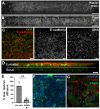
Fig. 2.. Establishment of a human cervical MPS incorporating cervical cells in the epithelial channel and BJ fibroblasts in the basal channel.
(A-B) Epifluorescence microscopy top-view images of the cervical MPS epithelium with A2EN cells in the epithelial channel cultured under (A) static conditions or (B) continuous flow conditions at 5 μL/min for 7 days, stained with Hoechst DNA stain (white). Scale bar, 1 mm. (C) XY confocal images of a A2EN cervical epithelium stained for E-cadherin (red) and nuclei (SYTOX-Green, green), after 7 days of static culture. Merged corresponding monochrome split images shown. Scale bar, 20 μm. (D) XZ side-view as in C. Dashed line is the membrane. Scale bar, 20 μm (E) Functional barrier permeability assessment of an empty insert (no cells) and a fully confluent A2EN cervical epithelium co-cultured with BJ fibroblasts assessed using a 150 KDa FITC-dextran permeability assay Data were analyzed with an unpaired t-test. (F-G) Top-view of the epithelial channel with primary cervical cells (HCxEC) stained for mucin 5B (MUC5B, green), F-actin (red), and nuclei (Hoechst, blue) at day 11 of culture, under static conditions. Scale bars are 10 and 20 μm, respectively.
Fig. 3.
Fig. 3.. Microbiota consortia grow in the MPS device and do not elicit negative epithelial response.
(A) Growth of the optimal and non-optimal microbiota consortia after 48 hours in the insert on A2EN and primary cervical (HCxEC) cells. Stability of the growth of optimal (B) and non-optimal (C) microbiota consortia in the insert on primary cervical cells (HCxEC) after 48 hours. (D) Bacteria comprising the optimal and non-optimal microbiota consortia (arrows) are alive after culture on epithelium. Scale bar, 10 μm. (E) Live/dead viability staining of HCxEC cells after 48 hours of growth without (i,ii) or with (iii-iv) each consortium and HCxEC cells treated with saponin (ii). Scale bar, 20 μm. (F) IL-8 and (G) IL-1β cytokine response of A2EN and HCxEC cells in the basal channel of the insert 48 hours after co-culture with optimal and non-optimal microbiota consortia. Significance determined by ANOVA * = p<0.05
Fig. 4.
Fig. 4.. C. trachomatis undergoes a complete developmental cycle in A2EN cervical cells co-cultured with BJ fibroblasts in a MPS device.
(A-B) Epifluorescence microscope images of the epithelial channel cultured under static conditions and infected with mCherry-expressing Ct. The mCherry channel is shown; each white dot represents Ct inclusions. (A) Low magnification of the entire channel at 48 hpi. Dotted lines indicate channel edge. Scale bar, 1 mm. (B) Representative images at higher magnification at 24 (left), 48 (middle), and 72 (right) hpi. Scale bar, 100 μm. (C) Epifluorescence microscopy merged images of A2EN epithelial cells in the apical channel of the MPS device, cultured under static conditions and infected with Ct expressing mCherry constitutively (red) and GFP under the control of a late developmental cycle promoter (green). Scale bar, 100 μm. (D) Quantification of the size of RB and EB containing inclusions at 24 and 48 hpi from three independent experiments. Mann-Whitney tests, **** p < 0.0001. (E) Quantification of recovered IFUs per insert at 48 hpi in response to increasing inoculum concentrations. Data represent three independent experiments and are presented as mean ± SEM.
Fig. 5.
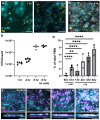
Fig. 5.. Protective capacity of the optimal microbiota consortium on HCxEC cells from Ct infection.
(A) Representative images of HCxEC cells co-cultured with BJ fibroblasts after 48 hours of co-culture with the optimal or non-optimal microbiota consortium. HCxEC nuclei are stained in blue, and chlamydiae are stained in green. Scale bar, 20 μm. (B) Quantification of infection with and without microbiota based on the number of cells with chlamydiae. (C) Representative images of secondary chlamydial infection in HeLa cells after HCxEC culture with or without microbiota. HeLa cell nuclei are stained in blue, and chlamydiae are stained in green. Scale bar, 20 μm. (D) Quantification of secondary infection in HeLa cells based on the number of cells with chlamydiae. **** p<0.0001
Fig. 6.
Fig. 6.. Ng infection of A2EN cells and consequent neutrophil transepithelial migration in the MPS device.
(A) Ng infection (yellow) at 1 hour and 8 hours post inoculation on A2EN and BJ fibroblast (cyan) laden inserts. Scale bar, 20 μm. (B) Quantification of Ng growth over time. Scale bar, 20 μm. Growth of Ng in cell culture medium in the MPS device without A2EN/fibroblast cells (no cells) is presented for comparison. (C) Infection of A2EN cells (cyan) with GFP+ (yellow) Ng that was prelabeled with Cell Trace Far Red (magenta). Cells were incubated for 6 hours under 2.5 μL/min flow conditions. As Ng replicates, magenta fluorescence is lost. (D) Quantification and (E) visualization of neutrophil (pink) migration through the insert in response to Ng (yellow) infection of the cervical epithelium (cyan), (timelapse movie: Supplemental Video S1). Ng was added for at t=−1 hr, PMN were added at t =0, images and measurements were taken at t = 1, 2, 4, 6 hr. Positive control, inserts to which the PMN chemoattractant fMLF was added for 6 hours; negative control, no fMLF addition and no Ng. Scale bar, 40 μm.

References

    1. CDC. Sexually Transmitted Infections Prevalence, Incidence, and Cost Estimates in the United States, April 2024.
    1. Chesson Harrell W., Mayaud Philippe, and Aral Sevgi O.. Sexually Transmitted Infections: Impact and Cost-Effectiveness of Prevention. In Holmes King K., Bertozzi Stefano, Bloom Barry R., and Prabhat Jha, editors, Major Infectious Diseases. The International Bank for Reconstruction and Development / The World Bank, Washington (DC), 3rd edition, 2017. ISBN 978-1-4648-0524-0978-1-4648-0525-7. - PubMed
    1. Global and regional STI estimates.
    1. Wiesenfeld Harold C., Hillier Sharon L., Krohn Marijane A., Landers Daniel V., and Sweet Richard L.. Bacterial vaginosis is a strong predictor of Neisseria gonorrhoeae and Chlamydia trachomatis infection. Clinical Infectious Diseases: An Official Publication of the Infectious Diseases Society of America, 36(5):663–668, March 2003. ISSN 1537-6591. doi: 10.1086/367658. - DOI - PubMed
    1. Tamarelle J., Thiébaut A. C. M., de Barbeyrac B., Bébéar C., Ravel J., and Delarocque-Astagneau E.. The vaginal microbiota and its association with human papillomavirus, Chlamydia trachomatis, Neisseria gonorrhoeae and Mycoplasma genitalium infections: a systematic review and meta-analysis. Clinical Microbiology and Infection: The Official Publication of the European Society of Clinical Microbiology and Infectious Diseases, 25(1):35–47, January 2019. ISSN 1469-0691. doi: 10.1016/j.cmi.2018.04.019. - DOI - PMC - PubMed

Publication types

LinkOut - more resources