[Preprint]. 2025 Jul 21:2025.07.16.665187.
doi: 10.1101/2025.07.16.665187.

Forced exercise modulates retinal inflammatory response and regulates miRNA expression to promote retinal neuroprotection during degeneration

Affiliations

Forced exercise modulates retinal inflammatory response and regulates miRNA expression to promote retinal neuroprotection during degeneration

Hayden Haupt et al. bioRxiv. .

Abstract

Background: Our labs have demonstrated exercise is protective in animal models of retinal degeneration (RD). Inflammation drives RD progression, and is regulated by the recruitment and reactivity of glia cells as well as through small non-coding RNAs, microRNAs (miRNAs). Here, we explore the effects of treadmill exercise on the recruitment and reactivity of retinal inflammatory cells within the neural retina and miRNA expression in a light-induced retinal degeneration model (LIRD) that exhibits phenotypes found in patients with RD.

Methods: Male 6-week-old BALB/c mice were randomly assigned to either active or inactive groups. Active groups were exercised by treadmill 1 hour a day for two weeks at a speed of 10m/min, meanwhile inactive groups were placed on static treadmills for the same duration. Light induced retinal degeneration (LIRD) was induced during the second week of exercise using light exposure of 5000 lux, control animals were kept at 50 lux. Retinal function was assessed using electroretinography (ERG) 5 days after LIRD. Retinas were collected 1-day and 5-days post-LIRD, sagittal sections were stained for inflammatory markers (GFAP and Iba1), TUNEL (cell death), and photoreceptor nuclei (outer nuclear layer; ONL) were quantified. RNA was extracted and miRNA expression quantified with GeneChip miRNA 4.0 array.

Results: Active+LIRD mice demonstrated significant preservation of retinal function, evidenced by higher a-wave and b-wave amplitudes in ERG 5-days post-LIRD, compared to inactive+LIRD mice. Retinal sections from active+LIRD mice had fewer Iba1+ cells and decreased GFAP labeling 5-days post-LIRD compared to inactive+LIRD mice. Active+LIRD mice had fewer ONL TUNEL+ cells compared to inactive+LIRD mice. Inactive+LIRD mice showed a decline in ONL counts 1-day post-LIRD with significant loss 5-days post-LIRD compared to active+LIRD mice. In active groups, exercise promoted significant differences in miRNA expression, such as miR-302b, miR-192-5p, miR-187 compared to inactive groups.

Conclusions: Our results indicate that treadmill exercise preserved photoreceptor density, slowed and or prevented apoptosis in the ONL, and decreased the presence/recruitment of inflammatory cells in the neural retina. Altered miRNA expression profiles in active groups are associated with cell survival (miR-302b), oxidative stress regulation (miR-192-5p) and photoreceptor homeostasis (miR-187). These results reveal how exercise alters the retinal inflammatory response over the course of 1-day to 5-days, providing insight into exercise-based therapies and treatments for RD and neuroinflammatory diseases.

Keywords: Exercise; Retinal Neuroprotection; Retinal inflammation; miRNA.


Conflict of interest statement

Competing interests Not applicable.

Figures

Figure 1.
Figure 1.. Active+LIRD mice have conserved retinal function 5 days post-LIRD.
To confirm treadmill exercise preserved retinal function as demonstrated in our laboratories previously, electroretinography (ERG, A,B) recordings, quantifying a- and b-wave amplitudes were performed to noninvasively measure retinal function. Representative ERG waveforms shown were collected from maximum dark-adapted stimuli (1.5 log cd s/m2,C). Active+LIRD mice had significant preservation of a-wave (A) and b-wave (B) amplitudes compared to inactive+LIRD mice. A- and b-waves show rod photoreceptor and inner retina function, respectively. Two-way ANOVA with Tukey’s multiple comparison analysis was performed. N=10 per group, **p<0.001, values are mean±SEM.
Figure 2.
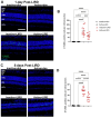
Figure 2.. Treadmill exercise preserves photoreceptor nuclei 1 and 5 days post-LIRD.
Retinal sections from all experimental groups (A, C) were used to quantify photoreceptor nuclei present in the outer nuclear layer (ONL). Morphometric analyses were constructed by plotting the quantification of the ONL nuclei at 1 and 5 days post-LIRD (B, D). 1 day post LIRD, photoreceptor nuclei numbers start to decline in both LIRD groups compared to dim groups and after 5 days, there is a significant decrease in inactive+LIRD animals compared to active+LIRD animals (****p<0.0001). Retinal layers are as follows: outer segment (OS), inner segment (IS), outer nuclear layer (ONL), inner nuclear layer (INL) and ganglion cell layer (GCL). N=9 per group, 3 retinal sections quantified per animal, ****p<0.0001. Scale bar= 25μm, values are mean±SEM.
Figure 3.
Figure 3.. Active+LIRD mice have decreased photoreceptor apoptosis at 1- and 5 days post-LIRD.
Quantification of TUNEL-positive cells (green) in the outer nuclear layer (ONL) revealed increased cell death occurred in inactive+LIRD animals compared to active+LIRD animals at 1- and 5-days post-LIRD. N=12 per group, 3 retinal sections quantified per animal, ***p<0.001, ****p<0.0001. Scale bar= 25μm, values are mean±SEM; blue, DAPI.
Figure 4.
Figure 4.. Exercise partially suppressed LIRD-induced astrocyte and Müller glia activation.
Glial fibrillary acidic protein (GFAP, red) labeling in retinal sections from all experimental groups 1 and 5 days post-LIRD revealed active+LIRD mice had substantially less reactive astrocyte and Müller glia compared to inctive+LIRD mice. N=9 per group, 3 retinal sections quantified per animal, **p<0.01, ***p<0.001, ****p<0.0001. Scale bar= 50μm, values are mean±SEM; blue, DAPI.
Figure 5.
Figure 5.. Active+LIRD mice exhibit fewer Iba1+ cells.
Figure 5. Ionized calcium-binding adaptor molecule-1 (Iba1) immunofluorescence in retinal sections from experimental groups showed active+LIRD mice had decreased presence of microglia in the outer plexiform (OPL), inner plexiform (IPL) and ganglion cell layer (GCL) at 1day post-LIRD and the ONL and GCL at 5days post-LIRD compared to inactive+LIRD mice. N=9 per group, 3 retinal sections quantified per animal, *p<0.05, **p<0.01, ****p<0.0001. Scale bar= 50μm, values are mean±SEM; blue, DAPI.
Figure 6.
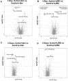
Figure 6.. miRNA analyses reveal exercise promotes cell survival, oxidative stress regulation and photoreceptor homeostasis.
Microarray analysis revealed exercise alters miRNA expression comparing dim and LIRD groups at 1- and 5-days. Altered miRNA expression profiles in active groups are associated with cell survival (miR-302b), oxidative stress regulation (miR-192–5p) and photoreceptor homeostasis (miR-187).

References

    1. Allen RS, Hanif AM, Gogniat MA, et al. TrkB signalling pathway mediates the protective effects of exercise in the diabetic rat retina. Eur J Neurosci. 2018;47(10):1254–1265. doi: 10.1111/ejn.13909 - DOI - PMC - PubMed
    1. Chu-Tan JA, Kirkby M, Natoli R. Running to save sight: The effects of exercise on retinal health and function. Clin Exp Ophthalmol. 2022;50(1):74–90. doi: 10.1111/ceo.14023 - DOI - PubMed
    1. Bales KL, Chacko AS, Nickerson JM, Boatright JH, Pardue MT. Treadmill exercise promotes retinal astrocyte plasticity and protects against retinal degeneration in a mouse model of light-induced retinal degeneration. J Neurosci Res. 2022;100(9):1695–1706. doi: 10.1002/jnr.25063 - DOI - PMC - PubMed
    1. Bales KL, Karesh AM, Hogan K, et al. Voluntary exercise preserves visual function and reduces inflammatory response in an adult mouse model of autosomal dominant retinitis pigmentosa. Sci Rep. 2024;14(1):6940. doi: 10.1038/s41598-024-57027-9 - DOI - PMC - PubMed
    1. Chu-Tan JA, Cioanca AV, Wooff Y, et al. Voluntary exercise modulates pathways associated with amelioration of retinal degenerative diseases. Front Physiol. 2023;14:1116898. doi: 10.3389/fphys.2023.1116898 - DOI - PMC - PubMed

Publication types

LinkOut - more resources