[Preprint]. 2025 Jul 26:2025.07.23.664455.
doi: 10.1101/2025.07.23.664455.

Human iPSC derived alveolar macrophages reveal macrophage subtype specific functions of itaconate in M. tuberculosis host defense

Affiliations

Human iPSC derived alveolar macrophages reveal macrophage subtype specific functions of itaconate in M. tuberculosis host defense

Adam Krebs et al. bioRxiv. .

Abstract

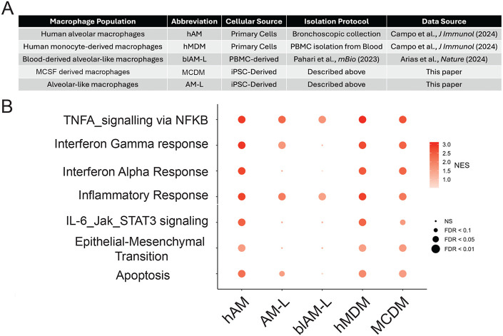
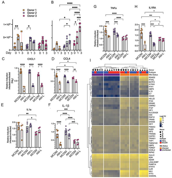
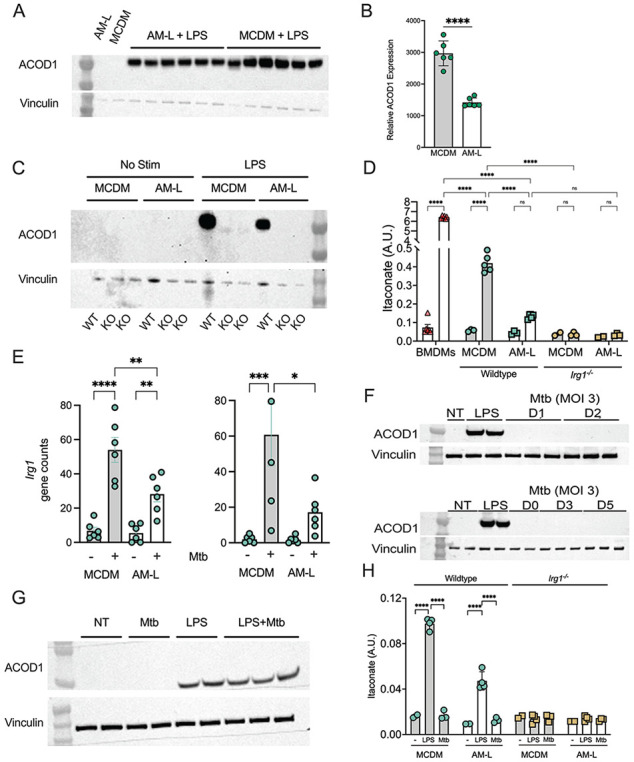
Mycobacterium tuberculosis (Mtb) must survive within multiple macrophage populations during infection, including alveolar macrophages (AM) and recruited inflammatory macrophages. In mice, itaconate, produced in macrophages by ACOD1 mediated decarboxylation of aconitate, has direct antimicrobial activity, modulates inflammatory cytokines, and is required for resistance to M. tuberculosis (Mtb) infection. The role of itaconate in human macrophages is less clear and whether itaconate mediates distinct effects in macrophage subtypes is unknown. Here, we investigated the role of itaconate in human iPSC-derived macrophages, either induced by GM-CSF to resemble alveolar macrophages (AM-Like cells), or treated with M-CSF to generate control macrophages (MCDM cells). Both types of human macrophages produce substantially less itaconate than mouse macrophages and AM-Ls produced 4-fold less itaconate than MCDMs. Surprisingly, ACOD1 deficient AM-L macrophages, but not MCDM macrophages, were permissive for Mtb growth. Moreover, itaconate functioned to dampen the Mtb induced inflammatory response in MCDMs, but not AM-L macrophages, affecting both the Type I IFN and TNF pathways. These results indicate that itaconate is involved in human macrophage responses to TB, with distinct roles in different macrophage subsets. These results also show that genetically tractable hiPSC-derived macrophages are a robust and versatile model to dissect cellular host pathogen interactions.


Conflict of interest statement

Conflict of interest MSG reports consulting fees and equity from Vedanta Biosciences and consulting fees from Fimbrion. KADM reports consulting fees from EuropaDX for bioinformatic tool development not related to this project.

Figures

Figure 1
Figure 1. iPSC derived macrophages phenocopy the alveolar and monocyte derived macrophage transcriptional state
(A) qPCR assessment of transcription factors that define tissue-resident alveolar macrophages (PPAR gamma, KLF4, CEBPB, ATF5) at each stage of the iPSC differentiation process (see Figure S1A), normalized to GAPDH. Data is derived from three independent iPSC donors, each signified as a different symbol. Unpaired t-test used to assess for significant differences between MCDM and AM-L populations. ns not significant * P value < 0.05 ** P value < 0.01. (B) RNA levels encoding the transcription factors PPAR gamma, KLF4, CEBPB, and ATF5 in primary AMs, isolated by bronchoalveolar lavage (hAM), and monocyte derived macrophages (hMDM) derived from reanalysis of RNA sequencing data published in (26). C) iPSC derived AM-Ls and MCDMs resemble human alveolar and monocyte derived macrophages in basal transcriptional state. The heat map clusters the 146 most significant differentially expressed genes between iPSC derived GM-CSF (AM-L) and M-CSF (MCDM) cells from three donors (green shades) and in 6 donors (purple shades) profiled in (26). Also see Figure S2 for an expanded 736 gene set.
Figure 2
Figure 2. Comparison of macrophage types with M. tuberculosis infection
(A) Table of macrophage populations assessed, with abbreviations, sources, method of isolation and production, and references. (B) Normalized enrichment scores (NES) are plotted for each hallmark pathway induced by Mtb infection for each macrophage type listed in (A).
Figure 3
Figure 3. Inter donor conservation of macrophage TB responses
(A,B) MCDM (A) and AM-L (B) macrophages from three iPSC donors were infected with Mtb at a target multiplicity of infection (MOI) of 3. Colony forming units (CFUs) were quantified immediately post and 1- and 3-days post infection. Each donor is depicted with a different color. (C-H) iPSC derived alveolar macrophages are hypoinflammatory Cytokine and chemokine responses of iPSC derived macrophages on day 3 of a Mtb infection. In each graph, MCDM cells are the shaded bars and AM-L cells are clear bars, with donor identify as depicted in (A). Each Y axis is the log2 fold change of the indicated cytokine of infected/uninfected determined by Luminex (see methods) for CXCL-1 (C), CCL4 (D), IL-1a (E), IL-1B (F), TNF (G), and IL-1RA (H). (I) Heatmap depicting log2 fold change of hallmark type I interferon gene expression in uninfected or Mtb infected MCDM or AM-L macrophages. Partitioning is based on k-means clustering. For all panels, two-way ANOVA was used to assess variance of the samples based on infection status and within a macrophage population. * P value <0.05 ** P value <0.01 ***P value <0.0001, ****P value <0.00001.
Figure 4
Figure 4. Human macrophages are feeble producers of itaconate with Mtb infection.
(A, B) Expression of ACOD1 protein in iPSC derived macrophage subtypes. Anti-ACOD1/Vinculin immunoblot of MCDM or AM-L cells following 6-hours of stimulation with LPS (100ng/mL) (A) and normalized quantitation of ACOD1 protein levels (B). **** P value < 0.0001 by unpaired t-test. (C) Production of irg1−/− macrophages. MCDM and AM-L macrophages were differentiated from CRISPR edited iPSCs (see Figure S4) or isogenic control cells. Anti-ACOD1 immunoblot of MCDM and AM-L cells derived from Irg1 deficient iPSCs or isogenic controls stimulated with LPS as in (A,B). (D) Human macrophage production of itaconate. GC-MS quantification of itaconate produced by mouse BMDMs, MCDM, AM-L, or irg−/− MCDMs and AM-Ls following 6 hours of LPS stimulation. Itaconate levels are graphed as arbitrary units (A.U) by normalization to total protein levels. Two-way ANOVA was used to assess variance of the samples within the same treatment group and based on stimulation status. **** P value <0.0001. (E) Gene counts of Irg1 mRNA following 4 hours or 24 hours of Mtb infection of MCDM or AM-L macrophages. Data is derived from three separate donors in biologic duplicates. *** P value < 0.005 **** P value <0.0001 by two-way ANOVA. (F) Anti-ACOD1/Vinculin immunoblot of wildtype MCDM cells following no treatment (NT), LPS stimulation (100ng/mL), or Mtb infection at MOI of 3 and assayed at days 1 and 2 (top panel), or days 0, 3, and 5 (bottom panel). (G) Anti-ACOD1/Vinculin immunoblot of wildtype MCDM cells following no treatment (NT), 4 hours of LPS stimulation (100ng/mL), or infection with Mtb at a multiplicity of 10 for 4 hours, or both. (H) GC-MS quantification of itaconate produced by MCDM and AM-L macrophages following 4 hours of infection with Mtb (MOI10) or LPS (100ng/mL). Itaconate levels were normalized to total protein levels.
Figure 5
Figure 5. Macrophage type-specific roles of itaconate on Mtb control and inflammatory responses
(A) Mtb colony forming units (CFUs) quantified from wild type (green symbols) and Irg1−/− (gold symbols) MCDM (shaded bars) and AM-L macrophages (clear bars), infected at an MOI of 3, by day of infection (B) Relative concentration of CXCL1 or CCL4 produced by WT or Irg1 −/− MCDM (shaded bars) and AM-L (clear bars) macrophages on day 3 of a Mtb infection. (C) Relative concentration of IL1-β and IL-1RA produced by WT and Irg1−/− MCDM and AM-L macrophages on day 3 of a Mtb infection. (D) Itaconate negatively regulates the Type I interferon response to Mtb in MCDM but not AM-L macrophages. Heatmap depicting z-score of hallmark Type I interferon gene expression in MCDM and AM-L macrophages in the presence or absence of Mtb. Partitioning is based on k-means clustering. (E) Hyperinduction of the Type I IFN pathway in itaconate deficient macrophages is due to phagosomal permeabilization by esx-1. RT-qPCR quantifying Trim5, Isg20, and Cxcl11 transcripts, normalized to GAPDH, in MCDM macrophages in response to infection by wildtype Mtb or MtbΔrv3877 (lacking the esx-1 secretion system) for four hours. ** P value <0.01 **** P value <0.0001 by two-way ANOVA. Error bars are SD.

References

    1. Chandra P, Grigsby SJ, and Philips JA. Immune evasion and provocation by Mycobacterium tuberculosis. Nature Reviews Microbiology. 2022;20(12):750–66. - PMC - PubMed
    1. Brites D, and Gagneux S. Co-evolution of Mycobacterium tuberculosis and Homo sapiens. Immunological Reviews. 2015;264(1):6–24. - PMC - PubMed
    1. Cohen SB, Gern BH, Delahaye JL, Adams KN, Plumlee CR, Winkler JK, et al. Alveolar Macrophages Provide an Early Mycobacterium tuberculosis Niche and Initiate Dissemination. Cell Host & Microbe. 2018;24(3):439–46.e4. - PMC - PubMed
    1. Huang L, Nazarova EV, Tan S, Liu Y, and Russell DG. Growth of Mycobacterium tuberculosis in vivo segregates with host macrophage metabolism and ontogeny. Journal of Experimental Medicine. 2018;215(4):1135–52. - PMC - PubMed
    1. Pisu D, Huang L, Grenier JK, and Russell DG. Dual RNA-Seq of Mtb-Infected Macrophages In Vivo Reveals Ontologically Distinct Host-Pathogen Interactions. Cell Reports. 2020;30(2):335–50.e4. - PMC - PubMed

Publication types