Case Reports
. 2025 Jul 23;30(3):180.
doi: 10.3892/etm.2025.12930. eCollection 2025 Sep.

A rare dermatological manifestation of follicular spicules in a patient with multiple myeloma and end-stage renal disease on hemodialysis: A case report

Affiliations
Case Reports

A rare dermatological manifestation of follicular spicules in a patient with multiple myeloma and end-stage renal disease on hemodialysis: A case report

Xiaobing Li et al. Exp Ther Med. .

Abstract

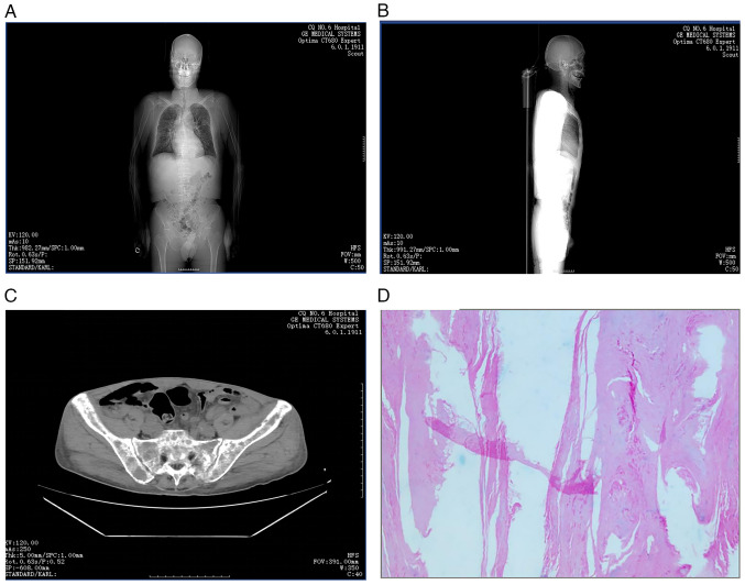
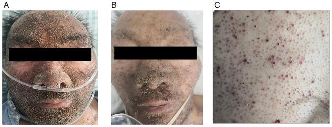
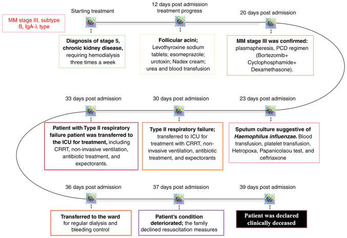
Multiple myeloma (MM) is a hematological malignancy characterized by the clonal expansion of malignant plasma cells within the bone marrow, leading to diverse systemic complications. While cutaneous manifestations of MM are uncommon, follicular spicules represent a highly specific but rare dermatological finding associated with MM. The present study presents the case of a 59-year-old man with MM who exhibited follicular acanthosis and hyperkeratosis as cutaneous indicators of the malignancy. The patient, with a history of stage 5 chronic kidney disease undergoing maintenance hemodialysis, was diagnosed with follicular spicules of MM (FSMM) through clinical evaluation and laboratory investigations. The present case highlights the clinical importance of recognizing rare cutaneous manifestations in MM, which can serve as critical diagnostic clues. Improved awareness of FSMM may facilitate a timely diagnosis and optimize patient management, ultimately contributing to improved clinical outcomes in this challenging population.

Keywords: chronic kidney disease; dermatological manifestations; end-stage renal disease; follicular acanthosis; follicular spicules; hemodialysis; hyperkeratosis; multiple myeloma.


Conflict of interest statement

The authors declare that they have no competing interests.

Figures

Figure 1
Figure 1
Karyotype analysis revealing complex chromosomal abnormalities in advanced MM. Conventional karyotyping revealed multiple numerical and structural abnormalities (red arrows) characteristic of high-risk MM: i) Hyperdiploidy with tetraploid cells (83-85<4n>,XXY), including an additional X chromosome. ii) Loss of Y chromosome (-Y), a common finding in hematological malignancies. iii) High-level polysomy of chromosome 1 (+1,+1), totaling four copies, indicating aggressive disease. iv) Dicentric chromosome [dic(1;10)(p13;q26)], suggesting genomic instability. v) Pseudo-isodicentric chromosome [psu idic(1)(p21)], reflecting complex chromosomal rearrangement. vi) Additional material on chromosomes 7 and 8 [add(7)(q32),add(8)(p23)x2]. vii) Possible unidentified material on chromosome 17 [?add(17)(q22)x2]. ix) Monosomy of chromosomes 19, 20, 21 and 22 (green arrows). xi) One to two marker chromosomes (+1-2mar). xii) Mosaic karyotype: Four abnormal clones with complex karyotype and 16 normal male karyotypes (46,XY), consistent with clonal evolution. MM, multiple myeloma.
Figure 2
Figure 2
SPE and immunofixation confirming monoclonal gammopathy. (A) SPE demonstrating an M-spike in the γ region. (B) Immunofixation electrophoresis revealing a monoclonal IgA λ pattern, confirming the presence of IgA-λ type multiple myeloma. (C) Immunofixation electrophoresis further confirming the monoclonal IgA λ pattern. AU, arbitrary units; SPE, serum protein electrophoresis. ELP, electrophoresis reference; G, IgG; A, IgA; M, IgM; K, κ light chain; L, λ light chain; D, IgD; E, IgE.
Figure 3
Figure 3
Immunohistochemical characterization of bone marrow biopsy. (A) Congo red staining showed amyloid deposits with birefringence under polarized light, confirming amyloidosis. (B) Scattered CD3+ T cells. (C) CD20+ B cells. (D) Diffuse CD56 positivity indicating natural killer cell involvement. (E) Diffuse CD138 staining marking plasma cell infiltration. (F) Negative κ light chain staining. (G) Diffuse positive staining of λ light chains, confirming monoclonality. (H) Multiple myeloma oncogene 1-positivity indicated plasma cell differentiation. (I) Hematoxylin and eosin staining revealed diffuse infiltration by atypical lymphoid/plasma cells. (J) Positive staining for amyloid deposits in bone marrow biopsy. All images were captured under a light microscope at x400 magnification.
Figure 4
Figure 4
Flow cytometric immunophenotyping of bone marrow cells from a patient with PCM. (A) Lymphocyte analysis showing a decreased CD4/CD8 ratio. (B and C) Quantification of CD57⁺ LGLs, demonstrating a normal proportion of this subset. (D) Marked expansion of plasma cells detected within the bone marrow aspirate. (E) Dual-parameter plot of CD38 and CD45 expression revealing a distinct CD38bright+/CD45dim+ population, accounting for 33.19% of total nucleated cells, consistent with malignant plasma cell infiltration. (F) Intracellular immunoglobulin light chain staining within the CD38bright+ population indicating prominent λ light chain restriction (97.37%) with minimal κ expression (0.03%), confirming clonal proliferation and supporting the diagnosis of PCM. LGLs, large granular lymphocytes; NC, nucleated cell; PCM, plasma cell myeloma; A, allophycocyanin-Alexa Fluor 700; APC, allophycocyanin; CD38bri+, CD38 bright positive; DNT, double negative T cells; DPT, dual-parameter plot; ECD, energy-coupled dye; H, height parameter; KO525, Krome Orange 525; Lym, lymphocytes; NK, natural killer cells; PC5.5, phycoerythrin-cyanine 5.5; PE, phycoerythrin; SSC, side scatter.
Figure 5
Figure 5
Comprehensive imaging and histopathological evaluation. (A) An anterior scout image from CT demonstrated diffuse skeletal involvement, including multiple hypodense lesions in the skull, ribs, spine, scapulae and pelvis, consistent with lytic lesions seen in multiple myeloma. Bilateral pulmonary infections and pleural thickening were also visible. (B) A lateral scout image further illustrated spinal abnormalities, including compression of the T12 vertebral body and suspected Hsu-Mo nodule at T1, as well as degenerative spinal changes. (C) An axial pelvic CT revealed lytic bone lesions, bilateral pathological rib fractures and pelvic effusion. (D) Histopathological examination of a skin lesion showed squamous epithelium with marked hyperkeratosis (hematoxylin and eosin staining; original magnification, x100), supporting dermatopathological involvement.
Figure 6
Figure 6
Clinical progression of cutaneous manifestations and therapeutic response. (A) Pre-treatment image of the face showing widespread scaling and crusted lesions. (B) Post-treatment image of the same area with notable resolution of lesions following systemic corticosteroid therapy. (C) Chest lesions showing follicular keratotic papules and erythematous pinpoint eruptions, consistent with follicular spicules of multiple myeloma.
Figure 7
Figure 7
Timeline of clinical course and interventions in a patient with IgA-λ type MM. The patient was diagnosed with stage III MM (subtype B, IgA-λ type) and regular hemodialysis was initiated. In March 2024, follicular acanthosis was noted; treatment included levothyroxine, esomeprazole, urotropine, urea cream, nadifloxacin cream and blood transfusion. In April 2024, the patient developed type II expiratory failure and was admitted to the ICU for CRRT, non-invasive ventilator support, and symptomatic management. Despite intensive care, the patient succumbed to disease-related complications. CRRT, continuous renal replacement therapy; ICU, Intensive Care Unit; MM, multiple myeloma.

Similar articles

References

    1. Rudnicka L, Chrostowska S, Kamiński M, Waśkiel-Burnat A, Michalczyk A, Rakowska A, Olszewska M. The role of trichoscopy beyond hair and scalp diseases. A review. J Eur Acad Dermatol Venereol: Mar 15, 2023 (Epub ahead of print). - PubMed
    1. Michael A, Fuller T, Brogan S, Murphy MC. An intrathecal pump misadventure in two acts: An unrecognized partial pocket fill followed by an unusual withdrawal syndrome two months later. J Palliat Med: Jan 30, 2025 (Epub ahead of print). - PubMed
    1. Chang YC, Peng CY, Chi KY, Song J, Chang Y, Chiang CH, Gao W, Chiang CH. Cardiovascular outcomes and mortality in diabetic multiple myeloma patients initiated on proteasome inhibitors according to prior use of glucagon-like peptide 1 agonists. Eur J Prev Cardiol: Jan 29, 2025 (Epub ahead of print). - PubMed
    1. Jia J, Yin J, Geng C, Liu A. Characteristics and outcomes of secondary acute lymphoblastic leukemia (sALL) after multiple myeloma (MM): SEER data analysis in a single-center institution. Cancer Pathog Ther. 2024;3:76–80. doi: 10.1016/j.cpt.2024.06.007. - DOI - PMC - PubMed
    1. Rapparini L, Pileri A, Robuffo S, Agostinelli C, La Placa M. Cutaneous involvement in multiple myeloma. A rare entity. Clin Case Rep. 2025;13(e9476) doi: 10.1002/ccr3.9476. - DOI - PMC - PubMed

Publication types

LinkOut - more resources