

Reversible Diffusion-Restricting White Matter Lesions in Sickle Cell Disease During Pain Crises: A Case Report
- PMID: 40777729
- PMCID: PMC12325237
- DOI: 10.1177/19418744251367181
Reversible Diffusion-Restricting White Matter Lesions in Sickle Cell Disease During Pain Crises: A Case Report
Abstract
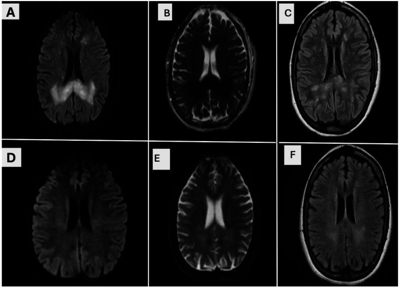
Sickle cell disease (SCD) is a chronic hemoglobinopathy characterized by recurrent vaso-occlusive events and significant neurological morbidity. While ischemic and hemorrhagic strokes are well-known complications, reversible diffusion-restricting white matter lesions are exceedingly rare and underreported. We present the case of an 18-year-old male with homozygous SCD (HbSS) who developed acute neurological deterioration during a vaso-occlusive pain crisis. MRI revealed symmetric areas of restricted diffusion and FLAIR hyperintensities in the splenium of the corpus callosum and periventricular white matter, typically associated with cytotoxic edema and irreversible injury. Remarkably, the patient experienced near-complete neurological recovery with aggressive disease-targeted therapy, including hydroxyurea, voxelotor, and serial exchange transfusions. Follow-up MRI at 4 months showed complete resolution of the prior abnormalities. This case underscores the importance of recognizing potentially reversible diffusion-restricting lesions in SCD and challenges the conventional interpretation of restricted diffusion as a marker of permanent injury. Early recognition, comprehensive management, and serial neuroimaging may improve neurological outcomes in similar cases. Clinicians should maintain a high index of suspicion for reversible white matter injury when evaluating patients with sickle cell disease presenting with acute neurological symptoms. Incorporating serial neuroimaging and a multidisciplinary treatment approach is essential for timely diagnosis and optimizing neurological recovery in this vulnerable population.
Keywords: cytotoxic edema; diffusion-weighted imaging; encephalopathy; exchange transfusion; hydroxyurea; periventricular white matter; reversible white matter lesions; sickle cell disease; splenium; vaso-occlusive crisis; voxelotor.
© The Author(s) 2025.
Conflict of interest statement
The authors declared no potential conflicts of interest with respect to the research, authorship, and/or publication of this article.
Figures
Similar articles
-
Sickle Cell Disease.2003 Sep 15 [updated 2025 Feb 13]. In: Adam MP, Feldman J, Mirzaa GM, Pagon RA, Wallace SE, Amemiya A, editors. GeneReviews® [Internet]. Seattle (WA): University of Washington, Seattle; 1993–2025. 2003 Sep 15 [updated 2025 Feb 13]. In: Adam MP, Feldman J, Mirzaa GM, Pagon RA, Wallace SE, Amemiya A, editors. GeneReviews® [Internet]. Seattle (WA): University of Washington, Seattle; 1993–2025. PMID: 20301551 Free Books & Documents. Review.
-
Hydroxyurea (hydroxycarbamide) for sickle cell disease.Cochrane Database Syst Rev. 2022 Sep 1;9(9):CD002202. doi: 10.1002/14651858.CD002202.pub3. Cochrane Database Syst Rev. 2022. PMID: 36047926 Free PMC article.
-
A Multifaceted Crisis: Sickle Cell Disease Complicated by Pulmonary Embolism, Avascular Necrosis, and Numb Chin Syndrome.Cureus. 2025 Jun 8;17(6):e85556. doi: 10.7759/cureus.85556. eCollection 2025 Jun. Cureus. 2025. PMID: 40630370 Free PMC article.
-
Interventions for chronic kidney disease in people with sickle cell disease.Cochrane Database Syst Rev. 2023 Aug 4;8(8):CD012380. doi: 10.1002/14651858.CD012380.pub3. Cochrane Database Syst Rev. 2023. PMID: 37539955 Free PMC article.
-
Hydroxyurea (hydroxycarbamide) for sickle cell disease.Cochrane Database Syst Rev. 2017 Apr 20;4(4):CD002202. doi: 10.1002/14651858.CD002202.pub2. Cochrane Database Syst Rev. 2017. Update in: Cochrane Database Syst Rev. 2022 Sep 1;9:CD002202. doi: 10.1002/14651858.CD002202.pub3. PMID: 28426137 Free PMC article. Updated.
References
-
- DeBaun MR, Kirkham FJ. Central nervous system complications and management in sickle cell disease. Blood. 2016;127(7):829-838. - PubMed
-
- Al-Jafar HA, Alroughani R, Abdullah TA, Al-Qallaf F. Neurological complications in sickle cell disease. Int J Clin Exp Neurol. 2016;4(1):9-18.
Publication types
LinkOut - more resources
Full Text Sources