Review
. 2025 Jan 2;5(4):1384-1400.
doi: 10.1016/j.fmre.2024.12.015. eCollection 2025 Jul.

Programmable DNA-based biomaterials for bone tissue engineering

Affiliations
Review

Programmable DNA-based biomaterials for bone tissue engineering

Xiaoyue Xu et al. Fundam Res. .

Abstract

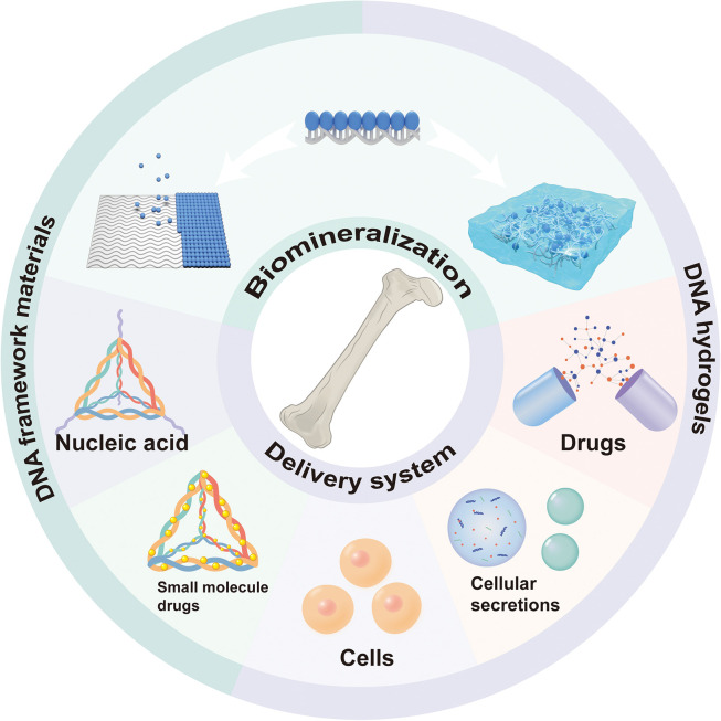
Bone defects are a common pathology in bone tissue diseases, and existing therapeutic interventions have significant limitations, highlighting the need for innovative strategies and advanced biomaterials. DNA, traditionally recognized as a prominent genetic material, also possesses exceptional properties as a biological material, making it an ideal nanoscale building block for creating various DNA-based biomaterials, such as DNA framework materials and DNA hydrogels. DNA-based biomaterials offer notable advantages, including structural versatility, biocompatibility, and, crucially, programmability, which position them as promising candidates for bone tissue engineering. This review explores recent advancements in the use of DNA-based biomaterials for bionic mineralization and drug delivery systems, as well as their future potential in this field.

Keywords: Bone tissue engineering; DNA framework materials; DNA hydrogels; DNA nanotechnology; Programmable biomaterials.


Conflict of interest statement

The authors declare that they have no conflicts of interest in this work.

Figures

Fig 1
Fig. 1
A timeline of DNA-based biomaterials relevant to bone tissue engineering. DNA-based biomaterials are used in bone tissue engineering mainly as bionic mineralization templates and delivery systems. Although not a comprehensive account, discoveries and major developments are listed. QCM, quartz crystal microbalance; CaP, calcium phosphate; HAP, hydroxyapatite; SBF, simulated body fluid; TDF, tetrahedral DNA framework; ADSCs, adipose-derived stem cells; nSi, silicate nanodisks; Dex, dexamethasone; pAsp, polyaspartic acid; dsDNA, double-stranded DNA; BMSCs, bone marrow mesenchymal stem cells. TLR4, toll-like receptor 4; VEGF, vascular endothelial growth factor.
Fig 2
Fig. 2
Schematic representation of DNA-based biomaterials for bone tissue engineering.
Fig 3
Fig. 3
CaP mineralization of DNA framework materials. (A) Schematic of CaP mineralization of DNA framework material templates with tetrahedral DNA as a detailed example. (B) TEM, FESEM, and AFM images of DNA templates after mineralization, using TDF as an example. Scale bar, 100 nm on the left and 30 nm on the right. (C) Schematic of immunostimulatory oligonucleotides (CpG) delivered using TDF and combined with the mineralization process, and AFM images of TDF-CpG (left) and mineralized TDF-CpG (TDF-CpG-CaP) (right). Solid circles showed TDF-CpGs with typical hollow structures; dashed circles showed collapsed TDF-CpGs. Upon mineralization, the hollow structures of TDF were filled in, but the tail-like structures of CpG remained. Scale bars: 100 nm; AFM height scale bar, 4 nm. (D) Different samples of stimulated macrophage RAW-264.7 expression level of tumor necrosis factor-α. TDF-CpG-CaP showed better immunostimulatory activity. (A-D) Adapted with permission [60]. Copyright 2019, CELL PRESS. (E-G) Biomimetic mineralization of DNA templates using particle attachment crystallization strategy. (E) Schematic of particle attachment crystallization strategy for biomimetic mineralization of DNA templates. (F) Rectangular DNA origami’s (I-origami) size schematic (top), AFM images after mineralization (m-I-origami) (middle), and height statistics (bottom). (G) Schematic of streptavidin pre-immobilized on I-origami that remained bound to biotinylated transferrin after mineralization. Adapted with permission [62]. Copyright 2020, AMER CHEMICAL SOC.
Fig 4
Fig. 4
CaP mimetic mineralization of surface-modified DNA templates. (A-B) Mineralization of polyaspartate-modified DNA templates. (A) Schematic of the constructed polyaspartate covalently linked three-stranded double helix DNA structure (3sDH-pAsp). (B) AFM images of 3sDH-pAsp incubated under mineralization conditions for 30 min and 3 h Adapted with permission [61]. Copyright 2019, ROYAL SOC CHEMISTRY. (C-E) Mineralization of SSEE peptide-modified DNA templates. (C) AFM images of SSEE peptide-functionalized DNA nanotubes after 20–30 min incubation under mineralized conditions. (D) AFM images of SSEE peptide-functionalized DNA rectangular origami after 20–30 min incubation under mineralized conditions. (E) Raman spectral characterization of HAP (control) and SSEE-functionalized DNA rectangular origami incubated under mineralized conditions for 24 h Confirmation of HAP by the appearance of a weak peak at 960 cm-1. Adapted with permission [63]. Copyright 2021, AMER CHEMICAL SOC.
Fig 5
Fig. 5
CaP mineralization of DNA hydrogels. (A-B) DNA hydrogels were mineralized by soaking in the mineralization solution for 16 h. (A) Schematic of the self-assembled DNA hydrogel. (B) SEM images of DNA hydrogels after 16 h of mineralization. Adapted with permission [78]. Copyright 2023, NATL ACAD SCIENCES. (c-g) Autonomous mineralized DNA hydrogel with improved osteogenic microenvironment for bone repair. (C) Schematic of mineralized DNAzyme hydrogel preparation. Autonomous mineralization used byproducts of rolling circle amplification. (D) SEM images of non-mineralized (left) and mineralized (right) DNA hydrogels. (E) The stability of MDH was verified by PAGE electrophoresis and grayscale analysis. (F) DCFH fluorescence assay for reactive oxygen species content in BMSCs under different treatments. The greater intensity of green fluorescence indicated higher intracellular reactive oxygen species content. (G) Micro-CT images after implantation of different hydrogels in rat bone defects. Adapted with permission [79]. Copyright 2023, WILEY-V C H VERLAG GMBH. NH, normal DNA Hydrogel, without mineralization reaction and DNA-Hemin; MH, mineralized DNA Hydrogel, without DNA-Hemin; MDH, mineralized DNAzyme hydrogel; N-BMSC, BMSC in the normal state; Os-BMSC, BMSC in the oxidative stress state.
Fig 6
Fig. 6
TDF as a delivery system in bone tissue engineering. (A-D) TDF delivery of microRNA. (A) Schematic of MiR@TDF complex formation by TDF loading MiR335–5p. (B) MiR@TDF promoted osteogenic differentiation of BMSCs. Alkaline phosphatase assay (top), alizarin red staining of calcium nodules (middle), Oil red O staining for lipid droplets (bottom). Scale bar = 20 μm. Adapted with permission [104]. Copyright 2021, Wiley-VCH. (C) Schematic of the process of TDF loading MiR2861 through sticky ends. (D) Schematic of enzymatic cleavage to release MiR2861 in the extracellular environment. Adapted with permission [108]. Copyright 2021, WILEY-V C H VERLAG. (E-G) TDF delivery of small molecule drugs applied to bone tissue engineering. (E) Clindamycin (CLI) was loaded onto TDF through electrostatic adsorption. (F) Confocal microscopy images of BMSCs uptake of TDF, TDF-CLI, and ss-DNA. Cy5 labeled TDF, TDF-CLI, and ss-DNA. (G) Bacterial colony counting data of different samples treated for 24 h at 0.25 minimum inhibitory concentration. Adapted with permission [113]. Copyright 2022, ELSEVIER SCIENCE SA.
Fig 7
Fig. 7
DNA hydrogels as cell carriers in bone tissue engineering. (A-D) Specific capture and release of BMSCs from DNA hydrogels generated by double-rolling circle amplification. (A) Schematic of the formation of double-rolling circle amplified DNA network structure, specific capture of BMSCs and nuclease-triggered release. The Apt19S aptamer has a high affinity for ALPL proteins on the membranes of BMSCs and ensures cell-specific anchoring. (B) Microscopic images of nuclease-triggered released BMSCs (left) and after 24 h of culture (right). Live and dead cells were stained with calcein-AM (green) and propidium iodide (PI, red), respectively. (C) Fluorescence microscopy images of cell mixtures of BMSCs and SMCs before (left) and after cell capture (right). BMSCs were stained with CM-Dil (red), and SMCs were stained with CM-DiO (green). SMCs, smooth muscle cells. (D) Statistics of the capture frequency of BMSCs in (C). This method was able to pick out BMSCs at a frequency of up to 81%. Adapted with permission [136]. Copyright 2020, AMER CHEMICAL SOC. (E-G) DNA hydrogels that provided efficient protection for BMSCs. (E) Schematic of the preparation of DNA hydrogels loaded with BMSCs. (F) Viability of BMSCs stained with calcein-AM (green, live) and PI (red, dead) after bidirectional rubbing in conventional liquid media or DNA hydrogels. (G) Percentage survival of BMSCs after bidirectional rubbing. Adapted with permission [137]. Copyright 2021, WILEY-V C H VERLAG GMBH.
Fig 8
Fig. 8
Sustained release of VEGF from BPNS-DNA hydrogel to promote vascularized bone regeneration. (A) Schematic of incorporation of BPNSs loaded with VEGF into DNA hydrogel combined with 3D printed polycaprolactone scaffold to promote vascularized bone regeneration. (B) Release profiles of VEGF in two types of DNA hydrogel scaffolds. (C) Immunohistochemical staining images of OCN and CD31 after 4 weeks of repairing cranial defects in rats with different samples, the yellow-brown area is the positive expression area of OCN and CD31. Adapted with permission [149]. Copyright 2022, KEAI PUBLISHING LTD.
Fig 9
Fig. 9
DNA hydrogels for controlled release of exosomes are applied in bone tissue engineering. (A-D) Exosomes were directly physically encapsulated in a dynamic bio-responsive PEG/DNA hybrid hydrogel for vascularized bone regeneration in diabetes mellitus (DM) rats. (A) Schematic of the synthesis and behavior of SCAP-Exo-loaded PEG/ DNA hybrid hydrogel in response to MMP-9 stimulation. (B) Exosome release profile of this hydrogel in PBS and MMP-9 solution. (C) Immunofluorescence staining images of CD31 (angiogenic marker, red) after bone defects in DM rats with different sample treatments. Scale bar = 100 μm. (D) Micro-CT images of bone defect areas in DM rats with different sample treatments. Adapted with permission [157]. Copyright 2022, AMER CHEMICAL SOC. (E) Schematic of sustained or light-controlled release of exosomes achieved by tethering exosomes in hydrogel. Exosomes were functionalized by ATRP initiator and cholesterol-modified DNA double strands (Chol-dsDNA-iBBr), and the polymer chains were grafted directly from the exosomes to form exosome-tethered hydrogels. Modification of the DNA strand with a photocleavable group, p-nitrophenyl, enabled the photocontrolled release of exosomes. Adapted with permission [158]. Copyright 2022, AMER CHEMICAL SOC.
Fig 10
Fig. 10
Sustained release of Dex from nSi-DNA hydrogels. (a) Schematic of composite DNA hydrogels prepared by a two-step gel method. In the first step, double-stranded DNA was denatured and cooled to form a pre-gel, and A-type network points were formed by complementary base pairing between DNA strands; in the second step, silicate nanodisks (nSi) were introduced to form B-type network points through electrostatic interactions between nSi and DNA strands. (b) SEM images of DNA hydrogel. The addition of nSi reduced the pore size of the hydrogel. Scale bar = 100 μm. (c) Release of Dex from DNA hydrogels with different nSi contents. Adapted with permission [165]. Copyright 2018, AMER CHEMICAL SOC.

Similar articles

References

    1. Nauth A., Schemitsch E., Norris B., et al. Critical-size bone defects: Is there a consensus for diagnosis and treatment? J. Orthop. Trauma. 2018;32:S7–S11. - PubMed
    1. Panetta N.J., Gupta D.M., Slater B.J., et al. Tissue engineering in cleft palate and other congenital malformations. Pediatr. Res. 2008;63:545–551. - PubMed
    1. Keating J.F., Simpson A.H.R.W., Robinson C.M. The management of fractures with bone loss. J. Bone Joint Surg. Br. 2005;87:142–150. - PubMed
    1. Rioja A.Y., Daley E.L.H., Habif J.C., et al. Distributed vasculogenesis from modular agarose-hydroxyapatite-fibrinogen microbeads. Acta Biomater. 2017;55:144–152. - PMC - PubMed
    1. Henkel J., Woodruff M.A., Epari D.R., et al. Bone regeneration based on tissue engineering conceptions — A 21st century perspective. Bone Res. 2013;1:216–248. - PMC - PubMed

LinkOut - more resources