. 2024 Dec 30;5(4):1440-1450.
doi: 10.1016/j.fmre.2024.12.014. eCollection 2025 Jul.

A metal-drug self-delivery nanomedicine alleviates tumor immunosuppression to potentiate synergistic chemo/chemodynamic therapy against hepatocellular carcinoma

Affiliations

A metal-drug self-delivery nanomedicine alleviates tumor immunosuppression to potentiate synergistic chemo/chemodynamic therapy against hepatocellular carcinoma

Chen Guo et al. Fundam Res. .

Abstract

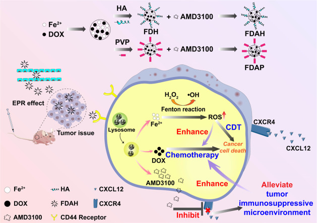
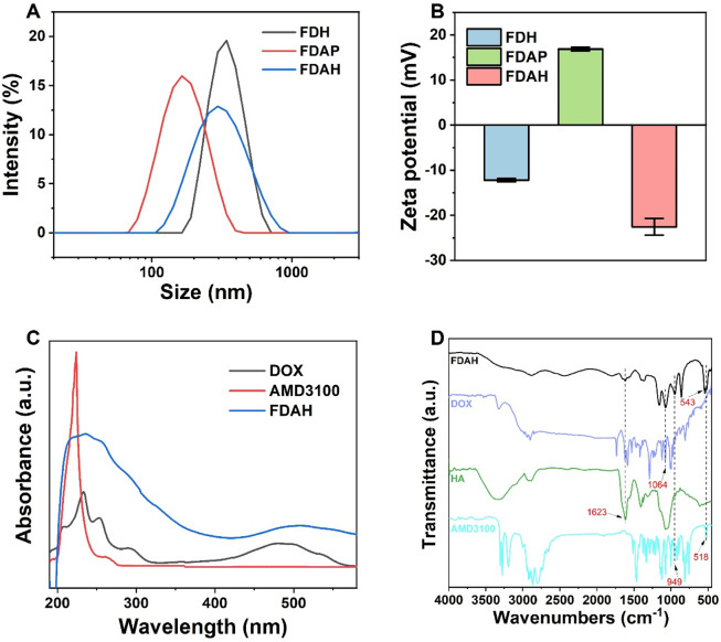
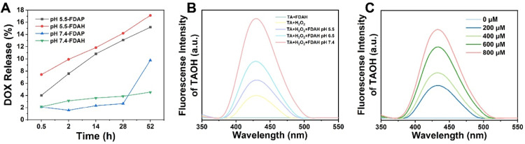
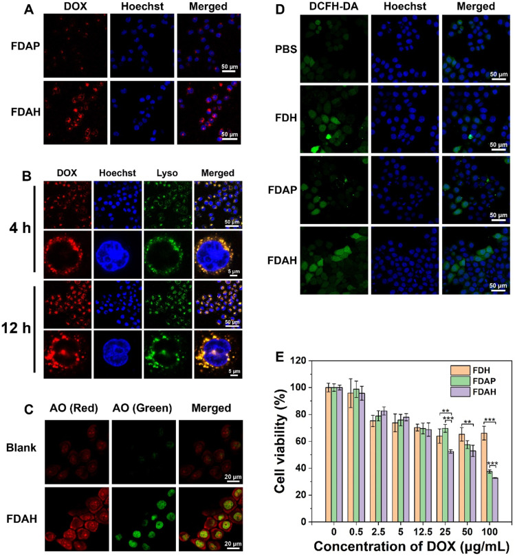
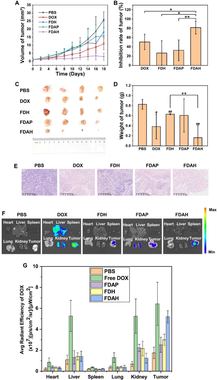
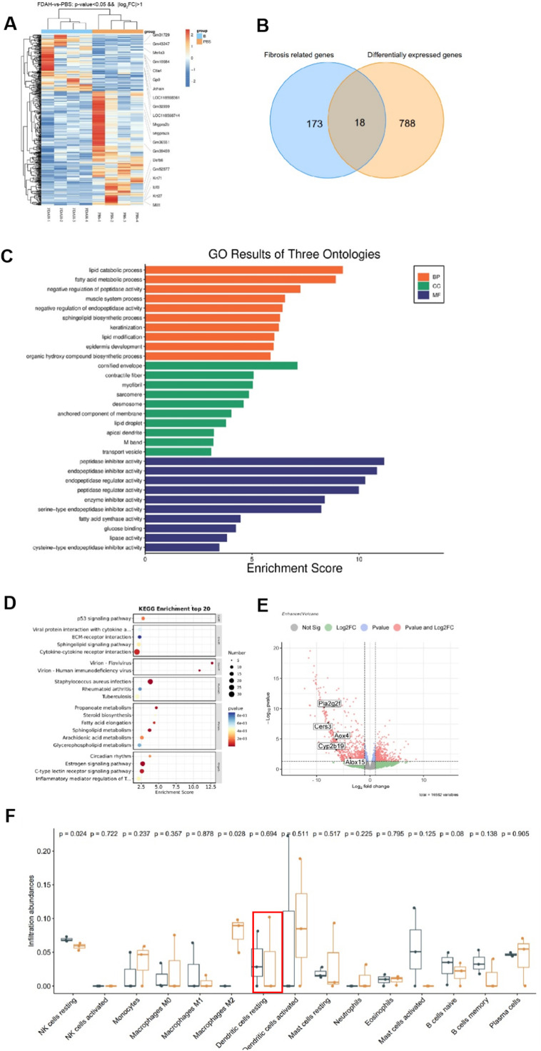
Hepatocellular carcinoma (HCC) is the most common primary liver cancer with a poor prognosis. Chemotherapy is one of the first-line clinical therapeutic strategies for HCC. Still, the effectiveness of chemotherapy is hampered by the tumor immunosuppressive microenvironment and drug resistance caused by insufficient delivery. Herein, we developed a metal-drug self-delivery nanomedicine (FDAH) to improve the chemo/chemodynamic therapeutic efficacy of HCC. The core of FDAH is an iron-based nanoparticle chelated with two clinical drugs, Doxorubicin (DOX) and Plerixafor (AMD3100). Additionally, the nanomedicine is externally modified with a hyaluronic acid (HA) shell, which can prolong the circulation time of the nanoparticles in the bloodstream after intravenous administration. After entering the bloodstream, the nanomedicine reaches the tumor tissue through the EPR effect and is phagocytosed by the tumor cells via HA/CD44-specific interaction. Iron ion-mediated chemodynamic therapy is mediated by the Fenton reaction to generate ROS, causing an imbalance of redox homeostasis within the tumor cells and enhancing the sensitivity of tumor cells to DOX. In addition, AMD3100 intervenes in the CXCL12/CXCR4 axis to influence the infiltration level of immune cells and promote DOX chemotherapy in tumor cells. This work suggests that alleviating immunosuppression via a metal-drug self-delivery system of the CXCR4 inhibitor can effectively improve the DOX chemotherapy and iron ions-mediated chemodynamic therapy.

Keywords: Chemodynamic therapy; Chemotherapy; Hepatocellular carcinoma; Immunosuppressive microenvironment; Metal-drug self-delivery nanomedicine.


Conflict of interest statement

The authors declare that they have no conflicts of interest in this work.

Figures

Image, graphical abstract
Graphical abstract
Scheme 1
Scheme 1
Enhanced FDAH therapy for HCC. Scheme of enhancing synergistic chemo/chemodynamic therapy of FDAH in an HCC tumor model by alleviating the tumor’s immunosuppressive microenvironment.
Fig 1
Fig. 1
Characterization of nanoparticles. (A) The hydrodynamic diameter of FDH, FDAP, and FDAH. (B) The zeta potential of FDH, FDAP, and FDAH. (C) UV–Vis absorption spectrum of DOX, AMD3100, and FDAH. (D) The FT-IR spectrum of FDAH, DOX, HA, and AMD3100.
Fig 2
Fig. 2
Drug release and nanoparticles-induced production of •OH through Fenton reaction. (A) The DOX release of FDAP and FDAH at different pH. FDAP and FDAH were incubated in NaAc/HAc buffer (pH 5.5) or PBS (pH 7.4) at 37 °C for various durations. The solutions were then centrifuged at 14,000 rpm for 10 min, and the UV–Vis absorption at 480 nm of the supernatant was recorded to calculate the DOX release amount based on a standard curve. An equal volume of buffer was added to the supernatant for further incubation. (B) Fluorescence spectra of TAOH after FDAH incubation at 37 °C for 20 min at different pH. The generation of •OH in the Fenton reaction catalyzed by FDAH was evaluated at different pH levels. Assay buffer containing 0.5 mM TA, 0.5 mM H2O2, and FDAH (50 µg/mL) was incubated for 20 min at 37 °C in the dark. Fluorescence emission spectra of TAOH were recorded at 315 nm excitation, with the intensity at 428 nm correlating positively with hydroxyl group levels. (C) Fluorescence spectra of TAOH after FDAH incubation at 37 °C for 20 min at different H2O2 concentrations.
Fig 3
Fig. 3
Intracellular distribution, intracellular ROS generation, and cytotoxicity of the nanoparticles. (A) CLSM images of HepG2 cells after incubating with FDAP and FDAH for 12 h HepG2 cells were seeded at a density of 2 × 10⁴ cells per well in a confocal dish and incubated at 37 °C with 5% CO2 for 24 h to allow adherence. Following this, FDAP or FDAH was added with a DOX concentration of 5 µg/mL and incubated for 12 h. The cells were then stained with 10 mM Hoechst 33,342 for 10 min and rinsed three times with PBS. Imaging was performed using CLSM, and the fluorescence intensity of DOX was quantified using ImageJ. (B) The CLSM images of colocalization of DOX in FDAH and Lyso-Tracker green in HepG2 cells after incubating with FDAH for 4 h and 12 h FDAH containing DOX at a concentration of 5 µg/mL was incubated with the cells for either 4 or 12 h. The cells were then washed three times with PBS buffer. Following this, Lysotracker Green and Hoechst 33,342 were used to stain the lysosomes and cell nuclei, respectively. Imaging was performed using a CLSM, and Pearson’s correlation coefficient for colocalization was calculated using NIS-Elements Viewer. (C) The CLSM images of the distribution of AO in HepG2 cells with or without FDAH treatment. HepG2 cells were incubated with or without FDAH at a DOX concentration of 5 µg/mL for 12 h. The cells were then washed three times with PBS buffer, and acridine orange (AO) at a concentration of 2 µg/mL was added directly to the cells, followed by incubation for 15 min at 37 °C with 5% CO2. After rewashing three times with PBS buffer, the medium was replaced with PBS containing 3% FBS. The cells were subsequently visualized using a confocal laser scanning microscope (CLSM). (D) The intracellular ROS in HepG2 cells was determined by DCFH-DA after the treatments of different nanoparticles for 24 h DCFH-DA at a concentration of 20 µM was added, followed by incubation for 20 min at 37 °C with 5% CO2. After staining with 10 mM Hoechst 33,342 for 10 min and rinsing three times with PBS, the cells were imaged using a CLSM. The fluorescence intensity of DCFH-DA was quantified using ImageJ. (E) The cytotoxicity to HepG2 cells of FDH, FDAP, and FDAH (mean ± SD, n = 4). **, P < 0.01; ***, P < 0.001 between indicated groups. The survival of HepG2 cells after treatment with different nanoparticles was evaluated using the MTT assay.
Fig 4
Fig. 4
Antitumor Effects of the nanoparticles in vivo. (A) Tumor growth profiles after the different treatments (mean ± SD, n = 4). The subcutaneous tumor model of hepatocellular carcinoma (HCC) was established by injecting 1 × 10⁷ HepG2 cells subcutaneously into the right leg of female BALB/c nude mice aged 4–5 weeks. Once the tumor volume reached approximately 50 mm³, the mice were randomly divided into five groups: PBS, DOX, FDH, FDAP, and FDAH. Nanoparticles were intravenously administered every two days at an equivalent dose of 10 mg/kg of DOX. Tumor growth and body weight were monitored every two days. (B) The inhibition rate of the tumor after the different treatments (mean ± SD, n = 4). (C) Photos of the tumors excised from the mice after the different treatments. (D) The weight of the tumors excised from the mice after the different treatments (mean ± SD, n = 4). #, P < 0.05; ##, P < 0.01 between indicated group and PBS group; *, P < 0.05; **, P < 0.01 between indicated groups. (E) H&E staining of the excised tumors after the different treatments. Scale bar = 200 µm. (F) Fluorescence imaging of excised tumors and organs of mice upon free DOX, PBS, FDH, FDAP, and FDAH. Six hours after the first injection, one mouse from each group was randomly selected for euthanasia, and the heart, liver, spleen, lungs, kidneys, and tumors were collected and imaged using an ex vivo imaging system with an excitation wavelength of 500 nm and emission wavelength of 580 nm. (G) Fluorescence intensity quantification of excised tumors and organs in mice by free DOX, PBS, FDH, FDAP and FDAH. Six hours after the first injection, one mouse from each group was randomly selected for euthanasia, and the heart, liver, spleen, lungs, kidneys, and tumors were collected and imaged using an ex vivo imaging system with an excitation wavelength of 500 nm and emission wavelength of 580 nm.
Fig 5
Fig. 5
Transcription and metabolomics analysis of HCC tumors after FDAH treatment. (A) Heat map of differential expression genes (DEGs) between PBS and FDAH-treated group. (B) The intersection of the DEGs with genes related to hepatic fibrosis. (C) Gene Oncology (GO) enrichment analysis of DEGs between PBS and FDAH-treated group. (D) Kyoto Encyclopedia of Genes and Genomes (KEGG) pathway enrichment analysis of DEGs between PBS and FDAH-treated group. (E) Volcano plot of the DEGs between PBS and FDAH-treated group in transcriptome and metabolome co-analysis. (F) The immune cell infiltration degree in each group. Blue is the PBS group, and yellow is the FDAH group (mean ± SD, n = 3).

Similar articles

References

    1. Singal A.G., Kanwal F., Llovet J.M. Global trends in hepatocellular carcinoma epidemiology: Implications for screening, prevention and therapy. Nat. Rev. Clin. Oncol. 2023;20:864–884. - PubMed
    1. Lu Y.-F., Zhou Q.-M., Yang X.-Y., et al. Acid-etched layered double hydroxides armed with dual “Doorkeepers” for Immunomodulatory-NIR III photodynamic therapy of hepatocellular carcinoma. Chem. Eng. J. 2024;481
    1. Wang M., Zhang J., Tang J., et al. A GM-CSF and DOX co-delivery nanoplatform modulates macrophage polarization to promote tumor suppression. JCIS Open. 2023;9
    1. Chen Y., Ramjiawan R.R., Reiberger T., et al. CXCR4 inhibition in tumor microenvironment facilitates anti-programmed death receptor-1 immunotherapy in sorafenib-treated hepatocellular carcinoma in mice. Hepatology. 2015;61:1591–1602. - PMC - PubMed
    1. Chen Y., Huang Y., Reiberger T., et al. Differential effects of sorafenib on liver versus tumor fibrosis mediated by stromal-derived factor 1 alpha/C-X-C receptor type 4 axis and myeloid differentiation antigen-positive myeloid cell infiltration in mice. Hepatology. 2014;59:1435–1447. - PMC - PubMed

LinkOut - more resources