Modulation of inflammatory response by electromagnetic field in Neuronal and Microglial cells
- PMID: 40778000
- PMCID: PMC12330400
- DOI: 10.26502/jsr.10020453
Modulation of inflammatory response by electromagnetic field in Neuronal and Microglial cells
Abstract
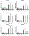
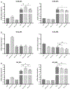
Neuroinflammation plays a key role in the development of CNS pathologies. This event encompasses a series of mechanisms involving the immune system and its cellular and molecular components. While it is necessary to activate the innate immune system during the early response to pathogens or traumas, persistent inflammation hinders neuronal recovery and contributes to the development of long-term neuronal complications. In this way, the application of pharmacological and non-pharmacological treatments is crucial to achieving better recovery of patients. We recently observed that the application of a low frequency electromagnetic field (EMF) decreases the expression of pro-inflammatory markers in an animal model of Traumatic Brain Injury in swine. To characterize this effect in terms of individualized response of neurons and microglial cells, we performed an in vitro model of pro-inflammatory damage by treating two different cell lines with tumor necrosis factor-α and then stimulating the cells with two frequencies of EMF. Transcriptional expression of inflammatory mediators was analyzed 24 and 48 hours after. Our results showed that both cell lines are susceptible to EMF, responding to the treatment by reducing the levels of the target genes in study. These observations further support the anti-inflammatory effect of EMF in the function of neurons and microglial cells and thus enhancing the recovery following traumatic brain injury, as observed under in vivo conditions in both experimental animals and human. These findings lay the foundation and warrants further preclinical and clinical studies to determine the effective frequency and duration of EMF stimulation in the healing of brain injury.
Keywords: Electromagnetic field; HCN2 cells; HMC3 cells; IL-1β; Microglial cells; Neurons; TNF-α; Traumatic Brain Injury.
Conflict of interest statement
Competing interest All the authors have read the manuscript and declare no competing or conflict of interest. No writing assistance was utilized in the production of this manuscript.
Figures


Similar articles
-
Microglial Response to Inflammatory Stimuli Under Electromagnetic Field Exposure.Arch Clin Biomed Res. 2025;9(4):304-315. doi: 10.26502/acbr.50170467. Epub 2025 Jun 30. Arch Clin Biomed Res. 2025. PMID: 40855885 Free PMC article.
-
Systemic pharmacological treatments for chronic plaque psoriasis: a network meta-analysis.Cochrane Database Syst Rev. 2021 Apr 19;4(4):CD011535. doi: 10.1002/14651858.CD011535.pub4. Cochrane Database Syst Rev. 2021. Update in: Cochrane Database Syst Rev. 2022 May 23;5:CD011535. doi: 10.1002/14651858.CD011535.pub5. PMID: 33871055 Free PMC article. Updated.
-
Systemic pharmacological treatments for chronic plaque psoriasis: a network meta-analysis.Cochrane Database Syst Rev. 2017 Dec 22;12(12):CD011535. doi: 10.1002/14651858.CD011535.pub2. Cochrane Database Syst Rev. 2017. Update in: Cochrane Database Syst Rev. 2020 Jan 9;1:CD011535. doi: 10.1002/14651858.CD011535.pub3. PMID: 29271481 Free PMC article. Updated.
-
Systemic pharmacological treatments for chronic plaque psoriasis: a network meta-analysis.Cochrane Database Syst Rev. 2020 Jan 9;1(1):CD011535. doi: 10.1002/14651858.CD011535.pub3. Cochrane Database Syst Rev. 2020. Update in: Cochrane Database Syst Rev. 2021 Apr 19;4:CD011535. doi: 10.1002/14651858.CD011535.pub4. PMID: 31917873 Free PMC article. Updated.
-
The Black Book of Psychotropic Dosing and Monitoring.Psychopharmacol Bull. 2024 Jul 8;54(3):8-59. Psychopharmacol Bull. 2024. PMID: 38993656 Free PMC article. Review.
References
Grants and funding
LinkOut - more resources
Full Text Sources
