. 2025 Aug 8:14:RP107534.
doi: 10.7554/eLife.107534.

Long-term live imaging, cell identification and cell tracking in regenerating crustacean legs

Affiliations

Long-term live imaging, cell identification and cell tracking in regenerating crustacean legs

Çağrı Çevrim et al. Elife. .

Abstract

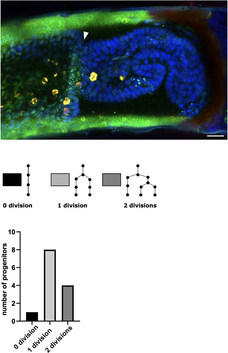
High-resolution live imaging of regeneration presents unique challenges due to the nature of the specimens (large mobile animals), the duration of the process (spanning days or weeks), and the fact that cellular resolution must be achieved without damage caused by lengthy exposures to light. Building on previous work that allowed us to image different parts of the process of leg regeneration in the crustacean Parhyale hawaiensis, we present here a method for live imaging that captures the entire process of leg regeneration, spanning up to 10 days, at cellular resolution. Our method includes (1) mounting and long-term live imaging of regenerating legs under conditions that yield high spatial and temporal resolution but minimise photodamage, (2) fixing and in situ staining of the regenerated legs that were imaged, to identify cell fates, and (3) computer-assisted cell tracking to determine the cell lineages and progenitors of identified cells. The method is optimised to limit light exposure while maximising tracking efficiency. Combined with appropriate cell-type-specific markers, this method may allow the description of cell lineages for every regenerated cell type in the limb.

Keywords: HCR; Parhyale hawaiensis; cell fate; cell lineage; developmental biology; hybridisation chain reaction; leg regeneration; live imaging.


Conflict of interest statement

ÇÇ, BL, MR, NL, JB, AA, MA No competing interests declared, KS KS is employed part-time by LPIXEL Inc

Figures

Figure 1.
Figure 1.. Morphology and regeneration of Parhyale legs.
Illustration of the distal-most podomeres of an intact Parhyale T4 or T5 leg, including the merus, carpus, and propodus (top), and of a leg amputated at the distal part of the carpus (bottom). In the amputated leg stump, the site where regeneration takes place is illustrated with a cartoon of the regenerating leg. Our live regeneration experiments focus on that region.
Figure 2.
Figure 2.. Live imaging capturing the phases of leg regeneration in Parhyale.
Phases of leg regeneration were observed by live imaging of nuclei labelled with H2B-mRFPruby (images from dataset li48-t5; Video 1). Proximal parts of the leg are to the left and the amputation site is on the right of each panel. (A) T5 leg imaged shortly after amputation, showing haemocytes adhering to the wound site (on the right of dashed line). (B) At 16 hpa, haemocytes have produced a melanised scab at the wound; epithelial cells are migrating below the scab. (C) At 32 hpa, the leg tissues have become detached from the scab (open arrow). (D) At 45 hpa, the new carpus–propodus boundary becomes visible (white arrow); dividing cells can be seen at the distal part of the leg stump (mitotic figures marked by circles). (E) At 57 hpa, the propodus–dactylus boundary becomes visible (black arrow); the carpus and propodus are well separated (white arrows). (F, G) In later stages, tissues in more proximal parts of the leg (to the left of the white arrows) retract, making space for the growing regenerating leg. hpa: hours post amputation. Scale bars, 20 µm.
Figure 2—figure supplement 1.
Figure 2—figure supplement 1.. Testing the effects of scanning speed and averaging on image quality.
Duplicate images were captured over a range of settings for scan speed (0.52–4.12 ms per pixel) and averaging (averaging 1–8 scans). Signal-to-noise and contrast ratios (SNR and CR, respectively) were determined as described in Ulman et al., 2017, using the Cell Tracking Challenge Fiji plug-in. (A) Images captured with different settings, indicating the SNR and CR measured in each case. (B) Plots showing the effects of scan speed and averaging on SNR and CR. The points highlighted in grey correspond to settings that result in the same amount (duration) of light exposure per pixel.
Figure 2—figure supplement 2.
Figure 2—figure supplement 2.. Apoptosis in legs that have not been subjected to live imaging.
(A) Quantification of apoptotic nuclei in the carpus in T4 or T5 limbs, 3 days post amputation. The legs were amputated at the distal part of the carpus, fixed 3 days post amputation, stained with DAPI and imaged on a confocal microscope. The number of apoptotic nuclei was counted within a 28-µm deep stack from the surface of the carpus. (B) Optical section through a carpus at 3 days post amputation. Nuclei that are undergoing apoptosis are highlighted in red circles. The data for making this figure are provided in Supplementary Data 7 at https://doi.org/10.5281/zenodo.15181497.
Figure 3.
Figure 3.. Overview of 22 live recordings of Parhyale leg regeneration.
For each recording, we indicate the span of the early phase of wound closure and proliferative quiescence prior to epithelial detachment from the scab (in blue), the early phases of cell proliferation and morphogenesis, up to the time when the first and second podomere boundaries become visible (in yellow and orange, respectively), and the late phases of regeneration, when the leg takes its final shape, cell proliferation gradually dies down and cells differentiate (in red). The total duration of this process varies from 3 to 10 days. Note that the fastest instances of regeneration occurred in young individuals (li51 and li52); the fastest, li51, was imaged at 29°C (marked by an asterisk). Datasets li-13 and li-16 were recorded until the molt; the other recordings were stopped before molting. Further details on each recording are given in Supplementary Data 1 available at https://doi.org/10.5281/zenodo.15181497.
Figure 3—figure supplement 1.
Figure 3—figure supplement 1.. Variation in the duration of early and late phases of regeneration in relation to the overall speed of leg regeneration.
For each of the 22 live recordings listed in Figure 3 and Supplementary Data 1 at https://doi.org/10.5281/zenodo.15181497, we defined the early phase as the period prior to epithelial detachment from the scab (wound closure phase, depicted in blue in Figure 3), and the late phase as the period between epithelial detachment and the appearance of both podomere boundaries (morphogenesis phase, depicted in yellow and orange in Figure 3). The later phase, leading to differentiation (red in Figure 3), was not included in this analysis, as the end of that phase could not be defined by an objective landmark. (A, B) Graphs showing the duration of the early and late phases of leg regeneration in relation to the overall duration of early and late phases. (A', B') Graphs showing the relative duration of the early and late phases of leg regeneration in relation to the overall duration of early and late phases. (C) Overview of the relative duration of different phases of leg regeneration, colour coded as in Figure 3.
Figure 4.
Figure 4.. Temporal pattern of cell divisions in regenerating Parhyale legs.
The number of cell divisions detected per time point is shown for five recordings of regenerating legs. The divisions were extracted from tracking data (for li13-t4) or detected using a semi-automated approach (see Methods). The detection of divisions is not exhaustive.
Figure 5.
Figure 5.. Tracking efficiency in relation to imaging depth.
Elephant’s performance in detecting nuclei at different depths was assessed in the datasets presented in Table 1A. Each column represents an independent replicate. Precision and recall of detection (see Methods) were scored at z intervals of 5 µm. The data for this figure are provided in Supplementary Data 8 at https://doi.org/10.5281/zenodo.15181497.
Figure 6.
Figure 6.. Identification of in situ stained cells in live image recordings.
(Α,Α’) Regenerated T5 leg at 189 hpa, in the last frame of a live recording (A) and after immunostaining with antibodies for Prospero and acetylated alpha tubulin (A'). (Β,Β’) Regenerated T5 leg imaged at 97 hpa, in the last frame of a live recording (B) and after hybridisation chain reaction (HCR) with probes for orthologue of spineless mostly nuclear dots corresponding to nascent transcripts, (B'). The same cells can be identified in the live recordings and in immunofluorescence or HCR stainings, as highlighted by coloured circles. Note that not all corresponding cells are visible in these optical sections, due to slight differences in mounting and tissue distortion.
Figure 6—figure supplement 1.
Figure 6—figure supplement 1.. In situ staining of Parhyale legs by hybridisation chain reaction (HCR).
(A) Leg segment at late embryonic stages (after cuticle deposition) stained with HCR probes for futsch (magenta) and nompA (green); the image also shows weak nuclear staining with DAPI and cuticle autofluorescence (grey). (B) Optical section through an adult leg stained with HCR probes for futsch, marking putative neurons in the interior of the leg (magenta, with weak autofluorescence in the cuticle), and nuclei stained with DAPI (grey). (C) Surface view of adult Parhyale leg stained with HCR probes for futsch (magenta) and nompA (green), and nuclei stained with DAPI (grey). Scale bars, 20 µm.
Figure 7.
Figure 7.. Tracking the progenitors of spineless-expressing cells in the distal carpus.
Snapshots of live recording of a regenerating Parhyale T5 leg at 0, 18, 60, 76, 85, and 97 h post amputation (left) and corresponding illustrations of the same legs (right). Coloured circles highlight cells that contribute to spineless-expressing cells in the distal part of the carpus; each colour highlights the lineage of a distinct progenitor cell. In the bottom-right panel, spineless-expressing nuclei (identified by HCR) are marked by filled circles, whereas spineless-non-expressing nuclei derived from the same progenitors are marked by open circles. The images show single optical sections as they appear in the Mastodon user interface; nuclei that are only partly captured in the current optical section appear as smaller circles. Distal parts of the leg oriented towards the right.
Figure 7—figure supplement 1.
Figure 7—figure supplement 1.. Spineless expression in a regenerating Parhyale leg.
(Top) T5 Parhyale leg at 97 h post amputation (same leg as in Figure 7) stained with HCR probes for spineless (in green) and futsch (in red), and with DAPI (in blue). Nascent transcripts of spineless are visible in a band of cells at the distal end of the carpus (arrowhead), and in other cells in the carpus, propodus, and dactylus. Autofluorescence is visible in the cuticle (in green) and in granular cells (in both red and green channels, appearing yellow). No futsch-stained neurons are visible in this optical slice. Scale bar, 20 µm. (Bottom) Topology of the lineage trees generating the tracked spineless-positive cells, and number of progenitors tracked with each tree topology.
Author response image 1.

Update of

  • doi: 10.1101/2024.09.11.612529
  • doi: 10.7554/eLife.107534.1

Similar articles

References

    1. Aharoni D, Khakh BS, Silva AJ, Golshani P. All the light that we can see: a new era in miniaturized microscopy. Nature Methods. 2019;16:11–13. doi: 10.1038/s41592-018-0266-x. - DOI - PMC - PubMed
    1. Almazán A, Çevrim Ç, Musser JM, Averof M, Paris M. Crustacean leg regeneration restores complex microanatomy and cell diversity. Science Advances. 2022;8:eabn9823. doi: 10.1126/sciadv.abn9823. - DOI - PMC - PubMed
    1. Alwes F, Enjolras C, Averof M. Live imaging reveals the progenitors and cell dynamics of limb regeneration. eLife. 2016;5:73. doi: 10.7554/eLife.19766. - DOI - PMC - PubMed
    1. Barbosa JS, Sanchez-Gonzalez R, Di Giaimo R, Baumgart EV, Theis FJ, Götz M, Ninkovic J. Live imaging of adult neural stem cell behavior in the intact and injured zebrafish brain. Science. 2015;348:789–793. doi: 10.1126/science.aaa2729. - DOI - PubMed
    1. Browne WE, Price AL, Gerberding M, Patel NH. Stages of embryonic development in the amphipod crustacean,Parhyale hawaiensis. Genesis. 2005;42:124–149. doi: 10.1002/gene.20145. - DOI - PubMed

LinkOut - more resources