Review
. 2025 Aug 8;19(8):e0013219.
doi: 10.1371/journal.pntd.0013219. eCollection 2025 Aug.

Radiological features of chronic pulmonary histoplasmosis: Easily mistaken for tuberculosis

Affiliations
Review

Radiological features of chronic pulmonary histoplasmosis: Easily mistaken for tuberculosis

David W Denning et al. PLoS Negl Trop Dis. .

Abstract

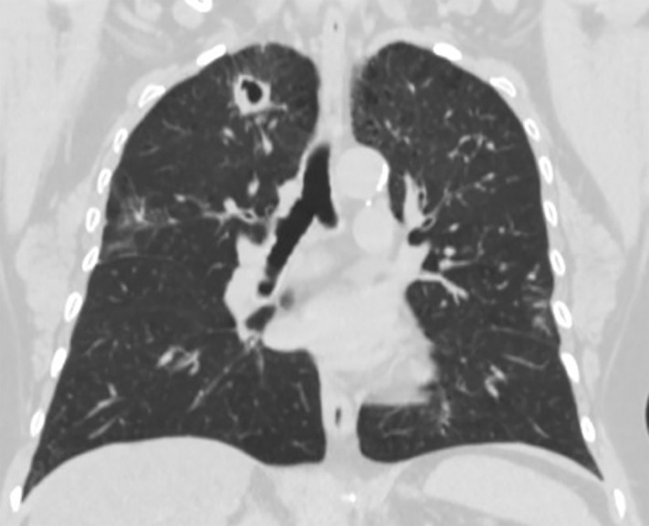
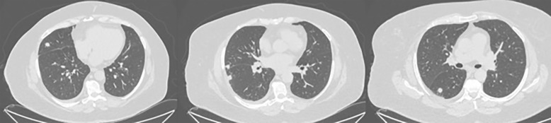
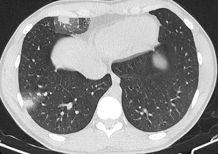
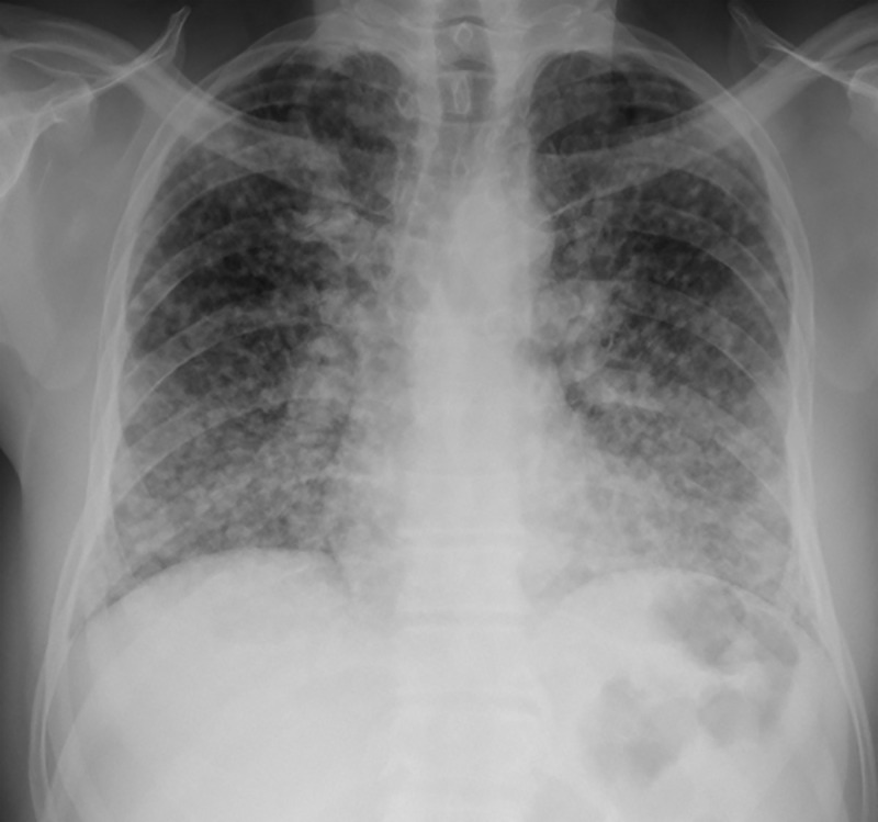
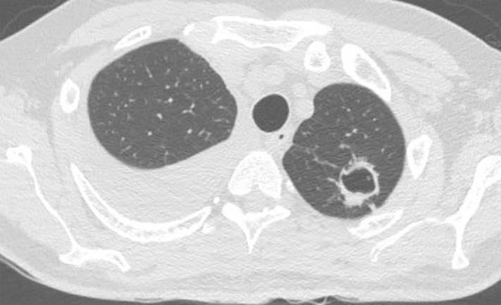
Histoplasma capsulatum var capsulatum is an endemic respiratory pathogen presenting in various forms including miliary histoplasmosis, acute and chronic pulmonary histoplasmosis, and acute or subacute disseminated disease. The differential diagnosis of chronic pulmonary histoplasmosis (CPH) is broad, encompassing bacterial, fungal and malignant aetiologies. PubMed was searched for relevant articles on the radiological characteristics of CPH and the most common differential diagnoses of tuberculosis and chronic pulmonary aspergillosis. The Fleischner Society Glossary of Terms for Thoracic Imaging was used to analyze the features. The contribution of culture, antibody and antigen and PCR to the diagnosis of CPH is summarized. Cavitation and pulmonary nodules are the most common features of CPH. Pleural effusion, pleural thickening, intrathoracic lymphadenopathy and bronchiectasis are not characteristic of CPH; uncommonly CPH can be complicated by an aspergilloma. Data on the radiologic features of CPH are derived primarily from the USA, Brazil, and China. CPH can be diagnosed by respiratory fungal culture (using extended culture times) or Histoplasma PCR (although data are scarce) and serum Histoplasma antigen and antibody. Data on bronchoscopy sampling for antigen are lacking. In patients with pulmonary cavitation without a confirmed diagnosis of tuberculosis or aspergillosis should be evaluated for CPH.


Conflict of interest statement

The authors have declared that no competing interests exist.

Figures

Fig 1
Fig 1. A 62-year-old woman with a history of COPD, asthma, and type 2 diabetes presented with chronic dyspnoea.
A chest CT revealed a pulmonary cavitation in the right upper lobe. Bronchoalveolar lavage culture grew Histoplasma capsulatum.
Fig 2
Fig 2. A 63-year-old woman with hypertension, obesity, and a history of smoking.
A chest CT showed multiple nodules in the lungs. The patient was diagnosed with pulmonary histoplasmosis after the biopsy and positive serology.
Fig 3
Fig 3. A 24-year-old man with a history of Crohn’s disease, currently on infliximab and azathioprine, presented with a two-week history of cough, fever, chills, night sweats, and dyspnea after visiting a cave.
A chest CT revealed two nodules with slightly irregular contours and a ground-glass halo, located in the peripheral region of the right lower and middle lobes. He was diagnosed with probable pulmonary histoplasmosis after positive serology.
Fig 4
Fig 4. A 55-year-old man with no previous comorbidities presented with a 3-week history of cough, expectoration, fatigue, fever, night sweats, dyspnoea, and weight loss.
A chest X-ray showed a miliary pattern. The patient was diagnosed with histoplasmosis after lung biopsy and positive serology.
Fig 5
Fig 5. A 48-year-old man with dermatomyositis, currently undergoing combination therapy with leflunomide and methotrexate, presented with chronic dyspnoea.
A chest CT scan revealed a right chronic pleural effusion (a rare finding in CPH) with associated thickened pleura and a cavitary lesion in the left upper lobe. He was diagnosed with pulmonary histoplasmosis following positive serology with high titers and confirmation via lung biopsy.
Fig 6
Fig 6. A. A 31-year-old woman with no comorbidities presented with facial plethora and oedema of the cervical region and upper limbs.
A CT angiography scan revealed a lesion measuring 5.4 × 3.0 × 4.4 cm, with approximately 75% reduction in the caliber of the superior vena cava. She was diagnosed with pulmonary histoplasmosis after positive serology results obtained by two different methods. The mediastinal biopsy revealed only fibrosis. B. PET/CT scan showed a mediastinal mass with high glycolytic activity.

Similar articles

References

    1. Lottenberg R, Waldman RH, Ajello L, Hoff GL, Bigler W, Zellner SR. Pulmonary histoplasmosis associated with exploration of a bat cave. Am J Epidemiol. 1979;110(2):156–61. doi: 10.1093/oxfordjournals.aje.a112800 - DOI - PubMed
    1. Antinori S, Giacomelli A, Corbellino M, Torre A, Schiuma M, Casalini G, et al. Histoplasmosis diagnosed in Europe and Israel: a case report and systematic review of the literature from 2005 to 2020. J Fungi (Basel). 2021;7(6). - PMC - PubMed
    1. Bongomin F, Kibone W, Atulinda L, Morgan B, Ocansey B, Storer ISR, et al. Frequency of fungal pathogens in autopsy studies of people who died with HIV in Africa: a scoping review. Clin Microbiol Infect. 2024;30(5):592–600. doi: 10.1016/j.cmi.2023.12.016 - DOI - PMC - PubMed
    1. Antinori S. Histoplasma capsulatum: more widespread than previously thought. Am J Trop Med Hyg. 2014;90(6):982–3. doi: 10.4269/ajtmh.14-0175 - DOI - PMC - PubMed
    1. Azar MM, Loyd JL, Relich RF, Wheat LJ, Hage CA. Current concepts in the epidemiology, diagnosis, and management of histoplasmosis syndromes. Semin Respir Crit Care Med. 2020;41(1):13–30. doi: 10.1055/s-0039-1698429 - DOI - PubMed

MeSH terms