. 2025 Aug 8;11(32):eadw8034.
doi: 10.1126/sciadv.adw8034. Epub 2025 Aug 8.

A two-step mechanism for RIG-I activation by influenza virus mvRNAs

Affiliations

A two-step mechanism for RIG-I activation by influenza virus mvRNAs

Emmanuelle Pitré et al. Sci Adv. .

Abstract

Influenza A virus (IAV) noncanonical RNAs are bound by retinoic acid-inducible gene I (RIG-I). However, innate immune activation is infrequent and it is not understood why noncanonical IAV RNAs activate RIG-I in a sequence- or RNA structure-dependent manner. We hypothesized that multiple events need to occur before IAV RNA synthesis activates RIG-I and investigated whether RIG-I activation is stimulated by the noncanonical or aberrant transcription of mini viral RNAs (mvRNA), an RNA that is overexpressed in highly pathogenic IAV infections. We find that mvRNAs can cause noncanonical transcription termination through a truncated 5' polyadenylation signal or a 5' transient RNA structure that interrupts polyadenylation. The resulting capped complementary RNAs stimulate the release of an mvRNA and complement RIG-I activation in trans. Overall, our findings indicate that sequential rounds of noncanonical or aberrant viral replication and transcription are needed for innate immune signaling by IAV RNA synthesis.


Figures

Fig. 1.
Fig. 1.. Noncanonical transcription of mvRNAs by the IAV RNA polymerase in vitro.
(A) Schematic showing the relation between canonical and noncanonical transcription and replication during IAV infection. (B) Molecular differences between IAV transcription and replication. (C) Overview of RNA molecules produced from mvRNA templates, and the primers used to differentiate among them. (D) Cap-snatching of a 32P-labeled capped, 20-nt-long RNA primer by the IAV RNA polymerase. Cleavage reactions were analyzed by 20% denaturing PAGE. (E) Representative image of the extension of a 32P-labeled capped, 11-nt-long RNA primer by the IAV RNA polymerase. Extension reactions were analyzed by 20% denaturing PAGE. The alternative initiation products CP and GP, as well as the realignment product (RP), are indicated. Asterisk indicates an unknown signal. (F) Representative image of a primer extension analysis of RNA extracted from capped, 11-nt-long RNA extension reactions using VNdT20.
Fig. 2.
Fig. 2.. Noncanonical transcription of mvRNAs by the IAV RNA polymerase in cell culture.
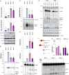
(A) Innate immune activation by IAV mvRNAs in HEK293T cells. Cells were transfected with plasmids encoding the RNA polymerase subunits, NP, a Renilla luciferase transfection control, a firefly-based IFN-β reporter, and the NP71.1, NP71.11, or NP71.2 mvRNAs. Twenty-four hours posttransfection, the luciferase and phosphorylated STAT1 (pSTAT1) levels were measured. PB1, NP, and tubulin protein expression was analyzed as control. (B) Steady-state mvRNA and 5S rRNA levels measured 24 hours posttransfection by primer extension using the NP− primer. (C) Steady-state total IAV capped RNA levels measured using the NP+ primer. (D) Steady-state cRNA and ccRNA levels measured using the NP5′ primer. (E) Steady-state cRNA and ccRNA levels in the presence of the WSN or A/Brevig Mission/1/1918 (H1N1) IAV RNA polymerase. (F) Schematic showing the removal of m7G from the 5′ end of viral capped RNAs using mRNA decapping enzyme (MDE), generating an RNA with a 5′ end monophosphate and the release of 7-methylguanosine disphosphate. The 5′ monosphosphate RNA is then cleaved by the exoribonuclease XRN1 in a 5′ → 3′ direction, generating ribonucleotide monophosphates. (G) Four micrograms of HEK293T cell RNA was subjected to enzymatic digestion using either MDE alone or a combination of MDE and XRN1. Viral RNA levels were detected using primer extension and quantified. In all figures, graphs show the mean of three independent experiments. Error bars indicate SD, and P values were determined using one-way ANOVA.
Fig. 3.
Fig. 3.. ccRNAs are enriched in RIG-I immunoprecipitation assays.
(A) RIG-I immunoprecipitation following the expression of different mvRNAs in the presence of the WSN RNA polymerase and NP in HEK293T cells. Viral RNA levels and the 5S rRNA loading control were analyzed 24 hours posttransfection by primer extension. PB1, c-myc-RIG-I, NP, and tubulin expression were analyzed by Western blot. RNA levels following immunoprecipitation are shown in the bottom panels. (B) Quantification of the mvRNA, cRNA, and ccRNA levels following RIG-I immunoprecipitation. (C) Quantification of the mvRNA levels following IFI16 immunoprecipitation. (D) Normalization of vRNA enrichment by vRNA length following IFI16 immunoprecipitation. (E) Quantification of the mvRNA levels following RIG-I or IFI16 immunoprecipitation. In [(B) to (E)], graphs show the mean of three independent experiments. Error bars indicate SD, and P values were determined using one-way ANOVA.
Fig. 4.
Fig. 4.. Transient RNA structures up-regulate ccRNA formation.
(A) Schematic of the putative mechanism for mvRNA generation using template switching between the 3′ end of the vRNA template and the 5′ U-stretch. (B) Primer extension analysis showing ccRNA, cRNA, and mvRNA levels in HEK293T cells expressing segment 2 mvRNAs. Graphs show quantification of ccRNA level of three biological repeats. Error bars indicate SD. P values were calculated using one-way ANOVA relative to PB1-C. (C) Graph of IFN-β promoter activity measured in the presence of DMSO or baloxavir. Error bars indicate SD. P values were calculated using two-way ANOVA with multiple corrections. (D) Structural alignment of the IAV RNA polymerase in a polyadenylation state (PDB 6T0S) with an AlphaFold 3 (AF) model in which base pairing between the entering and exiting RNA shifts the position of the PB1 β-ribbon. (E) Heatmap showing the stability of the t-loops in the mvRNAs analyzed. The position of the t-loop is aligned relative to the 5′ terminal end of each mvRNA to better indicate position 17 of U-tract on which polyadenylation occurs. (F) Schematic of RNA polymerase footprinting assay. In this assay, we expressed a tandem-affinity purified (TAP)–tagged IAV RNA polymerase and an mvRNA in HEK293T cells, copurified the mvRNA with the RNA polymerase, partially digested the mvRNA parts that are not protected by the RNA polymerase, and lastly mapped the protected footprint of the RNA polymerase using primer extension. (G) Representative image of an RNA polymerase footprinting assay in the presence of mvRNA NP71.1. Heatmap shows calculated t-loop stability for each position of the NP71.1 mvRNA. (H) Model showing the role of the PB1 β-ribbon in polyadenylation and displacement of the β-ribbon by a transient RNA structure.
Fig. 5.
Fig. 5.. ccRNAs trigger release of mvRNA template in a sequence-dependent manner.
(A) Schematic of assay to test the impact of ccRNA molecules on mvRNA template release during replication in vitro. Purified RNA polymerase is immobilized on magnetic beads and incubated with a radiolabeled mvRNA template. Excess template is washed away and ApG, NTPs, and an inactive RNA polymerase (PB1a) are added to start replication. A complementary ccRNA or noncomplementary ccRNA derived from a segment 6 mvRNA (NA71) is added to trigger template release. (B) Dot blot of RNA polymerase–bound and unbound fractions. Graph shows signal measured using radiography. (C) Cell-based complementation assay in which coexpression of mvRNAs leads to innate immune activation. PB1-E was titrated in combination with a fixed amount (250 ng) of PB1-C d10A at the following ratios: 1:20, 1:10, and 1.1. NP and tubulin expression were analyzed by Western blot. In [(B) and (C)], graphs show the mean of three independent experiments. Error bars indicate SD, and P values were determined using one-way ANOVA.
Fig. 6.
Fig. 6.. Model of a two-step mechanism that leads to innate immune activation.
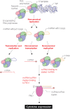
Model of RIG-I activation after noncanonical replication and transcription of t-loop–containing mvRNAs. Noncanonical replication produces two types of mvRNAs: mvRNAs that contain a t-loop that affects replication and/or transcription termination and mvRNAs that do not contain such a t-loop. When transcription termination is affected by t-loops, ccRNA molecules are produced. When t-loops affect replication, they can stall the RNA polymerase. ccRNAs trigger the release of mvRNAs on which the RNA polymerase has stalled, creating an RNA duplex with a triphosphate and cap. These duplexes contribute to RIG-I activation.

Similar articles

Cited by

References

    1. de Jong M. D., Simmons C. P., Thanh T. T., Hien V. M., Smith G. J., Chau T. N., Hoang D. M., Chau N. V., Khanh T. H., Dong V. C., Qui P. T., Cam B. V., Ha D. Q., Guan Y., Peiris J. S., Chinh N. T., Hien T. T., Farrar J., Fatal outcome of human influenza A (H5N1) is associated with high viral load and hypercytokinemia. Nat. Med. 12, 1203–1207 (2006). - PMC - PubMed
    1. de Wit E., Siegers J. Y., Cronin J. M., Weatherman S., van den Brand J. M., Leijten L. M., van Run P., Begeman L., van den Ham H. J., Andeweg A. C., Bushmaker T., Scott D. P., Saturday G., Munster V. J., Feldmann H., van Riel D., 1918 H1N1 influenza virus replicates and induces proinflammatory cytokine responses in extrarespiratory tissues of ferrets. J. Infect. Dis. 217, 1237–1246 (2018). - PMC - PubMed
    1. Kash J. C., Tumpey T. M., Proll S. C., Carter V., Perwitasari O., Thomas M. J., Basler C. F., Palese P., Taubenberger J. K., Garcia-Sastre A., Swayne D. E., Katze M. G., Genomic analysis of increased host immune and cell death responses induced by 1918 influenza virus. Nature 443, 578–581 (2006). - PMC - PubMed
    1. Te Velthuis A. J. W., Long J. C., Bauer D. L. V., Fan R. L. Y., Yen H. L., Sharps J., Siegers J. Y., Killip M. J., French H., Oliva-Martin M. J., Randall R. E., de Wit E., van Riel D., Poon L. L. M., Fodor E., Mini viral RNAs act as innate immune agonists during influenza virus infection. Nat. Microbiol. 3, 1234–1242 (2018). - PMC - PubMed
    1. Kobasa D., Jones S. M., Shinya K., Kash J. C., Copps J., Ebihara H., Hatta Y., Kim J. H., Halfmann P., Hatta M., Feldmann F., Alimonti J. B., Fernando L., Li Y., Katze M. G., Feldmann H., Kawaoka Y., Aberrant innate immune response in lethal infection of macaques with the 1918 influenza virus. Nature 445, 319–323 (2007). - PubMed

LinkOut - more resources