. 2025 Aug 1:S1534-5807(25)00446-0.
doi: 10.1016/j.devcel.2025.07.008. Online ahead of print.

Nuclear lamina phase separation orchestrates stress-induced transcriptional responses in plants

Affiliations

Nuclear lamina phase separation orchestrates stress-induced transcriptional responses in plants

Yu Tang et al. Dev Cell. .

Abstract

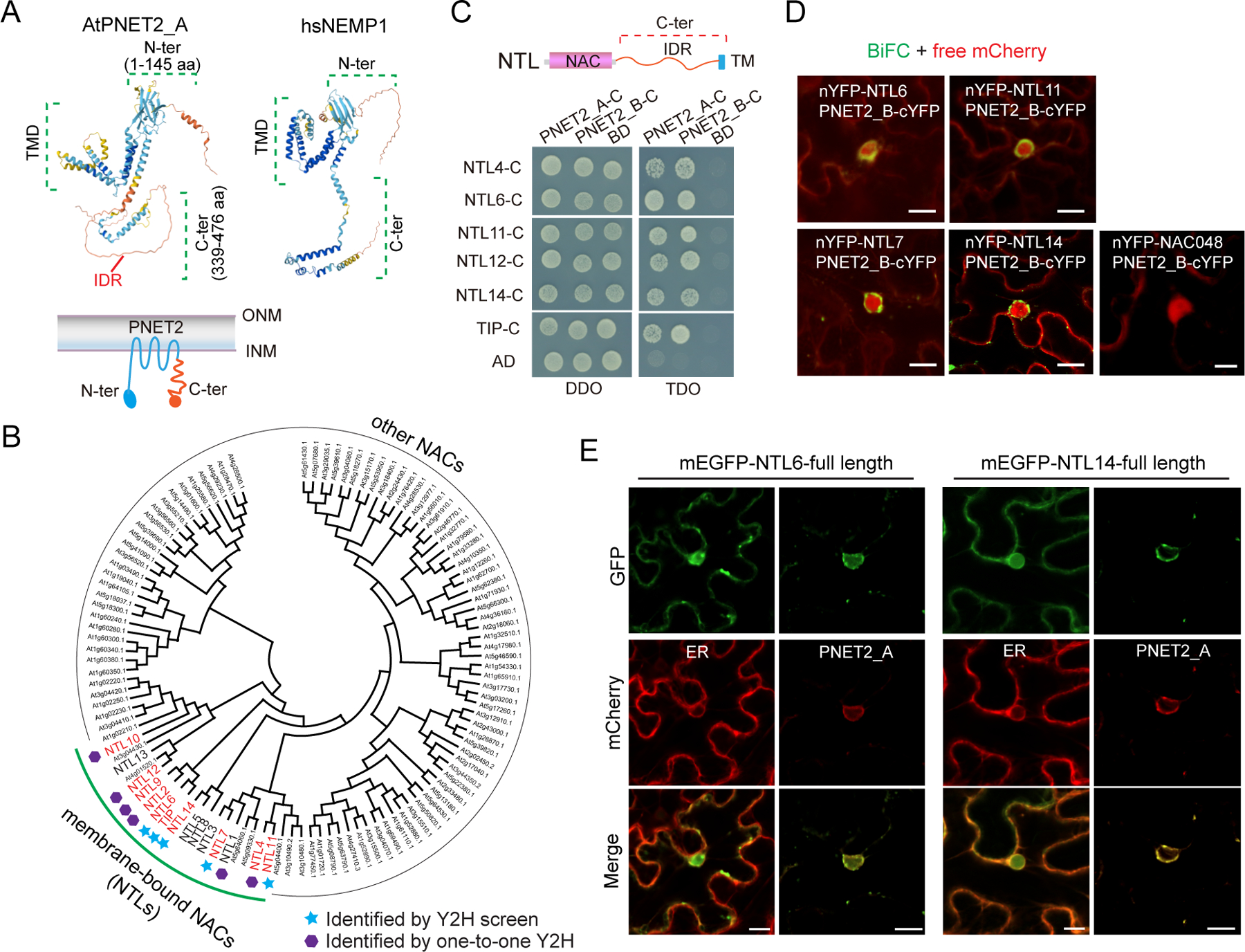
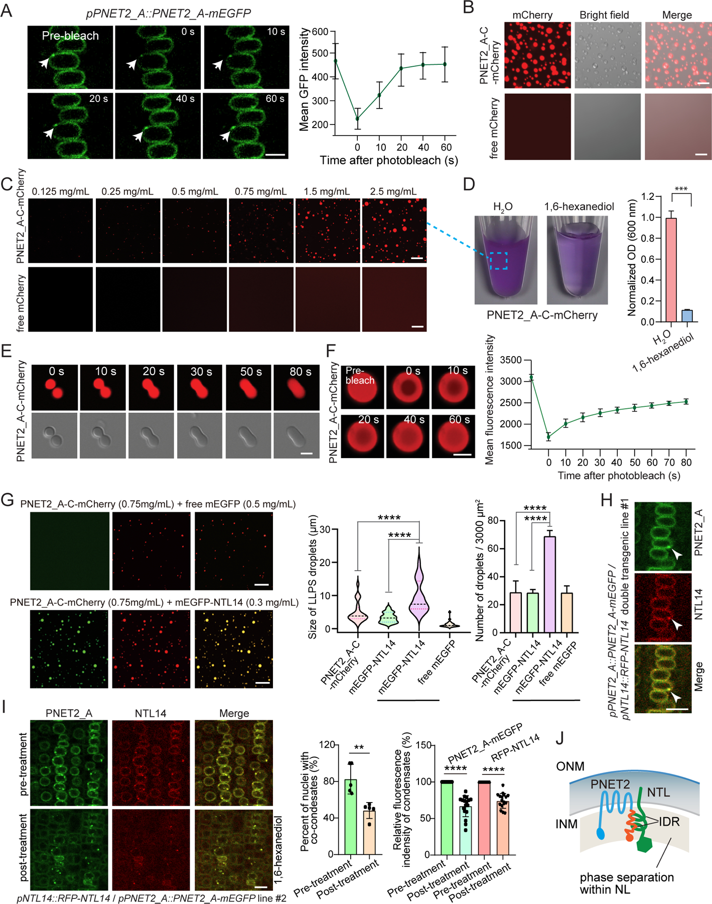
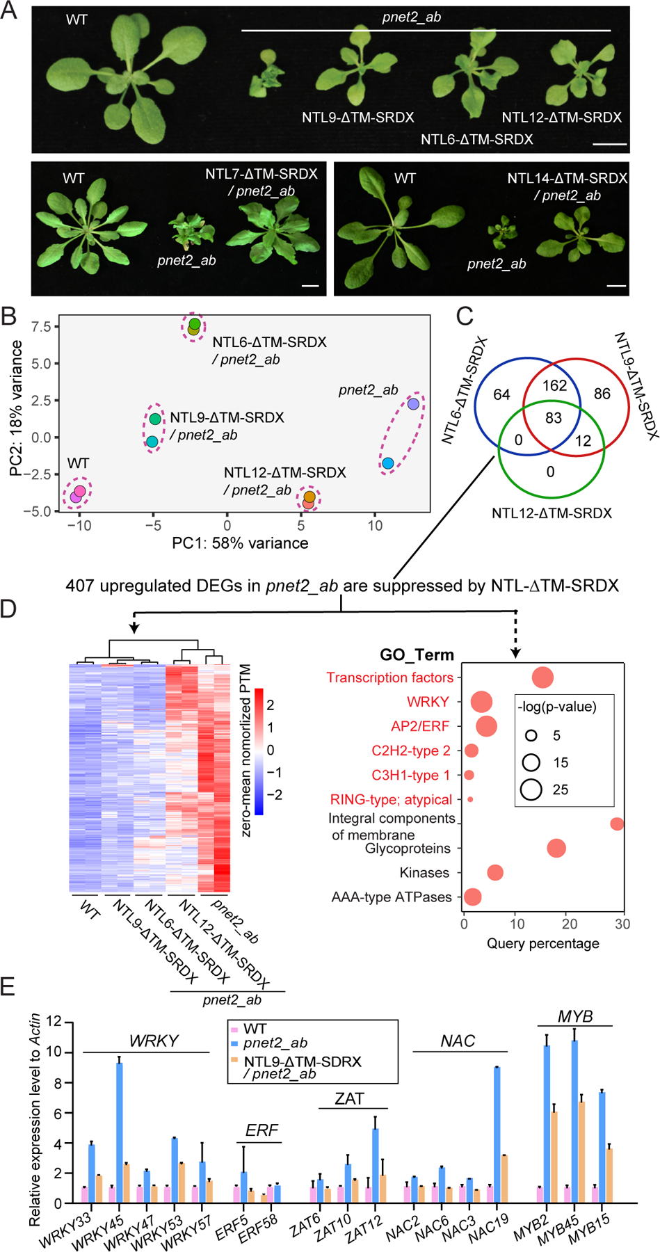
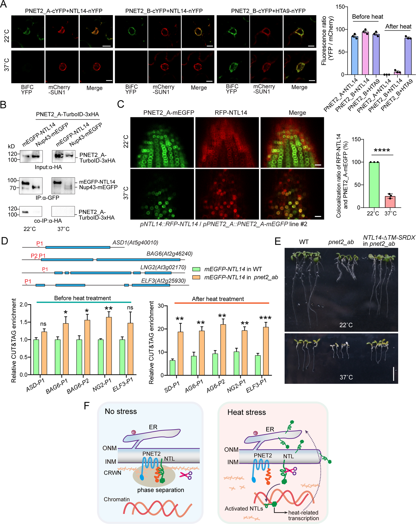
The nuclear lamina (NL), a perinuclear protein meshwork formed by nucleoskeleton and inner nuclear membrane (INM) proteins, is crucial for chromatin organization at the nuclear periphery and gene expression regulation in eukaryotic cells. However, NL-dependent transcriptional regulation remains poorly understood in plants due to the absence of most canonical NL proteins found in animals. Here, we report that the plant INM protein PLANT NUCLEAR ENVELOPE TRANSMEMBRANE 2 (PNET2) interacts with membrane-bound NAC (NAM, ATAF1/2, and CUC2) transcription factors, NTLs, via intrinsic disorder regions and promotes liquid-liquid phase separation within the NL. This compartmentalization effectively sequesters NTLs and restricts their transcriptional activity. In the absence of PNET2, NTLs become deregulated, triggering spontaneous and broad-spectrum stress responses. Importantly, we found that stress stimuli, such as heat shock, disrupt PNET2-NTL phase separation, releasing NTLs for target gene binding and transcriptional activation. These findings demonstrate a phase separation-based regulatory mechanism within the NL that controls membrane-bound transcription factor activity in response to environmental cues.

Keywords: NTL; PNET2; gene expression regulations; membrane-bound transcription factors; nuclear lamina; nuclear membrane; nucleoskeleton; phase separation; plant cells; stress responses.


Conflict of interest statement

Declaration of interests The authors declare no competing interests.

Figures

Figure 1.
Figure 1.. PNET2 interacts with membrane-bound NAC transcriptional factors at the nuclear lamina
(A) Predicted protein structure of Arabidopsis PNET2_A and its human homolog NEMP1 using AlphaFold (https://alphafold.com/). The folded N-terminus, multiple transmembrane domains, the intrinsically disordered region (IDR)-containing C-terminus were outlined. (B) Phylogenetic tree of the 112 NAC domain-containing proteins encoded by Arabidopsis, including 15 membrane-bound NTLs and 97 other NACs. NTLs identified by the yeast-two hybrid (Y2H) screen and one-to-one Y2H assays were marked with blue stars and purple hexagons, respectively. (C) The Y2H analysis using the PNET2 C-terminus as bait and NTL C-termini as prey. Zygote yeast cells were grown on DDO (–Leu–Trp) and TDO (–Leu–Trp–His + 3 mM 3-aminotriazole (3AT)) media. Empty vectors were used as negative controls. (D) Bimolecular fluorescence complementation (BiFC) assay between PNET2_B and full-length NTL proteins. Non-transmembrane NAC048 is included as a negative control. Indicated BiFC constructs were transiently coexpressed in N. benthamiana. Free mCherry was also coexpressed to mark the nuclei. (E) Transient co-expression of HEDL-mCherry (ER) or PNET2_A-mCherry with full-length GFP-NTL6/14 in N. benthamiana. Bars = 10 μm. See also Figure S1.
Figure 2.
Figure 2.. PNET2 and NTLs undergo liquid-liquid phase separation to promote coalescence
(A) Fluorescence Recovery After Photobleaching (FRAP) analysis using pPNET2_A::PNET2_A–mEGFP transgenic Arabidopsis seedlings. Root meristem regions were imaged. Arrows indicate the condensate bleached. FRAP recovery curve measuring GFP intensity is shown on the right (n = 8 condensates). Time 0 indicates the start of photobleaching. Bar = 10 μm. (B) Purified PNET2_A-C-mCherry protein forms distinct droplets under LLPS-promoting conditions (50 mM Tris pH 7.5, 100 mM NaCl, and 10% PEG 8,000). Bars = 5 μm. Images are representative of three independent experiments. (C) Formation of in vitro droplets by purified PNET2_A-C-mCherry protein at indicated concentrations. Bars = 5 μm. (D) The turbid solution containing PNET2_A-C-mCherry liquid droplets can be dissolved by treatment with 10% 1,6-hexanediol (n = 3 biological replicates). Quantification of solution turbidity measured at OD600 was shown on the right. (E) Time-lapse microscopy showing representative fusion events of two PNET2_A-C-mCherry droplets. Time 0 indicates the start of recording. Bar = 2 μm. (F) FRAP analysis of in vitro PNET2_A-C-mCherry droplets. Time 0 indicates the start of photobleaching. FRAP recovery curve is shown on the right (n = 13 droplets). Bar = 10 μm. (G) Co-condensation of PNET2_A-C-mCherry and mEGFP-NTL14 in vitro. LLPS was performed with 0.75 mg/mL PNET2_A-C-mCherry and 0.3 mg/mL mEGFP-NTL14 or 0.5 mg/mL free mEGFP. Quantification of LLPS droplet size (n = 30) and number (n = 50) is shown on the right. Bars = 5 μm. (H) Fluorescence imaging of root meristem cells from pPNET2_A::PNET2_A–mEGFP / pNTL14::RFP–NTL14 double transgenic seedlings (line #1). The PNET2_A-NTL14 co-condensate is indicated by arrowhead. Bar = 10 μm. (I) Root cells of pPNET2_A::PNET2_A–mEGFP / pNTL14::RFP–NTL14 double transgenic seedlings (line #2, higher expression) treated with 10% 1,6-hexanediol for 2 min. Measurements of condensate presence (n = 5 biological replicates, each containing 50 nuclei) and their relative fluorescent intensity (n = 20 nuclei) before and after treatment are shown on the right panel. Bar = 10 μm. (J) A model depicting that PNET2 sequesters NTLs within the nuclear lamina via IDR-driven LLPS. For line plots and bar plots, data are presented as mean ± SDM. For violin plots, the lower, middle, and upper dash lines indicate the 25th percentile, medium, and 75th percentile, respectively. Statistical analysis was performed using two-tailed Student’s t-tests. **p < 0.01, ****p < 0.0001. See also Figure S2.
Figure 3.
Figure 3.. NTLs function redundantly downstream of PNET2 to activate stress-related transcription
(A) Three-week-old (upper panel) and four-week-old (lower panels) soil-grown plants of indicated genotypes. Bars = 2 cm. (B) Principal component analysis (PCA) of RNA-seq data obtained from three-week-old WT, pnet2_ab, 35S::NTL6/9/12-ΔTM-SRDX / pnet2_ab plants (n = 2 biological replicates). (C) Venn diagram illustrating the number of upregulated differentially expressed genes (DEGs) in pnet2_ab that are suppressed by the expression of NTL6/9/12-ΔTM-SRDX. Suppression is defined as differential expression (35S::NTL6/9/12-ΔTM-SRDX / pnet2_ab plants vs pnet2_ab plants) with criteria of p.adj-value < 0.01 and fold-change > 2. (D) Heatmap (on the left) and GO enrichment analysis (on the right) of the 407 upregulated DEGs in pnet2_ab that are suppressed by NTL-ΔTM-SRDX expression. Representative GO terms are displayed in the bubble plot. (E) The relative expression level of stress-related transcription factor genes in 3-week-old WT, pnet2_ab, and 35S::NTL9-ΔC-SRDX / pnet2_ab plants measured by real-time qPCR. The gene expression level was normalized to that in WT. Actin was used as the reference gene. Data are represented as means ± SDM (n = 3 biological replicates). See also Figure S3.
Figure 4.
Figure 4.. PNET2 releases NTL14 for transcription activation in response to heat stress
(A) BiFC between PNET2_A/B and NTL14 at the nuclear periphery under normal condition and upon heat treatment. PNET2 interaction with histone protein is included as a positive control. BiFC YFP constructs and mCherry-SUN1 were transiently coexpressed in N. benthamiana. For heat stress treatment, the infiltrated N. benthamiana plants were incubated at 22°C for 24 h and then transferred to 37°C for another 24 h before imaging. Quantification of relative BiFC intensity to mCherry signal is shown on the right panel (n = 3 biological replicates, each containing 25 nuclei). Similar results have been obtained in at least two independent experiments. Bars = 10 μm. (B) Co-immunoprecipitation analysis showing PNET2_A interaction with NTL14 under normal and heat stress conditions. PNET2_A-TurboID-HA was transiently coexpressed with mEGFP-NTL14 in N. benthamiana. Nup43-mEGFP was used as a negative control. Protein extract was immunoprecipitated with GFP-Trap agarose beads before immunoblotting with anti-HA and anti-GFP antibodies. (C) Fluorescence imaging in Arabidopsis root tip cells using pPNET2_A::PNET2_A–mEGFP / pNTL14::RFP–NTL14 double transgenic seedlings before and after heat stress. Five-day-old transgenic seedlings were incubated at 22 °C (pre-treatment) or transferred to 37°C for 12 h (post-treatment) for imaging analysis. Quantification of RFP/GFP overlap is shown on the right (n = 3 biological replicates, each containing 10 nuclei). Bars = 10 μm. (D) CUT&Tag coupled qPCR analysis of NTL14 target genes before and after heat treatment. The upper panel showing the primer locations for qPCR analysis, and the lower panel showing relative enrichment for genes in T3 generation expressing GFP-NTL14 in WT and pnet2_ab mutant backgrounds (n = 6 biological replicates, each containing 15 seedlings). The enrichment level of each gene in WT plants before heat treatment is set to one. (E) Five-day-old seedlings of WT, pnet2_ab, 35S::NTL14-ΔTM-SRDX / pnet2_ab grown on MS-agar plates at 22 °C were exposed to 37 °C for 3 days. The heat-treated seedlings were recovered at 22 °C for another 3 days for survival analysis. Bars = 1 cm. (F) The working model of how PNET2 regulates NTLs at the nuclear lamina through phase separation. Data are presented as mean ± SDM. Statistical analysis was performed using two-tailed Student’s t-tests. *p < 0.05, **p < 0.01, ***p < 0.001, ****p < 0.0001. ns stands for not significant. See also Figure S4.

Similar articles

References

    1. Wong X, Melendez-Perez AJ, and Reddy KL (2022). The nuclear lamina. Cold Spring Harbor Perspect. Biol 14, a040113. 10.1101/cshperspect.a040113. - DOI - PMC - PubMed
    1. Greil F, Moorman C, and van Steensel B (2006). DamID: mapping of in vivo protein-genome interactions using tethered DNA adenine methyltransferase. Method enzymol 410, 342–359. 10.1016/s0076-6879(06)10016-6. - DOI - PubMed
    1. Karoutas A, and Akhtar A (2021). Functional mechanisms and abnormalities of the nuclear lamina. Nature Cell Biol 23, 116–126. 10.1038/s41556-020-00630-5. - DOI - PubMed
    1. Gaillard MC, and Reddy KL (2018). The Nuclear Lamina and Genome Organization. In Nuclear Architecture and Dynamics, Lavelle C, and Victor J-M, eds. (Academic Press; ), pp. 321–343. 10.1016/B978-0-12-803480-4.00014-4. - DOI
    1. Pawar S, and Kutay U (2021). The Diverse Cellular Functions of Inner Nuclear Membrane Proteins. Cold Spring Harbor Perspect. in Biol 13, a040477. 10.1101/cshperspect.a040477. - DOI - PMC - PubMed

LinkOut - more resources