. 2025 Aug 8;23(1):887.
doi: 10.1186/s12967-025-06750-6.

CDH3 promotes the progression of lung adenocarcinoma through driving epithelial-mesenchymal transition progress

Affiliations

CDH3 promotes the progression of lung adenocarcinoma through driving epithelial-mesenchymal transition progress

Wenlin Gong et al. J Transl Med. .

Abstract

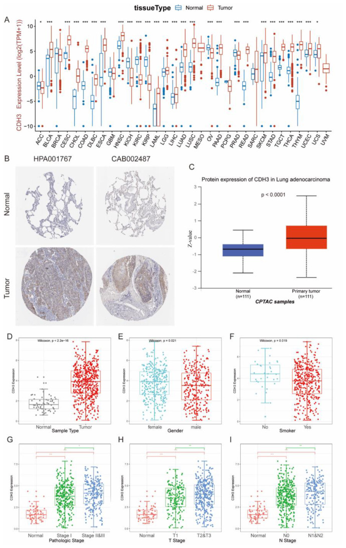
Background: Cadherin-3 (CDH3) participates in multiple oncogenic processes, but its biological role and mechanisms in lung adenocarcinoma (LUAD) remain inadequately understood.

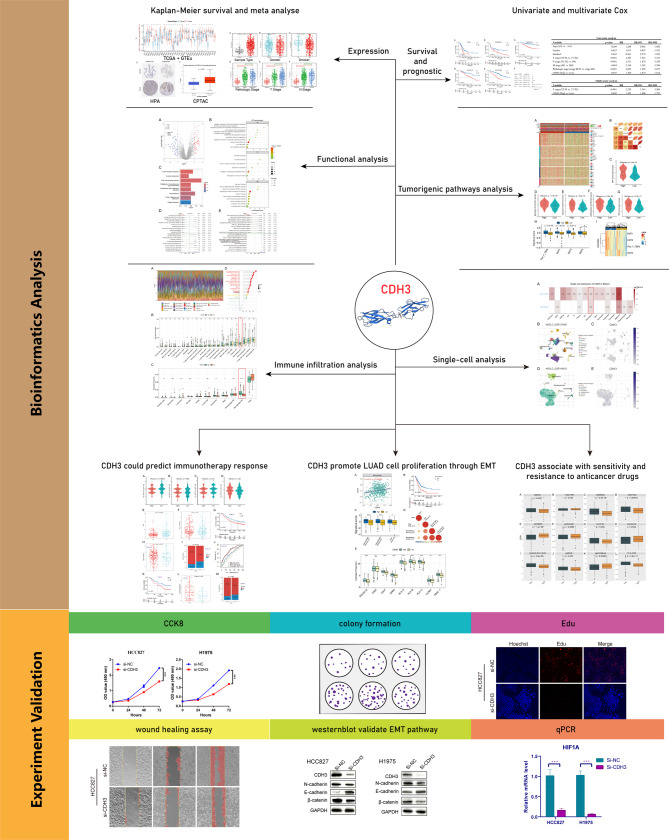
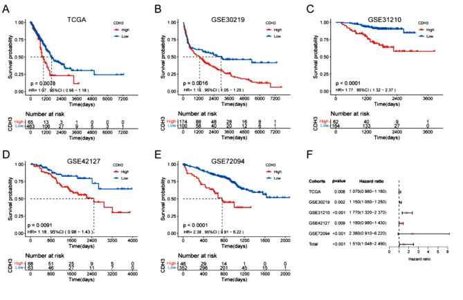
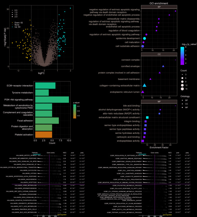
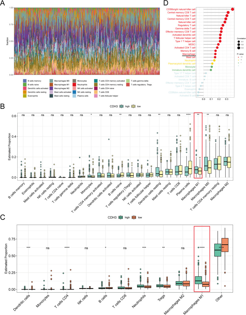
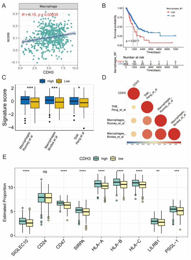
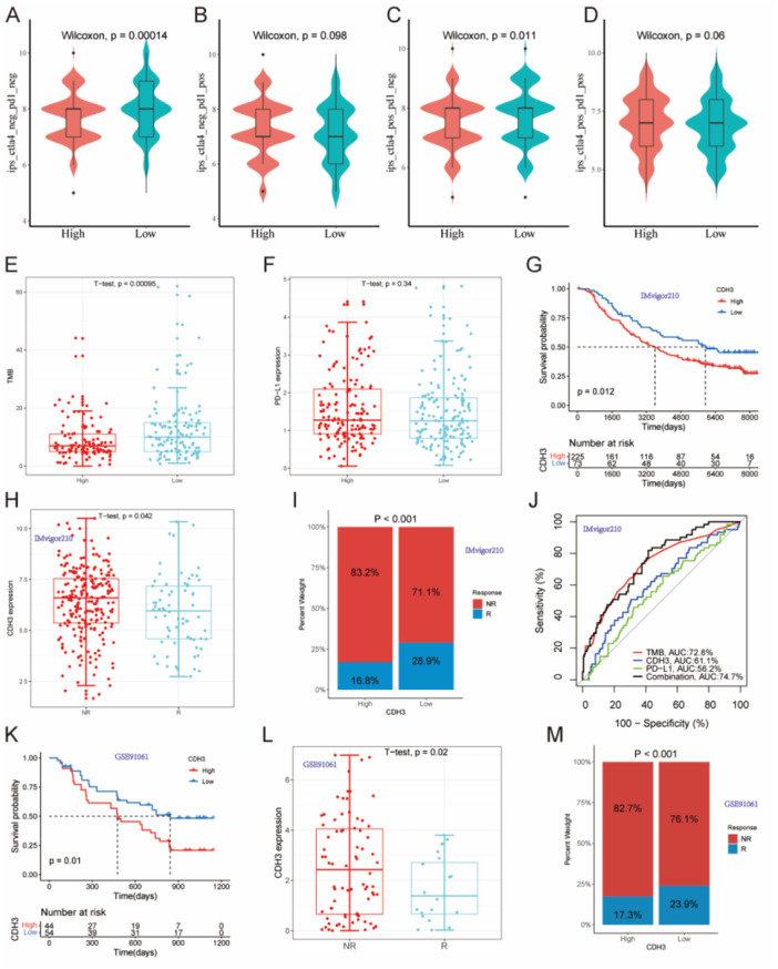
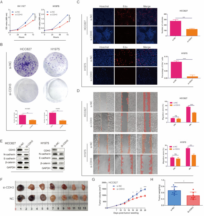
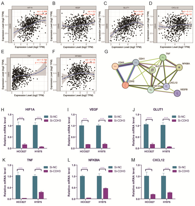
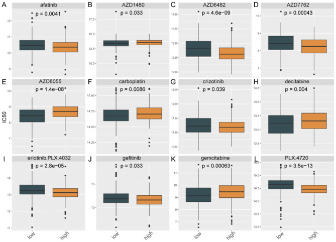
Methods: We analyzed CDH3 expression in LUAD versus adjacent normal tissues and assessed its association with patient outcomes. Bioinformatics, immune scoring, and single-cell analysis were used to explore CDH3's links to epithelial-mesenchymal transition (EMT), inflammatory responses, TNF pathway activation, glycolysis, hypoxia, and tumor-associated macrophage infiltration. Independent immunotherapy datasets (IMvigor210, GSE91061) were evaluated for CDH3's relationship to treatment response. In vitro CDH3 knockdown studies assessed effects on glycolysis, EMT, proliferation, and migration in LUAD cells, and in vivo tumor growth was measured post-knockdown.

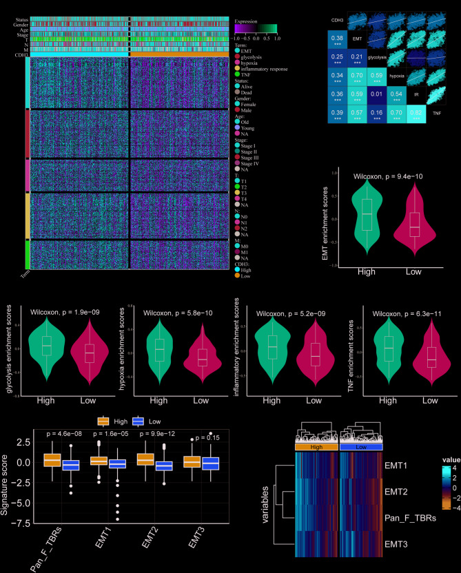
Results: CDH3 expression was significantly elevated in LUAD tissues and correlated with poorer patient survival. High CDH3 expression associated with EMT, inflammatory responses (including TNF pathway), glycolysis, and hypoxia. It also promoted tumor-associated macrophage infiltration and impaired anti-tumor immunity. Patients with low CDH3 expression showed improved immunotherapy response and prognosis. CDH3 knockdown suppressed LUAD cells glycolysis process, EMT, proliferation, and migration in vitro, and significantly repressed tumor growth in vivo.

Conclusions: CDH3 drives LUAD progression by promoting EMT, tumor growth, metastasis, and immunosuppression. It represents a promising diagnostic/prognostic biomarker and a novel therapeutic target for LUAD immunotherapy.

Keywords: CDH3; EMT; Immunotherapy; Lung adenocarcinoma; Tumor associated macrophages.


Conflict of interest statement

Declarations. Ethics approval and consent to participate: Not applicable. Consent for publication: Not applicable. Competing interests: The authors declare that the study was implemented without any financial or commercial correlations that could be established as underlying conflict of interest.

Figures

Fig. 1
Fig. 1
Study flowchart
Fig. 2
Fig. 2
CDH3 has diagnostic and prognostic value in LUAD patients. (A) Expression of CDH3 in paracancer and tumor tissues in TCGA and GTEx database. The protein expression of CDH3 in LUAD was obtained from the Human Protein Atlas (B) and CPTAC datasets (C). The expression level of CDH3 in LUAD based on (D) sample types, (E) gender, (F) smoking history, (G) different pathologic stages, (H) different T stages and (I) different N stages
Fig. 3
Fig. 3
The impact of CDH3 expression on prognosis of patients with LUAD. Overall survival (OS) analysis for patients with low or high CDH3 level in five independent cohorts: (A) TCGA (n = 528); (B) GSE30219 (n = 274); (C) GSE31210 (n = 226); (D) GSE37745 (n = 196); (E) GSE72094 (n = 398). (F) A meta-analysis based on related cohorts
Fig. 4
Fig. 4
Biological pathways of related to CDH3. (A) The volcano plot showed the 122 DEGs between patients with high CDH3 levels and low CDH3 levels. GO (B) and KEGG (C) enrichment analysis for these identified DEGs. GSEA hallmark term analysis (D) and GO term analysis (E) for high and low CDH3 expression groups. (|Log2FC|>1, adjusted P < 0.05)
Fig. 5
Fig. 5
The relationship between CDH3 and tumorigenic pathways. (A) EMT, glycolysis, hypoxia, inflammatory response and TNFα related gene profiles. (B) Correlation matrix was generated based on Pearson correlation analysis between CDH3 expression and the five tumorigenic pathways. Differences in EMT pathway (C), glycolysis (D), hypoxia (E), inflammatory response (F) and TNFα pathway (G) between high CDH3 and low CDH3 groups. The box chart shows the relationship between CDH3 and EMT score (H). The heat map shows the relationship between CDH3 and EMT score (I)
Fig. 6
Fig. 6
Immune landscape of high and low CDH3 expression groups. (A) Relative proportion of immune infiltration in high and low CDH3 expression groups by CIBERSORT algorithm. (B) Box plot visualizes the CIBERSORT scores significantly different immune cells. (C) Evaluated 11 immune cells by quanTIseq algorithm. (D) Evaluated 28 types of immune cells infiltration by ssGSEA algorithm. (*P < 0.05, **P < 0.01, ***P < 0.001, ****P < 0.0001)
Fig. 7
Fig. 7
Correlation between CDH3 and macrophage infiltration. (A) CDH3 expression significantly positively correlated with the infiltration of macrophage. (B)The impact of macrophage infiltration on prognosis of patients with LUAD. The box chart (C) and corplot (D) shows the relationship of CDH3 with macrophage score. (E) The box plot shows the relationship between CDH3 and tumor associated macrophage immune checkpoints. (*P < 0.05, **P < 0.01, ***P < 0.001, ****P < 0.0001)
Fig. 8
Fig. 8
The role of CDH3 in predicting immunotherapy benefit. (A-D) The violin diagram shows the differences in response index between CDH3-high and CDH3-low patients among four IPS subgroups in TCGA. The comparison of tumor mutation burden (TMB) (E), PD-L1 expression (F) between CDH3-high and CDH3-low patients in TCGA. Kaplan–Meier curves for patients with CDH3-high and CDH3-low in the IMvigor210 cohort (G) and Melanoma-GSE91061 cohort (K). The comparison of CDH3 expression in different anti-PD-L1 treatment response groups in the IMvigor210 cohort (H) and Melanoma-GSE91061 cohort (L). Treatment response rates of anti-PD-L1 immunotherapy in CDH3-high and CDH3-low groups in the IMvigor210 cohort (I) and Melanoma-GSE91061 cohort (M). (J) ROC curves evaluating the predictive accuracy of the TMB, CDH3, PD-L1, and combination of TMB, CDH3, and PD-L1 in the IMvigor210 cohort. NR represents progressive disease (PD)/stable disease (SD); R represents complete response (CR)/partial response (PR)
Fig. 9
Fig. 9
CDH3 knockdown suppressed LUAD malignant phenotype and inhibited pathological EMT. (A) Cell proliferation analysis in CDH3 knockdown HCC827 and H1975 cell lines using CCK8 assay (n = 3). (B) The quantification and representative images of colony formation assays for CDH3 knockdown HCC827 and H1975 cell lines (t-test, n = 3). (C) The quantification and representative images of EdU assays for CDH3 knockdown HCC827 and H1975 cell lines (t-test, n = 3). (D) The quantification and representative images of wound healing assays for CDH3 knockdown HCC827 and H1975 cell lines. (t-test, n = 3). (E) Western blot was used to evaluate the expression levels of the EMT-related markers in for CDH3 knockdown HCC827 and H1975 cell lines. (F-H)Subcutaneous tumors developed from CDH3 knockdown HCC827 cells and control cells. (*P < 0.05, **P  < 0.01, ***P < 0.001, ****P < 0.0001)
Fig. 10
Fig. 10
Inhibition of CDH3 can regulate the expression of tumorigenic pathways related biomarkers. CDH3 expression significantly positively correlated with (A) HIF1A, (B) VEGF, (C) GLUT1, (D) TNF, (E) NFKBIA and (F) CXCL12. (software: TIMER1.0, URL: https://cistrome.shinyapps.io/timer/ ). (G) PPI network confirmed the interaction relationship between CDH3 with these tumorigenic pathways biomarkers genes. Relative mRNA level of HIF1A (H), VEGF (I), GLUT1 (J), TNF (K), NFKBIA (L) and CXCL12 (M) were quantified using qPCR post-siRNA transfection in HCC827 and H1975 cell lines. The results were acquired from triplicated experiments. (*P < 0.05, **P < 0.01, ***P < 0.001, ****P < 0.0001)
Fig. 11
Fig. 11
Evaluation of CDH3 expression and IC50 for clinical medications. (A) afatinib, (B) AZD1480, (C) AZD6482, (D) AZD7762, (E) AZD 8055, (F) carboplatin, (G) crizotinib, (H) decitabine, (I) erlotinib.PLX.4032, (J) gefitinib, (K) gemcitabine, (L) PLX.4720

Similar articles

References

    1. Bray F, Laversanne M, Sung H et al. Global cancer statistics 2022: GLOBOCAN estimates of incidence and mortality worldwide for 36 cancers in 185 countries. CA Cancer J Clin 2024. - PubMed
    1. Zheng M. Classification and pathology of lung Cancer. Surg Oncol Clin N Am. 2016;25:447–68. - PubMed
    1. Allemani C, Matsuda T, Di Carlo V, et al. Global surveillance of trends in cancer survival 2000-14 (CONCORD-3): analysis of individual records for 37 513 025 patients diagnosed with one of 18 cancers from 322 population-based registries in 71 countries. Lancet. 2018;391:1023–75. - PMC - PubMed
    1. Nose A, Takeichi M. A novel Cadherin cell adhesion molecule: its expression patterns associated with implantation and organogenesis of mouse embryos. J Cell Biol. 1986;103:2649–58. - PMC - PubMed
    1. Berx G, van Roy F. Involvement of members of the Cadherin superfamily in cancer. Cold Spring Harb Perspect Biol. 2009;1:a003129. - PMC - PubMed

MeSH terms

LinkOut - more resources