

Developmental Changes in Neural Lateralization for Visual-Spatial Function? Evidence From a Line-Bisection Task
- PMID: 40781965
- PMCID: PMC12335016
- DOI: 10.1111/desc.70060
Developmental Changes in Neural Lateralization for Visual-Spatial Function? Evidence From a Line-Bisection Task
Abstract
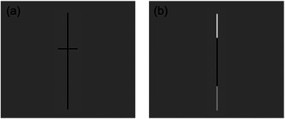
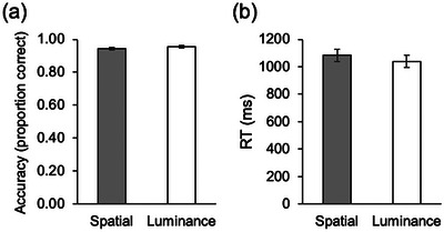
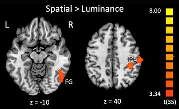
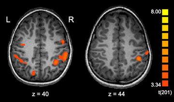
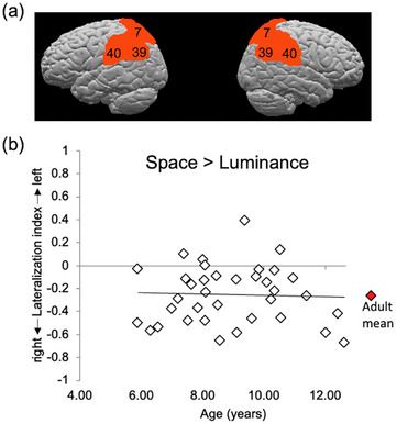
Studies of hemispheric specialization have traditionally cast the left hemisphere as specialized for language and the right hemisphere for spatial function. Much of the supporting evidence for this separation of function comes from studies of healthy adults and those who have sustained lesions to the right or left hemisphere. However, we know little about the developmental origins of lateralization. Recent evidence suggests that the young brain represents language bilaterally, with 4-6-year-olds activating the left-hemisphere regions known to support language in adults as well as homotopic regions in the right hemisphere. This bilateral pattern changes over development, converging on left-hemispheric activation in late childhood. In the present study, we ask whether this same developmental trajectory is observed in a spatial task, that is, strongly right-lateralized in adults-the line bisection (or "Landmark") task. We examined fMRI activation among children ages 5-12 years as they were asked to judge which end of a bisected vertical line was longer. We found that young children showed right-lateralized activation in the same parietal and posterior temporal areas as has been shown among adults, with no significant effects of age on lateralization within the age range we tested. We discuss potential underlying mechanisms and suggest that understanding the development of lateralization for a range of cognitive functions can play a crucial role in understanding general principles of how and why the brain comes to lateralize certain functions. SUMMARY: -Functional MRI was used to examine neural activation associated with a line bisection task in children ages 5-12 years. -Children showed right-lateralized activation in the same areas previously identified among adults. -There were no effects of children's age on the degree of right-lateralization. -This illustrates stable lateralization over development for the line bisection task and contrasts with findings of increasing lateralization over age in the domain of language.
© 2025 The Author(s). Developmental Science published by John Wiley & Sons Ltd.
Conflict of interest statement
The authors declare no conflicts of interest.
Figures
Similar articles
-
Developmental changes in neural lateralization for visual-spatial function: Evidence from a line-bisection task.Dev Sci. 2022 Jul;25(4):e13217. doi: 10.1111/desc.13217. Epub 2021 Dec 27. Dev Sci. 2022. Retraction in: Dev Sci. 2025 Sep;28(5):e70061. doi: 10.1111/desc.70061. PMID: 34913543 Free PMC article. Retracted.
-
Assessing the Early Lateralization of White Matter in the Infant Language Network.Hum Brain Mapp. 2025 Aug 1;46(11):e70286. doi: 10.1002/hbm.70286. Hum Brain Mapp. 2025. PMID: 40698875 Free PMC article.
-
Short-Term Memory Impairment.2024 Jun 8. In: StatPearls [Internet]. Treasure Island (FL): StatPearls Publishing; 2025 Jan–. 2024 Jun 8. In: StatPearls [Internet]. Treasure Island (FL): StatPearls Publishing; 2025 Jan–. PMID: 31424720 Free Books & Documents.
-
Signs and symptoms to determine if a patient presenting in primary care or hospital outpatient settings has COVID-19.Cochrane Database Syst Rev. 2022 May 20;5(5):CD013665. doi: 10.1002/14651858.CD013665.pub3. Cochrane Database Syst Rev. 2022. PMID: 35593186 Free PMC article.
-
A systematic review of speech, language and communication interventions for children with Down syndrome from 0 to 6 years.Int J Lang Commun Disord. 2022 Mar;57(2):441-463. doi: 10.1111/1460-6984.12699. Epub 2022 Feb 22. Int J Lang Commun Disord. 2022. PMID: 35191587
References
MeSH terms
LinkOut - more resources
Full Text Sources
Miscellaneous
