

Intraoperative ultrasonographic evaluation of inferior orbital rim reduction in non-comminuted zygomatic fractures: A case series of six patients
- PMID: 40782456
- PMCID: PMC12355127
- DOI: 10.1016/j.ijscr.2025.111771
Intraoperative ultrasonographic evaluation of inferior orbital rim reduction in non-comminuted zygomatic fractures: A case series of six patients
Abstract
Introduction and importance: Zygomatic fractures are common in maxillofacial trauma and often require surgical intervention for the restoration of facial symmetry. The inferior orbital rim significantly influences cheekbone asymmetry. The management of zygomatic fractures involves estimating the adequacy of reduction by comparing the malar projection on the fracture and healthy sides. This clinical approach is subjective and lacks precision.
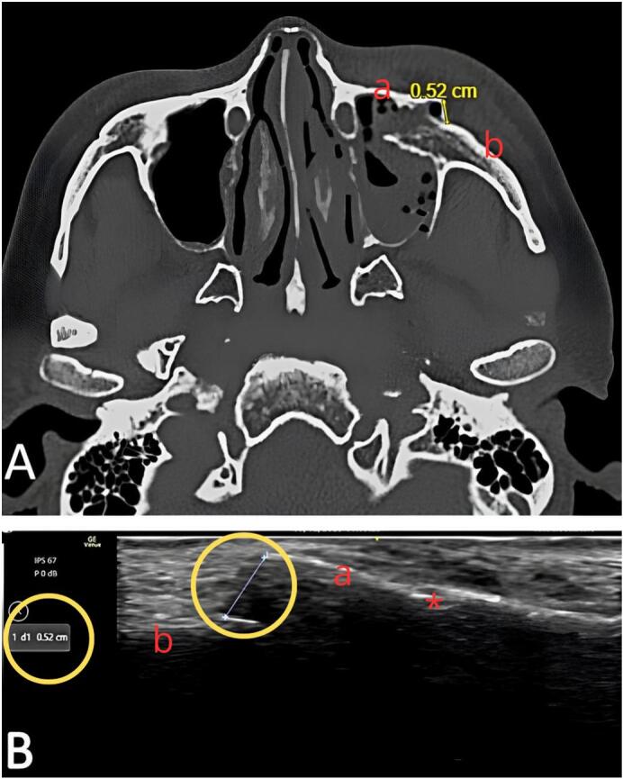
Presentation of case: Six consecutive patients with non-comminuted zygomatic fractures were included in the study. Ultrasound was performed pre- and post-reduction during the surgery to visualize the fracture site and measure the displacement by visualizing the periosteal discontinuity. The results were compared with those of pre- and post-operative CBCT scans.
Discussion: Ultrasound demonstrated comparable efficacy to CBCT for assessing fracture reduction. Unlike CBCT, ultrasound involves no radiation exposure and allows for real-time intra-operative assessment, potentially reducing the need for revision surgery.
Conclusion: Ultrasound is a precise and practical modality for evaluating zygomatic fracture reduction during J-hook procedures. It offers a radiation-free, inexpensive and readily available modality that improves intra-operative decision-making.
Keywords: Case report; Intra-operative ultrasonography; Orbital rim; Zygomatic fracture.
Copyright © 2025 The Authors. Published by Elsevier Ltd.. All rights reserved.
Conflict of interest statement
Declaration of competing interest The authors declare that they have no known competing financial interests or personal relationships that could have appeared to influence the work reported in this paper.
Figures


Similar articles
-
Elbow Fractures Overview.2025 Jul 7. In: StatPearls [Internet]. Treasure Island (FL): StatPearls Publishing; 2025 Jan–. 2025 Jul 7. In: StatPearls [Internet]. Treasure Island (FL): StatPearls Publishing; 2025 Jan–. PMID: 28723005 Free Books & Documents.
-
Zygomatic Arch Fracture.2024 Jan 26. In: StatPearls [Internet]. Treasure Island (FL): StatPearls Publishing; 2025 Jan–. 2024 Jan 26. In: StatPearls [Internet]. Treasure Island (FL): StatPearls Publishing; 2025 Jan–. PMID: 31751088 Free Books & Documents.
-
What Is the Patient-reported Outcome and Complication Incidence After Operative Versus Nonoperative Treatment of Minimally Displaced Tibial Plateau Fractures?Clin Orthop Relat Res. 2024 Oct 1;482(10):1744-1752. doi: 10.1097/CORR.0000000000003057. Epub 2024 May 9. Clin Orthop Relat Res. 2024. PMID: 38813973
-
Two-Point Versus Three-Point Fixation in Zygomaticomaxillary Complex (ZMC) Fractures: A Narrative Review.Cureus. 2025 Jun 9;17(6):e85659. doi: 10.7759/cureus.85659. eCollection 2025 Jun. Cureus. 2025. PMID: 40642666 Free PMC article. Review.
-
Rehabilitation for ankle fractures in adults.Cochrane Database Syst Rev. 2024 Sep 23;9(9):CD005595. doi: 10.1002/14651858.CD005595.pub4. Cochrane Database Syst Rev. 2024. PMID: 39312389
References
-
- Brucoli M., Boffano P., Broccardo E., Benech A., Corre P., Bertin H., Pechalova P., Pavlov N., Petrov P., Tamme T., Kopchak A., Hresko A., Shuminsky E., Dediol E., Tarle M., Konstantinovic V.S., Petrovic M., Holmes S., Karagozoglu K.H., Forouzanfar T. The “European zygomatic fracture” research project: The epidemiological results from a multicenter European collaboration. J. Craniomaxillofac. Surg. 2019 Apr;47(4):616–621. doi: 10.1016/j.jcms.2019.01.026. Epub 2019 Jan 30. PMID: 30765246. - DOI - PubMed
-
- Gong J., Zhao R., Zhang W., Li J., Yuan Z., Ma D. Role of intraoperative imaging in improving closed reduction of zygomatic arch fractures: a systematic review and Meta-analysis. J. Oral Maxillofac. Surg. 2023 Dec;81(12):1504–1516. doi: 10.1016/j.joms.2023.09.003. Epub 2023 Sep 12. PMID: 37775088. - DOI - PubMed
-
- Van Hout W.M., Van Cann E.M., Muradin M.S., Frank M.H., Koole R. Intraoperative imaging for the repair of zygomaticomaxillary complex fractures: a comprehensive review of the literature. J. Craniomaxillofac. Surg. 2014 Dec;42(8):1918–1923. doi: 10.1016/j.jcms.2014.07.012. Epub 2014 Aug 13. PMID: 25213198. - DOI - PubMed
LinkOut - more resources
Full Text Sources
Research Materials
