. 2025 Aug 9;16(1):7360.
doi: 10.1038/s41467-025-62723-9.

Opposing regulation of the K63-linked polyubiquitination of RIPK3 by SMURF1 and USP5 in necroptosis

Affiliations

Opposing regulation of the K63-linked polyubiquitination of RIPK3 by SMURF1 and USP5 in necroptosis

Chi Hyun Hwang et al. Nat Commun. .

Abstract

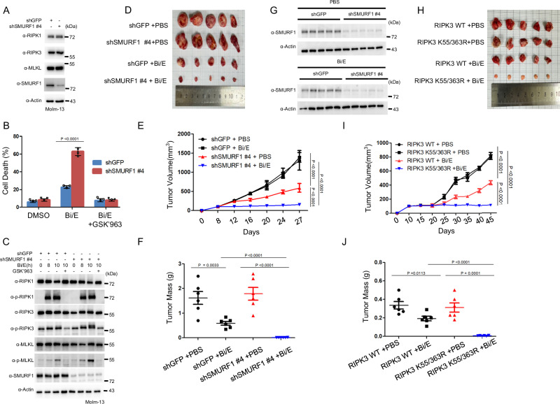
Receptor-interacting protein kinase 3 (RIPK3), a key regulator of necroptosis, is modulated by ubiquitination through various E3 ligases and deubiquitinases. However, the effects of different polyubiquitination processes on RIPK3 and necroptosis remain unclear. Using a proteomic approach, we identify SMAD Ubiquitination Regulatory Factor 1 (SMURF1) and Ubiquitin-specific peptidase 5 (USP5) as crucial regulators of RIPK3 within the necrosome during necroptosis. SMURF1 facilitates K63 polyubiquitination of RIPK3 at lysine 55 and 363, inhibiting necrosome formation and necroptosis. SMURF1 depletion accelerates necroptosis, while the reintroduction of functional SMURF1 reverses this. Conversely, USP5 acts as a deubiquitinase, removing K63 ubiquitin chains and promoting necroptosis. Reducing SMURF1, using a RIPK3 mutant defective in SMURF1-mediated ubiquitination, or overexpressing USP5 enhances necroptosis in leukaemia cells, leading to reduced tumour growth in xenograft models treated with birinapant and emricasan. These findings highlight the opposing regulation of K63-linked polyubiquitination of RIPK3 by SMURF1 and USP5 in necroptosis.


Conflict of interest statement

Competing interests: The authors declare no competing interests.

Figures

Fig. 1
Fig. 1. SMURF1 and USP5 are associated with the necrosome through RIPK3 during TNFα-induced necroptosis.
A Method that SDS-PAGE and Coomassie brilliant blue (CBB) staining of RIPK3-immunoprecipitated proteins from HEK 293T cells stably overexpressing FLAG-RIPK3. Icons of this figure created with BioRender.com. B Proteins that we identified by mass spectrometry (MS) analysis. Icons of this figure created with BioRender.com. C HT-29 cells were treated with 20 ng/mL TNFα, 2 μM Birinapant, and 20 μM Z-VAD-FMK (T/Bi/Z) for 2 h and 3 h. After treatment, the cell lysates were immunoprecipitated with RIPK3 antibody and analyzed using immunoblotting. D HT-29 cells were treated with T/Bi/Z for 2 h and 3 h. After treatment, the cell lysates were immunoprecipitated with SMURF1 antibody and analyzed using immunoblotting. E HT-29 cells were treated with T/Bi/Z for 2 h and 3 h. After treatment, the cell lysates were immunoprecipitated with SMURF1 antibody and analyzed using immunoblotting. F HT-29 cells were treated with T/Bi/Z for 3 h. The cell lysates were fractionated according to molecular size by gel-filtration chromatography and examined using immunoblotting. G HT-29 cells were treated with T/Bi/Z for 3 h. After treatment, the cells were fixed and stained using anti-SMURF1 and p-RPIPK3 antibodies, and DAPI. H HT-29 cells were treated with T/Bi/Z for 3 h. After treatment, the cells were fixed and stained using anti-USP5 and p-RIPK3 antibodies, and DAPI. Scale bars = 10 μm.
Fig. 2
Fig. 2. The E3 ligase function of SMURF1 controls necroptosis by suppressing necrosome formation.
A HT-29 cells were transduced with lentivirus to generate stable SMURF1-knockdown (KD) cell lines. The expression levels of RIPK1, RIPK3, MLKL, and SMURF1 in each cell line were analyzed by western blotting. B HT-29 cells depleted of SMURF1 using the indicated shRNAs were treated with T/Bi/Z in the presence or absence of 0.1 μM GSK'963 for 2.5 h. Flow cytometry was performed by counting 10,000 cells based on FSC (Forwarder Scatter) and SSC (Side Scatter), and calculating the percentage of cells in the Annexin V-FITC positive region. This experiment was independently repeated 3 times. Data are presented as means ± standard deviations (SDs), n = 3. Significance between groups was determined using two-sided Students’ test. C SMURF1 KD and control HT-29 cells were treated with T/Bi/Z to induce necroptosis. GSK'963 was used to confirm that this cell death is necroptosis. Cells were harvested at the indicated time points and analyzed by western blotting. D SMURF1 KD and control HT-29 cells treated with T/Bi/Z were lysed after 2 h. The necrosome was pulled down using an anti-RIPK3 antibody and protein-G agarose beads. The protein-bead conjugate was washed and analyzed by western blotting to assess the assembly of the RIPK1-RIPK3-MLKL complex. The asterisk indicates p-MLKL. E shSMURF1-infected cells, reconstituted with either SMURF1 WT or the E3 ligase-inactive form (C699A), were treated with T/Bi/Z in the presence or absence of 0.1 μM GSK'963 for 2 h. Flow cytometry was performed by counting 10,000 cells based on FSC (Forwarder Scatter) and SSC (Side Scatter), and calculating the percentage of cells in the Annexin V-FITC positive region. This experiment was independently repeated three times. Data are presented as means ± standard deviations (SDs), n  =  3. Significance between groups was determined using two-sided Students’ test. F shSMURF1-infected cells, reconstituted with either SMURF1 WT or the E3 ligase-inactive form (C699A), were treated with T/Bi/Z to induce necroptosis. GSK'963 was used to confirm that this cell death is necroptosis. Cells were harvested at the indicated time points and analyzed by western blotting. The asterisk indicates p-RIPK3. G shSMURF1-infected cells, reconstituted with either SMURF1 WT or the E3 ligase-inactive form (C699A), were treated with T/Bi/Z and lysed after 2 h. The necrosome was pulled down using an anti-RIPK3 antibody and protein-G agarose beads. The protein-bead conjugate was washed and analyzed by western blotting to examine the assembly of the RIPK1-RIPK3-MLKL complex.
Fig. 3
Fig. 3. SMURF1 mediates K63-linked polyubiquitylation of RIPK3.
A FLAG-RIPK1, RIPK3, MLKL and 3xMYC-SMURF1 were overexpressed in HEK 293T cells via PEI transfection. Cells were lysed and incubated with anti-FLAG antibodies and protein-G agarose beads to pull down 3xMYC-SMURF1.The protein-bead conjugate was washed and analyzed by SDS-PAGE. The immunoprecipitation of the SMURF1 and RIPK1, RIPK3, MLKL were assessed using western blotting. B FLAG-RIPK3 was transfected into HEK 293T cells along with HA-Ub, with or without 3xMYC-SMURF1 expression. After 20 h, cells were lysed in a 1% SDS buffer, and lysates diluted to 0.1% SDS were immunoprecipitated using an anti-FLAG antibody. RIPK3 ubiquitylation was detected by western blotting with an HRP-conjugated anti-HA antibody. C FLAG-RIPK3 (1–296, kinase domain; 294–518, C-term domain; 1–440 and 452–518 ΔRHIM domian) and 3xMYC-SMURF1 were overexpressed in HEK 293T cells via PEI transfection. Cells were lysed and incubated with anti-FLAG antibodies and protein-G agarose beads to pull down 3xMYC-SMURF1. The protein-bead conjugate was washed and analyzed by SDS-PAGE. The immunoprecipitation of the SMURF1 and RIPK3 domains was assessed using western blotting. The schematic diagram of RIPK3 shows the binding domain for SMURF1. Icons of this figure created with BioRender.com. D HA-SMURF1 (1–159, 119–731, 1–345, 346–731, 119–365) and FLAG-RIPK3 were overexpressed in HEK 293 T cells via PEI transfection. Cells were lysed and incubated with anti-HA antibodies and protein-G agarose beads to pull down the specific domain of HA-SMURF1. The protein-bead conjugate was washed and analyzed by SDS-PAGE. The immunoprecipitation of the RIPK3 and SMURF1 domains was assessed using western blotting. The schematic diagram of RIPK3 shows the binding domain for RIPK3. Icons of this figure created with BioRender.com. E HEK 293 T cells were transfected with FLAG-RIPK3, 3xMYC–SMURF1, and HA-Ub along with various ubiquitin mutants. After 20 h, cells were lysed in a 1% SDS buffer, and lysates diluted to 0.1% SDS were immunoprecipitated using an anti-FLAG antibody. RIPK3 ubiquitylation was detected by western blotting with an HRP-conjugated anti-HA antibody. F HEK 293T cells were transfected with FLAG-RIPK3, 3xMYC–SMURF1, and HA-Ub along with the K63R ubiquitin mutant. After 20 h, cells were lysed in a 1% SDS buffer, and lysates diluted to 0.1% SDS were immunoprecipitated using an anti-FLAG antibody. RIPK3 ubiquitylation was detected by western blotting with an HRP-conjugated anti-HA antibody. G To confirm the endogenous ubiquitination status of RIPK3, HT-29 cells expressing shGFP or shSMURF1 (#4 and #8) were treated with T/Bi/Z to induce necroptosis. After 3 h, SMURF1 KD and control HT-29 cells were lysed. Immunoprecipitation was performed using an anti-RIPK3 antibody and protein-G agarose beads. The protein-bead conjugate was washed, and ubiquitination was detected using an anti-Ub-HRP conjugated antibody and an anti-K63-Ub-HRP conjugated antibody.
Fig. 4
Fig. 4. SMURF1 mediates K63-linked ubiquitination of RIPK3 at Lys55/363 during TNFα-induced necroptosis.
A This is a schematic diagram illustrating the various ubiquitination enzyme sites on RIPK3 identified to date. Icons of this figure created with BioRender.com. B FLAG-RIPK3 WT, K55R, K197R, K302R, K363R, K364R, K518R, and K55/363R were transfected into HEK 293T cells with HA-Ub, with or without 3xMYC-SMURF1 expression. After 20 h, cells were lysed in a 1% SDS buffer, and lysates diluted to 0.1% SDS were immunoprecipitated using an anti-FLAG antibody. RIPK3 ubiquitination was detected by western blotting with an HRP-conjugated anti-HA antibody. C Adenovirus-mediated expression of RIPK3 WT, K55R, K363R, and K55/363R was induced in HeLa cells, which were then treated with T/Bi/Z to induce necroptosis. After 5 h, the cells were harvested and stained with propidium iodide (PI). Flow cytometry was performed by counting 10,000 cells based on FSC (Forwarder Scatter) and SSC(Side Scatter), and calculating the percentage of cells in the PI(Propidium Iodide) positive region. This experiment was independently repeated 3 times. Data are presented as means ± standard deviations (SDs), n = 3. Significance between groups was determined using two-sided Students’ test. D HeLa cells expressing RIPK3 WT, K55R, K363R, and K55/363R were treated with T/Bi/Z to induce necroptosis at the indicated time points. Cells were harvested at these time points and analyzed by western blotting. The asterisk indicates p-RIPK3. E HeLa cells expressing shGFP, shSMURF1, RIPK3 WT, and RIPK3 K55/363R were treated with T/Bi/Z to induce necroptosis. After 5 h, the cells were harvested and stained with propidium iodide (PI). Flow cytometry was performed by counting 10,000 cells based on FSC (Forwarder Scatter) and SSC (Side Scatter), and calculating the percentage of cells in the PI(Propidium Iodide) positive region. This experiment was independently repeated 3 times. Data are presented as means ± standard deviations (SDs), n = 3. Significance between groups was determined using two-sided Students’ test. F HeLa cells expressing shGFP, shSMURF1, RIPK3 WT, and RIPK3 K55/363R were treated with T/Bi/Z to induce necroptosis. After 4 h, the cells were harvested and analyzed by western blotting.
Fig. 5
Fig. 5. USP5 regulates TNFα-induced necroptosis through its deubiquitinase function.
A HT-29 cells were transduced with lentivirus to generate stable USP5-knockdown (KD) cell lines. The expression levels of RIPK1, RIPK3, MLKL, and USP5 in each cell line were analyzed by western blotting. B HT-29 cells depleted of USP5 using the indicated shRNAs were treated with T/Bi/Z in the presence or absence of 0.1 μM GSK'963 for 3.5 h. Flow cytometry was performed by counting 10,000 cells based on FSC (Forwarder Scatter) and SSC (Side Scatter), and calculating the percentage of cells in the Annexin V-FITC positive region. This experiment was independently repeated 3 times. Data are presented as means ± standard deviations (SDs), n = 3. Significance between groups was determined using two-sided Students’ test. C USP5 KD and control HT-29 cells were treated with T/Bi/Z to induce necroptosis. GSK'963 was used to confirm that this cell death is necroptosis. Cells were harvested at the indicated time points and analyzed by western blotting. D USP5 KD and control HT-29 cells treated with T/Bi/Z were lysed after 3.5 h. The necrosome was pulled down using an anti-RIPK3 antibody and protein-G agarose beads. The protein-bead conjugate was washed and analyzed by western blotting to assess the assembly of the RIPK1-RIPK3-MLKL complex. E shUSP5-infected cells, reconstituted with either USP5 WT or the deubiquitinase-defective mutant USP5 C335A, were treated with T/Bi/Z in the presence or absence of 0.1 μM GSK'963 for 3.5 h. Flow cytometry was performed by counting 10,000 cells based on FSC (Forwarder Scatter) and SSC (Side Scatter), and calculating the percentage of cells in the Annexin V-FITC positive region. This experiment was independently repeated 3 times. Data are presented as means ± standard deviations (SDs), n = 3. Significance between groups was determined using two-sided Students’ test. F shUSP5-infected cells, reconstituted with either USP5 WT or the deubiquitinase-defective mutant USP5 C335A, were treated with T/Bi/Z to induce necroptosis. GSK'963 was used to confirm that this cell death is necroptosis. Cells were harvested at the indicated time points and analyzed by western blotting. The asterisk indicates p-RIPK3 and p-MLKL. G shUSP5-infected cells, reconstituted with either USP5 WT or the deubiquitinase-defective mutant USP5 C335A, were treated with T/Bi/Z and lysed after 3.5 h. The necrosome was pulled down using an anti-RIPK3 antibody and protein-G agarose beads. The protein-bead conjugate was washed and analyzed by western blotting to examine the assembly of the RIPK1-RIPK3-MLKL complex.
Fig. 6
Fig. 6. SMURF1-mediated K63-linked polyubiquitination of RIPK3 is reversed by the deubiquitination process of USP5.
A HT-29 cells depleted of SMURF1, USP5 and Double Knocked down by the indicated shRNAs. B HT-29 cells depleted of SMURF1, USP5 and Double Knocked down by the indicated shRNAs were treated with T/Bi/Z in the presence or absence of 0.1 μM GSK’963 for 3 h. The cells were harvested and stained with annexin V-FITC. Flow cytometry was performed by counting 10,000 cells based on FSC (Forwarder Scatter) and SSC (Side Scatter), and calculating the percentage of cells in the Annexin V-FITC positive region. This experiment was independently repeated 3 times. Data are presented as means ± standard deviations (SDs), n   =   3. Significance between groups was determined using two-sided Students’ test. C Each HT-29 cells depleted of SMURF1, USP5 and Double Knocked down cells were treated with T/Bi/Z to induce necroptosis. GSK’963 (0.1 μM) was used to inhibit necroptosis. Cells were harvested and analyzed using western blotting. D Each HT-29 cells depleted of SMURF1, USP5 and Double Knocked down cells treated with T/Bi/Z were lysed after 3 h. The necrosome was pulled down using an anti-RIPK3 antibody and protein-G agarose beads. The protein-bead conjugate was washed and analyzed by western blotting to assess the assembly of the RIPK1-RIPK3-MLKL complex. E To confirm the endogenous ubiquitination status of RIPK3, HT-29 cells depleted of SMURF1, USP5 and Double Knocked down were treated with T/Bi/Z to induce necroptosis. After 3 h, SMURF1 KD and control HT-29 cells were lysed. Immunoprecipitation was performed using an anti-RIPK3 antibody and protein-G agarose beads. The protein-bead conjugate was washed, and ubiquitination was detected using an anti-Ub-HRP conjugated antibody and an anti-K63-Ub-HRP conjugated antibody.
Fig. 7
Fig. 7. Downregulation of SMURF1 or the K55/363R RIPK3 mutant sensitizes acute myeloid leukemia cells to necroptosis in xenograft models.
A Molm-13 cells were transduced with lentivirus to generate stable SMURF1-KD cell lines. The expression levels of RIPK1, RIPK3, MLKL, and SMURF1 in each cell line were analyzed by western blotting. B Molm-13 depleted of SMURF1 using the indicated shRNAs were treated with Bi/E in the presence or absence of 0.1 μM GSK'963 for 12 h. The cells were harvested and stained with PI. Flow cytometry was performed by counting 10,000 cells based on FSC (Forwarder Scatter) and SSC (Side Scatter), and calculating the percentage of cells in the PI (Propidium Iodide) positive region. This experiment was independently repeated 3 times. Data are presented as means ± standard deviations (SDs), n = 3. Significance between groups was determined using two-sided Students’ test. C SMURF1 KD and control Molm-13 cells were treated with Bi/E to induce necroptosis. GSK'963 was used to confirm that this cell death is necroptosis. Cells were harvested at the indicated time points and analyzed by western blotting. D Tumor xenografts were established by injecting control or SMURF1-knockdown (KD) Molm-13 cell lines into the flanks of mice (n = 6). This experimental setup aimed to examine the effect of SMURF1 depletion on tumor progression and response to treatment. E Following xenograft establishment, the mice received intraperitoneal injections of birinapant (2 mg/kg) and emricasan (1 mg/kg) every 4 days for a total of six treatments. Six independent mice were used for xenograft experiments in each experimental group, and treatments were administered independently for each group. Representative images of the resulting xenografts were taken to visually document tumor development. Tumor growth was systematically measured at 4-day intervals throughout the treatment period to monitor any differences between control and SMURF1 KD groups. Data are presented as means ± standard deviations (SDs). Significance was determined using the two-sided Students’ test. F At the end of the experiment, the tumors were excised, and their masses were recorded to evaluate the impact of SMURF1 depletion on tumor size. The tumor weight was measured from each of the six independent mice in each group. Data are presented as means ± standard deviations (SDs). Significance was determined using the two-sided Students’ test. G The tumors were lysed, and SMURF1 expression was analyzed using western blotting. H Additional tumor xenografts were generated using NB4 cell lines expressing either RIPK3 WT or the RIPK3 K55/363R mutant (n = 6). This setup aimed to assess the role of specific RIPK3 mutations in tumor growth and response to therapeutic agents. I Mice bearing these xenografts were treated with birinapant (2 mg/kg) and emricasan (1 mg/kg) via intraperitoneal injection every 5 days, for a total of eight treatments. Six independent mice were used for xenograft experiments in each experimental group, and treatments were administered independently for each group. Tumor growth was measured at 5-day intervals throughout the course of the experiment to track progression over time in both RIPK3 WT and K55/363R groups. Data are presented as means ± standard deviations (SDs). Significance was determined using the two-sided Students’ test. J Upon completion of the experiment, tumors were dissected, and their weights were measured to quantify differences between groups. The tumor weight was measured from each of the six independent mice in each group. Tumor weights were compared to determine any statistically significant variations attributable to SMURF1 KD or RIPK3 mutations.

Similar articles

References

    1. Cho, Y. et al. Phosphorylation-driven assembly of the RIP1-RIP3 complex regulates programmed necrosis and virus-induced inflammation. Cell137, 1112–1123 (2009). - PMC - PubMed
    1. Vandenabeele, P., Galluzzi, L., Vanden Berghe, T. & Kroemer, G. Molecular mechanisms of necroptosis: an ordered cellular explosion. Nat. Rev. Mol. cell Biol.11, 700–714 (2010). - PubMed
    1. Newton, K. et al. Activity of protein kinase RIPK3 determines whether cells die by necroptosis or apoptosis. Science343, 1357–1360 (2014). - PubMed
    1. Kaczmarek, A., Vandenabeele, P. & Krysko, D. V. Necroptosis: the release of damage-associated molecular patterns and its physiological relevance. Immunity38, 209–223 (2013). - PubMed
    1. Moriwaki, K. et al. The necroptosis adaptor RIPK3 promotes injury-induced cytokine expression and tissue repair. Immunity41, 567–578 (2014). - PMC - PubMed

MeSH terms

Substances

LinkOut - more resources