. 2025 Aug 10;16(1):7375.
doi: 10.1038/s41467-025-62487-2.

F-actin disassembly by the oxidoreductase MICAL1 promotes mechano-dependent VWF-GPIbα interaction in platelets

Affiliations

F-actin disassembly by the oxidoreductase MICAL1 promotes mechano-dependent VWF-GPIbα interaction in platelets

Jean Solarz et al. Nat Commun. .

Abstract

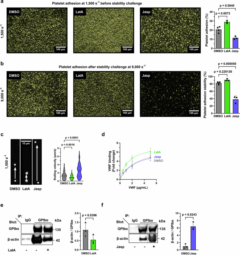
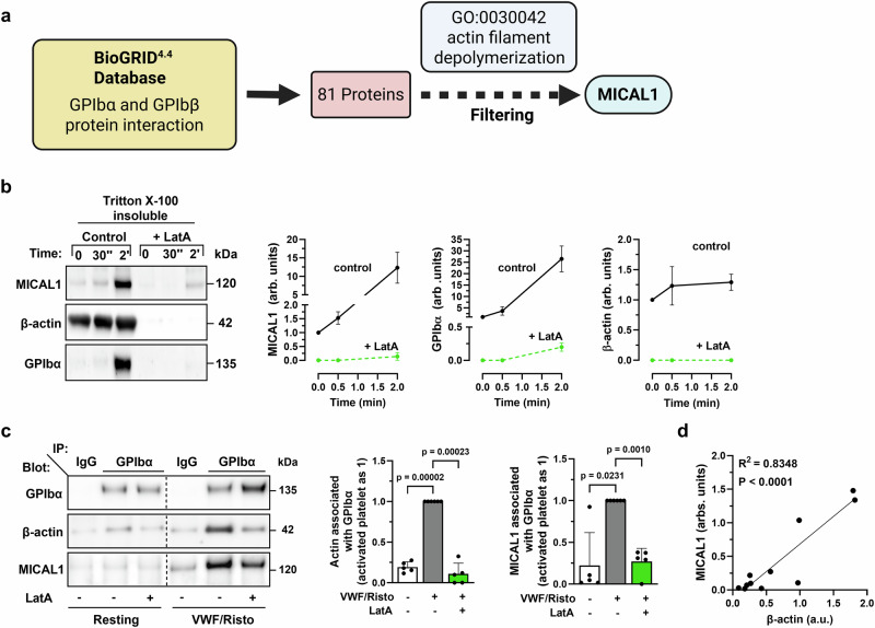
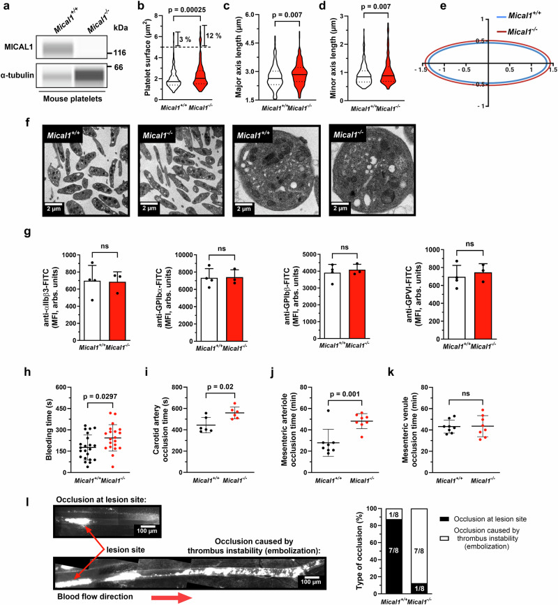
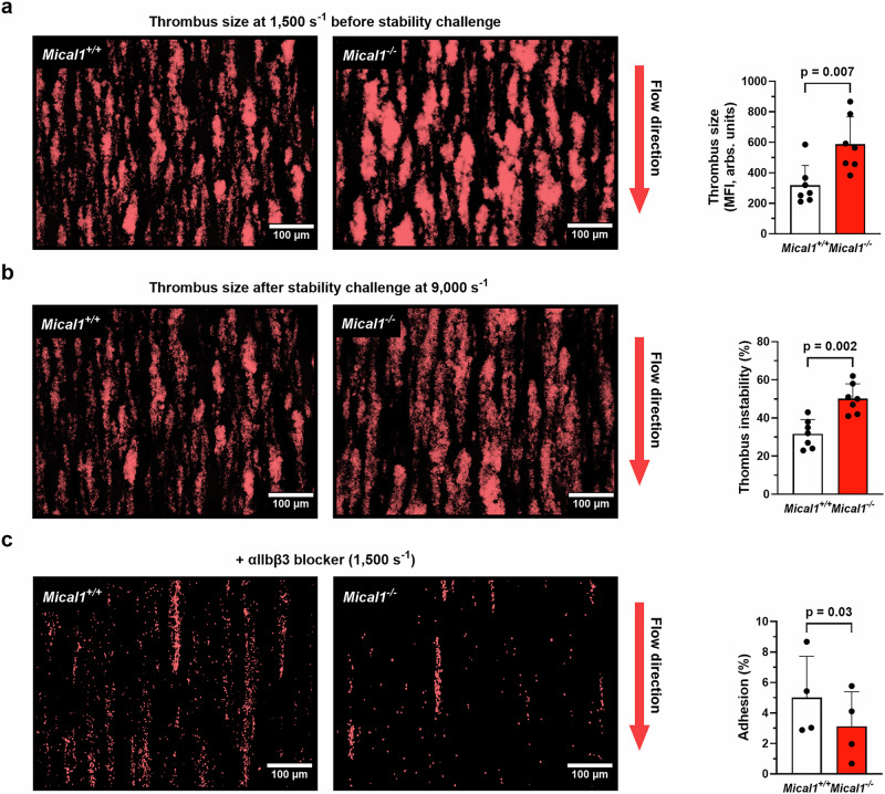
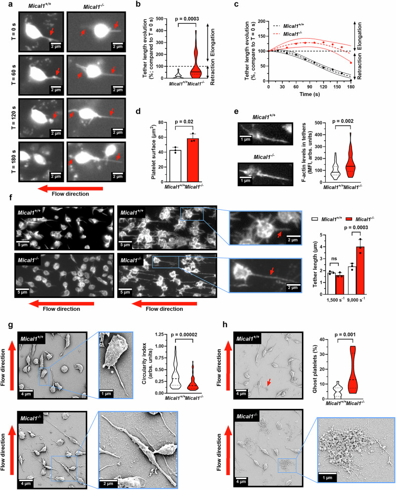
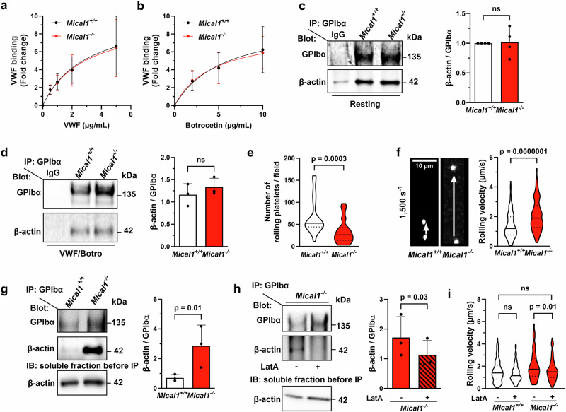
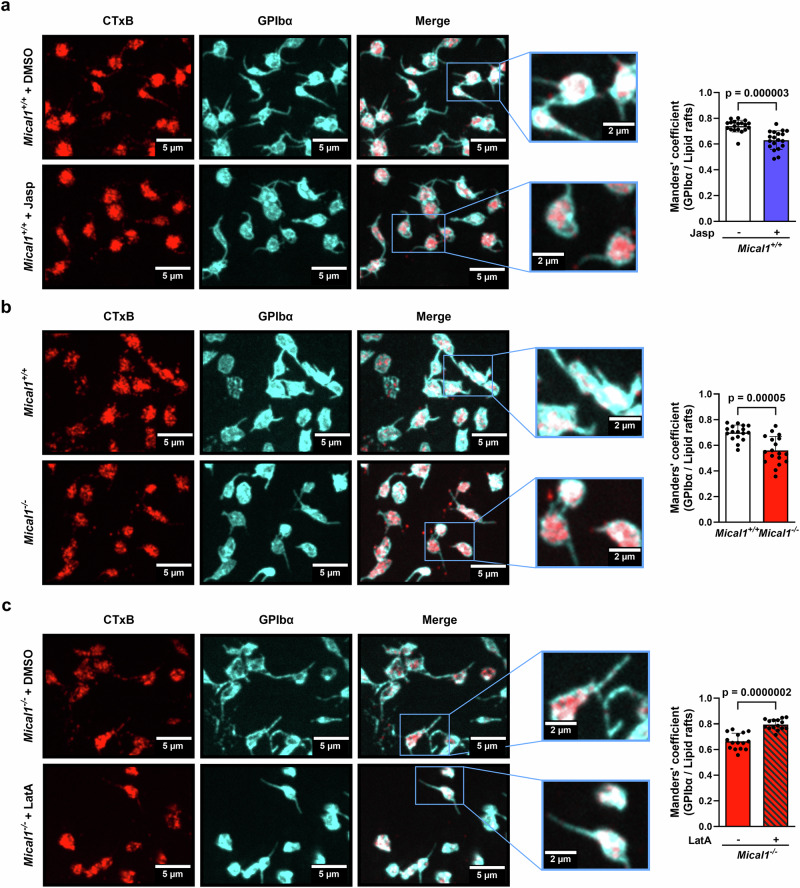
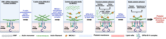
Mechano-dependent interactions are key to thrombus formation and hemostasis, enabling stable platelet adhesion to injured vessels. The interaction between von Willebrand factor (VWF) and the platelet receptor GPIb-IX-V is central to this process. While GPIbα connects to the actin cytoskeleton, whether actin dynamics are important for GPIbα function under hemodynamic, high shear conditions remains largely unknown. Here, we show that actin disassembly is critical for proper VWF-GPIbα binding under shear. Mechanistically, we identify the oxidoreductase MICAL1 as a shear-activated regulator that promotes local F-actin disassembly around the GPIb-IX-V complex. This enables its translocation to lipid rafts and reinforces VWF binding. MICAL1-deficient platelets display impaired adhesion, increased deformability under shear, and defective thrombus formation in vivo. Thus, MICAL1 drives shear-dependent actin remodeling that supports GPIb-IX-V mechanotransduction and platelet function. These findings uncover a role for actin oxidation in platelet adhesion, providing a connection between cytoskeletal redox control and platelet function during thrombus formation.


Conflict of interest statement

Competing interests: The authors declare no competing interests.

Figures

Fig. 1
Fig. 1. Platelet-VWF interaction under shear requires F-actin disassembly to promote efficient platelet adhesion and stability.
ac Mouse blood was treated with LatA (500 nM) or Jasp (1 µM) or DMSO before perfusion on recombinant mouse VWF (Supplementary Fig. 1). a Platelet adhesion (% of surface coverage) (mean ± SD, N: DMSO = 4, LatA = 3, Jasp = 3 independent experiments, one-way ANOVA with Tukey post-hoc test, F = 39.05, degree of freedom (df) = 7) and b platelets adhesion stability (mean ± SD, N: DMSO = 4, LatA = 3, Jasp = 3 independent experiments, one-way ANOVA with Tukey post-hoc test, F = 74.73, df = 7). Left panel: images showing platelet adhesion before (a) and after (b) the stability challenge. Scale bars = 100 µm. Platelet adhesion and stability were calculated as described in Supplementary Fig. 1. c Mouse platelet rolling velocity. Violin plots with the median represented by a central line and the interquartile range (25th–75th percentiles) indicated by the upper and lower lines. (N = 3 independent experiments DMSO = 133, LatA = 131, Jasp = 129 platelets; one-way ANOVA with Tukey post-hoc test: F = 75.84, df = 390). Supplementary Movies 1, 2 and 3. Left panel: images of platelet velocity with a time-lapse of 10 s. Scale bar = 10 µm. d Recombinant mouse VWF binding measured by flow cytometry. Washed mouse platelets were treated with either LatA (500 nM), Jasp (1 µM), or DMSO and activated with botrocetin (5 µg/mL) and a range of mouse VWF. (mean ± SD, N = 3 independent experiments). GPIbα immunoprecipitation (in static conditions) treated with either DMSO or LatA (500 nM) (e) or Jasp (1 µM) (f). Samples were blotted for GPIbα and β-actin. e Mean ± SD, N = 3 independent experiments, two-tailed paired Student’s t test, t = 6.297, df = 2. Left panel: representative western blots. f Mean ± SD, N = 3 independent experiments, two-tailed paired Student’s t test, t = 4.874, df = 2. Left panel: representative western blots. Vertical lines indicate a repositioned gel lane. In histograms, each symbol represents 1 individual. Independent experiments correspond to different mice. Source data are provided as a Source Data file.
Fig. 2
Fig. 2. F-actin induces the recruitment of MICAL1 to the GPIb-IX-V complex upon platelet activation.
a Identification of MICAL1 as a potential actin-depolymerizing protein interacting with GPIbα/GPIbβ thanks to the database BioGRID4.4 and the Gene Ontology “Actin filament depolymerization” (GO: 0030042). Created with BioRender.com. b Lysates of human platelets treated or not with LatA (15 µM) at 0 s (resting platelets), after 30 s or 2 min of activation (VWF 10 mg/mL/Risto 0.4 mg/mL) were separated into Triton X-100 soluble and insoluble fractions and blotted. Quantification of insoluble MICAL1 (mean ± SD, N = 4 independent experiments from 4 different donors, two-way ANOVA with Šídák post hoc test, F(2, 18) = 27.06, 2 min: p = 0.00000003), GPIbα (mean ± SD, N = 3 independent experiments from 4 different donors, two-way ANOVA, F(2, 12) = 49.40, 2 min: p = 0.00000005) and β-actin (mean ± SD, N = 4 independent experiments from 4 different donors, two-way ANOVA with Šídák post hoc test, F(2, 18) = 2.424, 2 min: p = 0.0000000004). Left panel: representative western blots. Right panels: MICAL1, GPIbα, β-actin quantification from western blots. c GPIbα immunoprecipitation from lysates from washed human platelets in resting or activated conditions (VWF 10 mg/mL/Risto 0.4 mg/mL) human platelets treated with LatA (15 µM) or DMSO as control and blotted for GPIbα, β-actin, (mean ± SD, N = 5 independent experiments from 4 different donors, one-way ANOVA with Šídák post hoc test, F = 202, df = 4) and MICAL1 (mean ± SD, N = 5 independent experiments from 4 different donors, one-way ANOVA with Šídák post hoc test, F = 19.2, df = 4). Left Panel: representative western blot. Right panel: β-actin and MICAL1 quantification from western blots. d Correlation of the presence of MICAL1 and β-actin associated to GPIbα after immunoprecipitation from experiments performed in (c). R2 and p were obtained through simple linear regression. Vertical dotted lines indicate a repositioned gel lane. Source data are provided as a Source Data file.
Fig. 3
Fig. 3. MICAL1 contributes to hemostasis and thrombosis under high shear.
a Mical1 expression in Mical1+/+ and Mical1−/− platelets analyzed by Wes (α-tubulin as loading control). Source data are provided. be Platelet ultrastructure assessed by transmission electron microscopy in two independent experiments. b Platelet surface area, c major axis, and d minor axis were measured (N = 200 platelets per genotype). Violin plots show median (central line) and interquartile range (25th–75th percentiles, upper and lower lines). Two-tailed unpaired Student’s t tests: b t = 3.693, df = 398; ct = 2.706, df = 398; d t = 2.699, df = 398. e Schematic of platelet axes (Created with BioRender.com). f Representative images of Mical1+/+ and Mical1–/− platelets. Scale bars: 2 µm (top), 0.2 µm (bottom). g Surface expression of αIIbβ3, GPIbα, GPIbβ, and GPVI by flow cytometry (mean fluorescence intensity ± SD; Mical1+/+: N = 4; Mical1−/−: N = 3). Two-tailed unpaired Student’s t tests: αIIbβ3, t = 0.1012; GPIbα, t = 0.09656; GPIbβ, t = 0.5239; GPVI, t = 0.5602; df = 5. h Tail bleeding time (mean ± SD, N = 22); two-tailed Mann–Whitney test: U = 134. i Time to carotid artery occlusion after FeCl3 injury (mean ± SD, N = 6); paired Student’s t test: t = 3.429, df = 5. Time to occlusion in mesenteric arterioles (j) and venules (k) following FeCl3 injury (mean ± SD, N = 8); unpaired t-tests: (j) t = 3.947, (k) t = 0.08440; df = 14, p = 0.93. l Type of occlusion in mesenteric arterioles (lesion site vs. distant embolic occlusion). Left: representative reconstructions. Scale bars: 100 µm. Right: percentage distribution (N = 8). ns not significant.
Fig. 4
Fig. 4. MICAL1 is required for thrombi stability under high shear.
ac Rhodamine 6G stained Mical1+/+ or Mical1−/− platelets in whole blood were perfused on type I collagen matrix. a Thrombus size at a shear rate of 1500 s−1 (mean fluorescence intensity (MFI) ± SD, N = 7 independent experiments; two-tailed unpaired Student’s t test, t = 3.227, df = 12), and b the percentage of thrombus instability at a shear rate of 9000 s−1 (see experimental protocol overview in Supplementary Fig. 1) (mean ± SD, N = 7 independent experiments; two-tailed Mann–Whitney, U = 2). Left panel: representative images showing a thrombus size before and b after stability challenge. c Platelet adhesion in the absence of thrombus formation was assessed using αIIbβ3 blocking antibody (Leo.H4) (mean ± SD, N = 4 independent experiments; two-tailed paired Student’s t test, t = 4.097, df = 3). Left panel: representative images showing platelet adhesion. In each graph, each symbol represents 1 individual. In all images, scale bars = 100 μm. Independent experiments correspond to different mice. Source data are provided as a Source Data file.
Fig. 5
Fig. 5. Control of F-actin disassembly by MICAL1 supports shear-dependent platelet adhesion.
a, b, fg Mical1+/+ or Mical1−/− whole blood was perfused on recombinant mouse VWF matrix. a Platelet adhesion (% of the surface coverage) (mean ± SD, N = 7 independent experiments from different mice, two-tailed unpaired Student’s t test, t = 4.420, df = 12), and b platelet adhesion stability (mean ± SD, N = 7 independent experiments, two-tailed unpaired Student’s t test, t = 3,813, df = 12). Left panels: representative images before (a) and after (b) stability challenge. Scale bars = 100 µm. c Integrin αIIbβ3 activation of Mical1+/+ or Mical1−/− platelets under flow (1500 s−1) (mean ± SD, N = 600 platelet, from 4 independent experiments corresponding to different mice, two-tailed unpaired Student’s t test, t = 12.30, df = 1198). Left panels: representative images showing JON/A staining. Scale bars = 10 µm. d F-actin levels in platelets (integrated density (IntDen) ± SD, N = 819 platelets from 3 independent experiments from different mice, two-tailed unpaired Student’s t test, t = 3.110, df = 4) (Left panels: representative images of F-actin. Scale bars = 5 µm). e GPIbα expression (integrated density (IntDen)) ± SD, N: Mical1+/+ = 1034; Mical1−/− = 1011 platelets from 3 independent experiments from different mice, two-tailed unpaired Student’s t test, t = 0.4695, df = 4). f, g Rescue experiment of platelet adhesion in the presence of LatA. Mical1+/+ or Mical1−/− blood treated with DMSO or LatA (125 nM) was perfused on recombinant mouse VWF matrix. f Platelet adhesion (% of the surface coverage) (mean ± SD, N: Mical1+/+/DMSO = 4, Mical1+/+/LatA = 4, Mical1−/−/DMSO = 3, Mical1−/−/LatA = 3 independent experiments from different mice, one-way ANOVA with Tukey post-hoc test, F = 2.007, df = 8) and g stable platelet adhesions were measured (mean ± SD, N: Mical1+/+/DMSO = 4, Mical1+/+/LatA = 4, Mical1–/−/DMSO = 3, Mical1−/−/LatA = 3 independent experiments from different mice, one-way ANOVA with Tukey post-hoc test, F = 0.9851, df = 8). Left panel: representative images showing platelet adhesion before (f) and after (g) stability challenge. Scale bars = 100 µm. ns not significant. Source data are provided as a Source Data file.
Fig. 6
Fig. 6. MICAL1 promotes stable platelet adhesion to VWF by increasing resistance to shear-induced morphological changes.
a, b Whole blood from Mical1+/+ or Mical1−/− mice was perfused over a recombinant mouse VWF-coated surface at 1500 s⁻¹. a Representative time-lapse images showing individual platelet tether formation. Red arrows indicate tethers. Scale bars, 2 µm. See Supplementary Movies 4 and 5. b Tether elongation was quantified as [(length at 3 min/length at 0 s) ×100]; mean ± SD, N = 30 tethers from 3 independent experiments (Welch’s t-test, t = 4.105, df = 31.52). c Tether length was tracked over time; N = 30 tethers from 3 experiments; mean ± 95% CI (two-way ANOVA with Šídák’s test, F(9, 504) = 4.375; p = 0.003 at 80 s; p < 0.001 from 100 to 160 s). df Unstained Mical1+/+ or Mical1−/− blood was perfused on VWF. Post-perfusion, coverslips were fixed and stained with phalloidin and anti-integrin β3. d Platelet surface area measured in 3 experiments (273 platelets/experiment); mean ± SD (Student’s t test, t = 3.938, df = 4). e F-actin quantification in tethers at 1500 s⁻¹ using phalloidin (Mical1+/+ = 39, Mical1−/− = 37 tethers; Welch’s t test, t = 3.261, df = 61.62). Left: representative images with actin-positive tethers (red arrows). Scale bars, 1 µm. f Tether length measured from at least 254 platelets/experiment (N = 3); mean ± SD (two-way ANOVA with Fisher’s LSD test, F(1,8) = 22.09). Left: representative β3-labeled tethers at 1500 and 9000 s⁻¹. Scale bars, 5 µm. g Scanning electron microscopy (SEM) images of platelets from 9000 s⁻¹ flow assays. Circularity index quantified in 2 experiments (n = 2 mice/condition pooled); Mical1+/+ = 65, Mical1−/− = 63 platelets (Student’s t test, t = 4.495, df = 126). Scale bars: 4 µm (overview), 1–2 µm (zoom). h Frequency of “ghost” platelets per field assessed by SEM in 2 experiments (Mical1+/+ = 743, Mical1−/− = 609 platelets; Student’s t test, t = 4.495, df = 126). Representative SEM images with zooms shown. Scale bars: 4 µm and 2 µm.Violin plots display medians (central line) and interquartile ranges (25th–75th percentiles). ns: not significant. Source data are provided as a Source Data file.
Fig. 7
Fig. 7. MICAL1 promotes F-actin disassembly in the GPIb-IX-V complex in a mechano-sensitive manner, increasing GPIbα association with VWF.
a, b VWF binding assessed by flow cytometry in Mical1+/+and Mical1−/− platelets. a Platelets were activated with 5 µg/mL botrocetin and increasing concentrations of recombinant mouse VWF. b Platelets were activated with 0, 5 or 10 µg/mL botrocetin in the presence of 10 µg/mL recombinant VWF. Data are mean ± SD from N = 3 (a) or 4 (b) independent experiments (two-way ANOVA with Šídák’s test; (a) F(4, 20) = 0.004839; (b) F(3,24) = 0.02602). c, d GPIbα immunoprecipitation from lysates of washed platelets. c Resting platelets; d platelets stimulated in static conditions with recombinant VWF (10 µg/mL) and botrocetin (5 µg/mL). Immunoblots show GPIbα and β-actin (mean ± SD, N = 4; Student’s t tests: c t = 0.1464, df = 6; d t = 0.9347, df = 4). e, f) Platelet rolling on VWF under shear (1500 s⁻¹). e Number of rolling platelets per field (Mical1+/+ = 30, Mical1−/− = 30 fields; t = 3.864, df = 58). f Rolling velocity (Mical1+/+ = 131, Mical1−/− = 129 platelets; t = 5.452, df = 258). Left: representative time-lapse images (10 s). Scale bar, 10 µm. See Supplementary Movies 6 and 7. g GPIbα immunoprecipitation from platelet lysates after flow. Soluble fractions were immunoblotted for β-actin as control. Mean ± SD from N = 3 experiments (ratio paired Student’s t test, t = 8.084, df = 2). h GPIbα immunoprecipitation after flow in Mical1−/− platelets treated with LatA (125 nM) or DMSO. Soluble fractions were immunoblotted for β-actin as control. Mean ± SD from N = 3 experiments (ratio paired Student’s t test, t = 5.888, df = 2). i Platelet rolling rescue experiment with LatA. Mical1+/+ and Mical1−/− platelets were treated with LatA (125 nM) or DMSO and perfused under flow. Velocity measured from N = 3 experiments (Mical1+/+ + DMSO = 183, +LatA = 141; Mical1−/− + DMSO = 134, +LatA = 138 platelets; one-way ANOVA with Tukey’s test, F = 10.05, df = 581). See Supplementary Movies 8–11. ns: not significant. Source data are provided as a Source Data file.
Fig. 8
Fig. 8. MICAL1 induced F-actin disassembly in the GPIb-IX-V complex enable GPIbα translocation into lipid rafts.
ac Colocalization of lipid rafts (CTxB, Red) and GPIbα (Green) in Mical1+/+ and Mical1−/− platelets after flow assays at 1500 s−1 from 3 independent experiments corresponding to different mice. Left panels: representative images (scale bars: 5 µm) and zoom (scale bars: 2 µm). a Manders’ overlap coefficient for Mical1+/+ platelets treated with either Jasp (2 µM) or DMSO as control (mean ± SD, N = 19 fields from 3 independent experiments for both conditions, two-tailed unpaired Student’s t test, t = 5.563, df = 36). b Manders’ overlap coefficient for Mical1+/+ vs Mical1−/− platelets (mean ± SD, N = 17 fields from 3 independent experiments for both genotypes, two-tailed unpaired Student’s t test, t = 4.642, df = 32). c Manders’ overlap coefficient for Mical1−/− platelets treated with either a rescue dose of LatA (125 nM) or DMSO as control (mean ± SD, N = 15 fields from 3 independent experiments for both genotypes, two-tailed unpaired Student’s t test, t = 6.792, df = 28).
Fig. 9
Fig. 9. F-actin disassembly by the oxidoreductase MICAL1 promotes mechano-dependent VWF-GPIbα interaction in platelets.
GPIbα interaction with VWF under high shear stress induces F-actin and MICAL1 recruitment in the GPIb-IX-V complex, which limits F-actin associated within the GPIb-IX-V complex. This process both facilitates receptor translocation to lipid rafts, which strengthens its interaction with VWF and promotes the retraction of platelet membrane tethers, resulting in increased platelet adhesion. Ultimately, GPIbα mechanotransduction activates platelets, increasing their resistance to deformation and enabling stable platelet adhesion and thrombus stability. Created in BioRender. Solarz, J. (2025) https://BioRender.com/hfvgvsz.

Similar articles

References

    1. Ruggeri, Z. M. The role of von Willebrand factor in thrombus formation. Thromb. Res.120, S5–S9 (2007). - PMC - PubMed
    1. Zhang, W. et al. Identification of a juxtamembrane mechanosensitive domain in the platelet mechanosensor glycoprotein Ib-IX complex. Blood125, 562–569 (2015). - PMC - PubMed
    1. Zhang, X. F., Zhang, W., Quach, M. E., Deng, W. & Li, R. Force-regulated refolding of the mechanosensory domain in the platelet glycoprotein Ib-IX complex. Biophys. J.116, 1960–1969 (2019). - PMC - PubMed
    1. Zhang, Y., Ehrlich, S. M., Zhu, C. & Du, X. Signaling mechanisms of the platelet glycoprotein Ib-IX complex. Platelets33, 823–832 (2022). - PMC - PubMed
    1. Dopheide, S. M., Maxwell, M. J. & Jackson, S. P. Shear-dependent tether formation during platelet translocation on von Willebrand factor. Blood99, 159–167 (2002). - PubMed

MeSH terms

LinkOut - more resources