. 2025 Aug 9;16(1):7357.
doi: 10.1038/s41467-025-62373-x.

DeepISLES: a clinically validated ischemic stroke segmentation model from the ISLES'22 challenge

Affiliations

DeepISLES: a clinically validated ischemic stroke segmentation model from the ISLES'22 challenge

Ezequiel de la Rosa et al. Nat Commun. .

Abstract

Diffusion-weighted MRI is critical for diagnosing and managing ischemic stroke, but variability in images and disease presentation limits the generalizability of AI algorithms. We present DeepISLES, a robust ensemble algorithm developed from top submissions to the 2022 Ischemic Stroke Lesion Segmentation challenge we organized. By combining the strengths of best-performing methods from leading research groups, DeepISLES achieves superior accuracy in detecting and segmenting ischemic lesions, generalizing well across diverse axes. Validation on a large external dataset (N = 1685) confirms its robustness, outperforming previous state-of-the-art models by 7.4% in Dice score and 12.6% in F1 score. It also excels at extracting clinical biomarkers and correlates strongly with clinical stroke scores, closely matching expert performance. Neuroradiologists prefer DeepISLES' segmentations over manual annotations in a Turing-like test. Our work demonstrates DeepISLES' clinical relevance and highlights the value of biomedical challenges in developing real-world, generalizable AI tools. DeepISLES is freely available at https://github.com/ezequieldlrosa/DeepIsles .


Conflict of interest statement

Competing interests: E.dl.R. (D.R., V.A., D.M.S., A.B.) was (are) employed by Icometrix. H.A. received compensation as a speaker from Bayer A.G. C.K. has financial interests in OPTICHO, which, however, did not support this work. The remaining authors declare no competing interests.

Figures
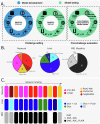
Fig. 1
Fig. 1. Overview of the ISLES’22 challenge and post-challenge experimental design, including the developed algorithmic solutions.
A Challenge and post-challenge phases and datasets. B Summary of algorithmic solutions stratified by network architecture, loss function, and input modalities. C Challenge leaderboard stratified by network architecture, loss function, and input modalities. CE Cross-entropy.
Fig. 2
Fig. 2. Test set performance metrics obtained by DeepISLES.
A Performance by imaging center. Data is grouped by the center where the images come from (#1, #2, or #3) and by a seen or unseen label indicating if images from the same center were used for training the models. B Performance by lesion size. C Performance by stroke phase (acute or sub-acute). D Performance by the stroke phase (acute or sub-acute) grouped by lesion size. E Performance by stroke pattern subgroups, including single vessel infarcts, scattered infarcts based on micro-occlusions, and single vessel infarcts with accompanying scattered infarcts. All boxplots are based on a sample size of N = 150. Boxes show the interquartile range (IQR; 25th–75th percentiles), the center line marks the median, whiskers span values within 1.5 × IQR, and points beyond are displayed as outliers. 5th, 50th, and 95th inter-rater variability percentiles are plotted in dashed lines for Dice and F1 score. SVI: single vessel infarct; SI: scattered infarcts based on micro-occlusions; SVI with SI: single vessel infarct with accompanying scattered infarcts. DSC Dice Similarity Coefficient; F1 score lesion-wise F1 score; AVD absolute volume difference; ALD absolute lesion count difference. y-axes are displayed using a non-linear scale to enhance data visibility. Source data are provided as a Source Data file.
Fig. 3
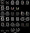
Fig. 3. Lesion ground truth (red) and DeepISLES predictions (green) for scans from the ISLES’22 test set.
Note that we selected the scans with median Dice scores (and not the best performing scans) to paint a realistic picture. Model outputs closely align with expert annotations across various types of stroke patterns and configurations. Results are grouped by healthcare center, imaging time, lesion size, stroke pattern, and vascular territory affected. MT mechanical thrombectomy; SVI single vessel infarct; SI scattered infarcts based on micro-occlusions. SVI with SI single vessel infarct with accompanying scattered infarcts. MCA middle cerebral artery; ACA anterior cerebral artery; PCA posterior cerebral artery.
Fig. 4
Fig. 4. Qualitative lesion segmentation results obtained in a Turing-like test.
Neuroradiologists prefer lesions delineated by DeepISLES over manual expert delineations (sample size N = 150). Score values range between 1 and 6 (worst and best quality scenarios, respectively). Boxes show the interquartile range (IQR; 25th-75th percentiles), the center line marks the median, whiskers span values within 1.5 × IQR, and points beyond are displayed as outliers.
Fig. 5
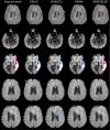
Fig. 5. Segmentation outcomes on the external Johns Hopkins dataset.
DeepISLES improves upon suboptimal segmentations generated by individual algorithmic approaches. Yellow arrows indicate false positives, while red circles highlight false negatives.
Fig. 6
Fig. 6. Algorithmic comparison on a subset of the Johns Hopkins dataset (sample size N = 417).
DeepISLES demonstrates exceptional generalizability outperforming DAGMNet, despite DAGMNet being specifically trained on the Johns Hopkins dataset. The inter-rater Dice line indicates the Dice coefficient obtained between manual delineations by two experts on a subset of scans (N = 220), as reported by Liu et al. AVD Absolute Volume Difference, ALD Absolute Lesion Count Difference. Boxes show the interquartile range (IQR; 25th–75th percentiles), the center line marks the median, whiskers span values within 1.5 × IQR, and points beyond are displayed as outliers. Source data are provided as a Source Data file.

References

    1. Czap, A. L. & Sheth, S. A. Overview of imaging modalities in stroke. Neurology97, S42–S51 (2021). - PMC - PubMed
    1. Goyal, M. et al. Challenging the ischemic core concept in acute ischemic stroke imaging. Stroke51, 3147–3155 (2020). - PubMed
    1. Giancardo, L. et al. Segmentation of acute stroke infarct core using image-level labels on CT-angiography. NeuroImage Clin.37, 103362 (2023). - PMC - PubMed
    1. Merino, J. G. & Warach, S. Imaging of acute stroke. Nat. Rev. Neurol.6, 560–571 (2010). - PubMed
    1. Chen, L., Bentley, P. & Rueckert, D. Fully automatic acute ischemic lesion segmentation in DWI using convolutional neural networks. NeuroImage Clin.15, 633–643 (2017). - PMC - PubMed

MeSH terms

LinkOut - more resources